 Detection of Leishmania DNA and identification of blood meal sources in sand flies (Psychodidae: Phlebotominae) from an Amazonian endemic area of cutaneous leishmaniasis
ABSTRACT
Cutaneous leishmaniasis is endemic in the state of Acre, western Amazonia, but natural vectors and reservoirs are poorly known. This study investigated Leishmania infection and blood meal sources in sand flies in the town of Sena Madureira. Sand flies were captured using CDC light and Shannon traps. Leishmania DNA was detected using PCR targeting hsp70; using PCR against cytb from engorged females’ blood samples. A total of 560 sand flies from 12 genera were collected, with Psychodopygus representing 56.6% of specimens.Leishmania DNA was detected in 12 samples, sequencing confirmed L. (Viannia) braziliensis infection in Nyssomyia fraihai and Psychodopygus hirsutus. Blood-meal testing identified cytb sequences matching human (5 flies), Tamandua tetradactyla (2 flies), and Cebus unicolor DNA (1 fly). This study provides new evidence ofLeishmaniacirculation in anthropic environments, highlighting potential risk for human infection and provides new methods for understanding leishmaniasis transmission.
Keywords:
cutaneous leishmaniasis; vectors; Leishmania sp.; wild animals
RESUMO
A leishmaniose cutânea é endêmica no estado do Acre, na Amazônia Ocidental, porém os vetores e reservatórios naturais ainda são pouco conhecidos. Este estudo investigou a infecção por Leishmania e as fontes de repasto sanguíneo em flebotomíneos no município de Sena Madureira. Os flebotomíneos foram capturados utilizando armadilhas luminosas CDC e armadilhas de Shannon. O DNA de Leishmania foi detectado por PCR direcionada ao gene hsp70; as fontes de sangue foram identificadas por PCR para o gene cytb a partir de amostras de fêmeas ingurgitadas. Ao todo, foram coletados 560 flebotomíneos pertencentes a 12 gêneros, sendo Psychodopygus responsável por 56,6% dos espécimes. O DNA de Leishmania foi detectado em 12 amostras, identificando L. (V.) braziliensis em Nyssomyia fraihai e Psychodopygus hirsutus. A análise de DNA mitocondrial em 12 flebotomíneos alimentados identificou sangue de Homo sapiens (n = 5), Tamandua tetradactyla (n = 2) e Cebus unicolor (n = 1). Este estudo fornece novas evidências da circulação de Leishmania em ambientes antrópicos, destacando o potencial risco de infecção humana, além de apresentar novas abordagens para a compreensão da transmissão da leishmaniose.
Palavras-chave:
leishmaniose cutânea; vetores; Leishmania sp.; animais silvestres
Cutaneous leishmaniasis (CL) is a neglected enzootic and zoonotic disease caused by protozoan parasites of the genus Leishmania and transmitted by sand flies. In Brazil, CL is a major public health concern in both rural and urban areas, where a variety of Leishmania species circulate among multiple vector species and vertebrate hosts (Brazil et al. 2015; Shaw 2025). In 2023, more than 50% of reported cases occurred in the Northern region of Brazil, with the state of Acre presenting the highest detection rate (691 cases per 100,000 inhabitants) and accounting for 76.9% of the cases reported in the region (Brasil 2024).
In Acre, several sand fly species are vectors of Leishmania (Viannia) braziliensis Vianna, 1911, L. (V.) guyanensis Floch, 1954, L. (V.) lainsoni Silveira et al., 1987, and L. (V.) shawi Lainson et al., 1989 (Ávila et al. 2018; Araujo-Pereira et al. 2020; Brilhante et al. 2022; Barroso et al. 2025). Continued study of phlebotomine sand flies, combined with Leishmania infection and blood meal analysis, is essential for guiding surveillance efforts in endemic areas (Shaw 2025). Thus, we investigated Leishmania infection and identified blood meal sources in sand flies collected from rural communities with confirmed human cases of CL in the municipality of Sena Madureira, Acre State, Brazil.
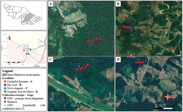
We collected sandflies in four rural locations, chosen based on accessibility, logistics, and reports of human infections: Cazumbá-Iracema - CI (09°08'30.8"S; 68°55'40.4"W; August 2022), Novo Amparo - NA (09°04'16.0"S; 68°27'02.0"W; March 2023), Joaquim José de Matos - JJM (09°23'35.6"S; 68°35'37.8"W; July 2023), and São José - SJ (09°22'28.6"S; 68°43'29.0"W; November 2023) (figure 1).
Geographic location with vegetation cover, of the sampling points in rural areas of the municipality of Sena Madureira, Acre State, Brazil.
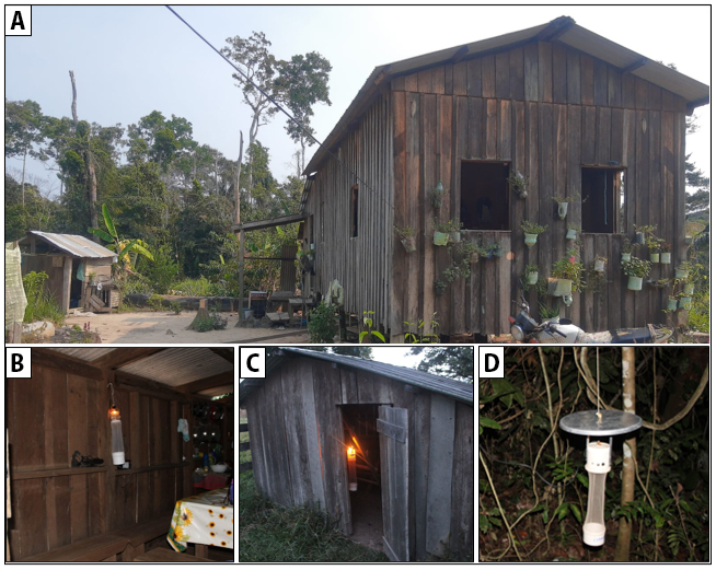
Sand flies were collected for two consecutive nights using CDC light traps (18:00-07:00) and a Shannon trap (18:00-20:00). On the first night, six CDC traps were installed in two houses with confirmed human CL cases (indoors, areas near animal shelters, and at the forest edge close to the households), along with a Shannon trap at the forest edge (figure 2). On the second night, five CDC traps and one Shannon trap were placed along trails in primary forest fragments.
Typical rural landscape in Acre State: (A) rural house showing the three ecotypes selected for trap placement; (B) intradomicile; (C) peridomicile area near domestic animal shelters; and (D) edge of forest fragment near residences.
At each locality, CDC sampling included 2 trap-nights (26 h) in each intradomicile, peridomicile, and forest-edge setting, and 5 trap-nights (65 h) in primary forest fragments. Shannon traps covered 2 trap-nights (4 h), one at the forest edge and one in primary forest fragments. Specimens were preserved in 70% ethanol, according to trapping localities and collection ecotype, and identified morphologically following Galati (2024).
Non-engorged females were tested individually when unique for a given species and collection site or grouped into pools of up to five specimens of the same species and locality when more than one individual was available. Engorged females were analyzed individually for blood meal identification and Leishmania infection detection.
DNA was extracted using the EXTRACTA®32 system with the Extracta® DNA/RNA Kit for Pathogens (MPTA-B01K). Leishmania DNA was detected by PCR targeting hsp70 (≈ 230 bp) (Graça et al. 2012), using L. (V.) guyanensis (MHOM/BR/1975/M4147; IOC/L0565) as positive control and Psychodopygus davisi (Root 1934) male sand flies and water as negative control. Blood meal sources were identified by PCR amplification and sequencing of a ~358-bp fragment of the mitochondrial cytb gene (Boakye et al. 1999), with Homo sapiens DNA as positive control and water as negative control. The IVS6 cacophony gene (≈220 bp) served as endogenous control in all reactions (Lins et al. 2006).
PCR reactions for hsp70 and cytb were performed in 50 µL containing 35 µL ultrapure water, 5.0 µL 10× buffer, 1.5 µL MgCl₂ (50 mM), 1 µL dNTPs (2 mM), 1 µL of each primer (10 pmol), 0.5 µL Taq DNA polymerase (5 U µL-1), and 5 µL extracted DNA. The IVS6 reaction was carried out in a final volume of 25 µL containing 17.5 µL ultrapure water, 2.5 µL 10× buffer, 0.75 µL MgCl₂ (50 mM), 0.5 µL dNTPs (2 mM), 0.5 µL of each primer (10 pmol), 0.25 µL Taq DNA polymerase (5 U µL-1), and 2.5 µL extracted DNA.
Amplifications were carried out in a CFX96 Touch thermocycler with initial denaturation at 94 °C (2 min), 35 cycles of 94 °C (30 s), 56 °C (45 s), 72 °C (45 s), and final extension at 72 °C (5 min). PCR products were visualized on 2% agarose gels, positive amplicons were purified (PureLink PCR Purification Kit) and Sanger sequenced (Fiocruz platform RTP01A). Data was analyzed in Geneious 2023.0.3, compared by BLAST, and deposited in GenBank (PV587120-PV587121). The study was authorized by the Instituto Chico Mendes de Conservação da Biodiversidade (ICMBio) (permit n. 68547-1) and approved by the IOC/Fiocruz Research Ethics Committee (CAAE: 57573822.2.0000.5248).
A total of 560 sand flies were collected, comprising 12 genera and 35 species (Table 1). Most were from JJM (23 species, 408 specimens) followed by CI (14 species, 68 specimens), SJ (13 species, 35 specimens) and NA (12 species, 49 specimens). Most specimens were in the genera Psychodopygus (56.6%), Trichophoromyia (14.3%) and Nyssomyia (12.0%). Two species were found indoors: Nyssomyia antunesi (Coutinho, 1939) and Pressatia sp. were found indoors in CI and SJ, respectively, and five species (Br. Pentacantha, Pr. choti, Ps. claustrei, Ps. davisi and Th. octavioi) were found in peridomiciliary areas. A total of 23 species were detected at the forest edge and 30 species in primary forest fragments.
Composition of the sand fly fauna in rural communities of the municipality of Sena Madureira, Acre, Brazil.
Twelve of the 420 females analyzed were engorged, from which we had 193 samples (12 individuals and 181 pools) that we submitted to PCR. Leishmania DNA testing was positive for 12 out of 193 samples (5.6%), with positives at all four sites, including forest edges (CI, SJ, NA), primary forest fragments, and peridomestic areas (JJM). Sequencing confirmed L. (V.) braziliensis in one engorged Ny. fraihai (Martins, Falcão & Silva, 1979) and one non-engorged Ps. hirsutus hirsutus (Mangabeira, 1942) (Table 2). Mitochondrial DNA testing of 12 blood-fed sand flies was positive for humans (Homo sapiens, n = 5), southern tamandua (Tamandua tetradactyla, n = 2), and capuchin monkey (Cebus unicolor, n = 1) (Table 3). The Ny. fraihai positive for L. braziliensis had fed on T. tetradactyla. Remaining sequences (hsp70 and cytb) had low quality or provided no database match.
Sand fly species that tested positive for Leishmania DNA in the municipality of Sena Madureira, Acre, Brazil.
Identification of blood meals in sand flies collected in the municipality of Sena Madureira, Acre, Brazil.
Among the 35 species recorded, Ps. carrerai (Barretto, 1946) and Ps. davisi predominated (27.5% of specimens), consistent with previous reports from the state (Araujo-Pereira et al. 2020; Ávila et al. 2018; Brilhante et al. 2022). Most specimens and over 50% of Leishmania infections were detected in forested areas, suggesting a predominantly sylvatic transmission cycle. Nevertheless, the presence of anthropophilic species in peridomestic environments suggests peridomestic transmission.
Previous studies have identified Ny. antunesi, Bi. flaviscutellata (Mangabeira 1942) Ps. hirsutus hirsutus, and Ps. ayrozai (Barretto & Coutinho 1940) as frequent species at forest edges and in primary forest, incriminating them as Leishmania vectors in the region (Ávila et al. 2018; Araujo-Pereira et al. 2020; Barroso et al. 2025). In the present study, Ps. hirsutus hirsutus from primary forest fragments tested positive for L. (V.) braziliensis, consistent with previous reports of this species in CL transmission in Acre. Ps. ayrozai was recorded both at the forest edge near households and in primary forest fragments, supporting its known association with zoonotic and enzootic transmission cycles in the Amazon, given its ecological generalist habitat and anthropo-opportunistic feeding behavior.
Although Ny. antunesi was uncommon, it was captured indoors, in forest edges, and in primary forest fragments, demonstrating ecological plasticity. Despite testing negative for Leishmania DNA in this study, previous research in Acre found L. (V.) braziliensis and L. (V.) guyanensis in this fly, implicating it in CL transmission (Ávila et al. 2018; Araújo-Pereira et al. 2020; Carneiro et al. 2023). Its frequent occurrence in forest fragments close to dwellings, including in Rio Branco, suggests adaptation to human-modified environments (Brilhante et al. 2022; Carneiro et al. 2023).
The detection of L. (V.) braziliensis in Ny. fraihai and Ps. hirsutus hirsutus, captured in primary forest fragments and forest edges, is a descriptive record of parasite detection in the sampled area. This species has also been reported in Acre in sand flies (Teles et al. 2016; Araujo-Pereira et al. 2020; Brilhante et al. 2021), human (Teles et al. 2015; Araujo-Pereira et al. 2018) and domestic dogs (Brilhante et al. 2019).
Blood meal analysis identified human DNA and DNA from two sylvatic species in phlebotomine species captured in primary forest fragments and forest edge, indicating human exposure across different ecotopes. Previous studies in the state have also reported avian species, multiple mammalian hosts, and mixed feeding involving H. sapiens and T. tetradactyla (Ávila et al. 2018; Araújo-Pereira et al. 2020; Barroso et al. 2025).
Ten samples positive for the hsp70 target could not be characterized due to low sequence quality, which may have led to underestimating Leishmania diversity. The use of pooled samples may also have influenced infection rate estimates, as it does not allow determination of individual infection status. The detection of L. (V.) braziliensis and the identification of blood meal sources demonstrate the efficacy of PCR-based methods for better characterizing parasite-host interactions, which is important for understanding disease dynamics.
ACKNOWLEDGEMENTS
The authors are thankful to Laboratório de Pesquisa e Diagnóstico Molecular em Doenças Infecciosas, Centro de Infectologia Charles Mérieux - CMIC/FUNDHACRE, Secretaria de Estado de Saúde do Acre - SESACRE, Núcleo de Gestão Integrada do Instituto Chico Mendes de Conservação da Biodiversidade - NGI/ICMBio de Sena Madureira, Acre, for their support in field and laboratory activities. This work was supported by the U.S. DoD Armed Forces Health Surveillance Division (AFHSD) and the Global Emerging Infections Surveillance Branch (ProMIS ID P0091_24_N6); Fundação Carlos Chagas Filho de Amparo à Pesquisa do Estado do Rio de Janeiro - FAPERJ (P.C. CNE E-26/200.487/2023 and R.P.B. APQ1 SEI-260003/006302/2024); Núcleo de Gestão Integrada do Instituto Chico Mendes de Conservação da Biodiversidade - NGI/ICMBio de Sena Madureira, Acre (SEI-02119.002027/2020-97); Coordenação de Aperfeiçoamento de Pessoal de Nível Superior - CAPES (L.S.S. nº 88887.636656/2021-00); and Fundação Oswaldo Cruz - FIOCRUZ -PAEF (E.C., P.C., L.C. grant No. IOC-023-FIO-18-2-63).
REFERENCES
- Araujo-Pereira, T.; Pereira, D.P.; Moreira, R.B.; Galdino, T.S.; Duarte, M.P.O.; Brazil, R.P.; et al. 2018. Molecular diagnosis of cutaneous leishmaniasis in an endemic area of Acre state in the Amazonian region of Brazil. Revista da Sociedade Brasileira de Medicina Tropical 51: 376-381.
- Araujo-Pereira, T.; Pita-Pereira, D.; Baia-Gomes, S.M.; Boité, M.; Silva, F.; Pinto, I.S.; et al. 2020. An overview of the sandfly fauna (Diptera: Psychodidae) followed by the detection of Leishmania DNA and blood meal identification in the state of Acre, Amazonian Brazil. Memórias do Instituto Oswaldo Cruz 115: e200157.
- Ávila, M.M.; Brilhante, A.F.; Souza, C.F.; Bevilacqua, P.D.; Galati, E.A.B.; Brazil, R.P. 2018. Ecology, feeding and natural infection by Leishmania spp. of phlebotomine sand flies in an area of high incidence of American tegumentary leishmaniasis in the municipality of Rio Branco, Acre, Brazil. Parasites & Vectors 11: 64.
- Barroso, E.P.; Souza, E.A.; Carneiro, A.C.G.; Ávila, M.M.; Júnior, Y.F.M.; Sousa, A.R.M.; et al. 2025. Sand fly fauna in urban parks in the Brazilian Western Amazon: Potential areas for American cutaneous leishmaniasis transmission. EcoHealth 22: 42-54.
- Boakye, D.A.; Tang, J.; Truc, P.; Merriweather, A.; Unnasch, T.R. 1999. Identification of bloodmeals in haematophagous Diptera by cytochrome B heteroduplex analysis. Medical and Veterinary Entomology 13: 282-287.
- Brazil, R.P.; Rodrigues, A.A.F.; Filho, J.D.A. 2015. Sand fly vectors of Leishmania in the Americas - a mini review. Entomology, Ornithology & Herpetology 4: 1000144.
- Brilhante, A.F.; Lima, L.; Ávila, M.M.; Medeiros-Sousa, A.R.; Souza, J.F.; Santos, N.P.; et al. 2021. Remarkable diversity, new records and Leishmania detection in the sand fly fauna of an area of high endemicity for cutaneous leishmaniasis in Acre State, Brazilian Amazonian Forest. Acta Tropica 223: 106103.
- Brilhante, A.F.; Lima, L.; Zampieri, R.A.; Nunes, V.L.B.; Dorval, M.E.C.; Malavazi, P.F.N.; et al. 2019. Leishmania (Viannia) braziliensis type 2 as probable etiological agent of canine cutaneous leishmaniasis in Brazilian Amazon. PLoS ONE 14: e0216291.
- Brilhante, A.F.; Zampieri, R.A.; Souza, E.A.D.; Carneiro, A.C.G.; Barroso, E.P.; Ávila, M.M.D.; et al. 2022. Preliminary observations of the urbanization and domiciliation of the American cutaneous leishmaniasis in Rio Branco, Acre, Western Amazon. Revista da Sociedade Brasileira de Medicina Tropical 55: e0359-2022.
- Carneiro, A.C.G.; Souza, E.A.; Barroso, E.P.; Ávila, M.M.; Melchior, L.A.K.; Rocha, R.D.C.; et al. 2023. Phlebotomine fauna (Diptera: Psychodidae) and infection by Leishmania spp. in forest fragments of a university campus, Western Amazon. Journal of Medical Entomology 60: 218-223.
- Galati, E.A.B. 2024. Phlebotominae (Diptera, Psychodidae): Classificação, morfologia, terminologia e identificação de adultos. 1 ed. São Paulo: Universidade de São Paulo. 140p.
- Graça, G.C.; Volpini, A.C.; Romero, G.A.; Neto, M.P.O.; Hueb, M.; Porrozzi, R.; et al. 2012. Development and validation of PCR-based assays for diagnosis of American cutaneous leishmaniasis and identification of the parasite species. Memórias do Instituto Oswaldo Cruz 107: 664-674.
- Lins, R.M.; Oliveira, S.G.; Souza, N.A.; Queiroz, R.G.; Justiniano, S.C.; Ward, R.D.; et al. 2002. Molecular evolution of the cacophony IVS6 region in sandflies. Insect Molecular Biology 11: 117-122.
- Shaw, J.J. 2025. A review of Leishmania infections in American phlebotomine sand flies - Are those that transmit leishmaniasis anthropophilic or anthropportunists? Parasite 32: 57.
-
Brasil. 2025. Painel epidemiológico da leishmaniose tegumentar americana (LTA). Ministério da Saúde. Available at: Available at: https://leishmanioses.aids.gov.br/ Accessed on 11 May 2025.
» https://leishmanioses.aids.gov.br/ - Teles, C.B.G.; Medeiros, J.F.; Santos, A.P.D.A.D.; Freitas, L.A.R.D.; Katsuragawa, T.H.; Cantanhede, L.M.; et al. 2015. Molecular characterization of American cutaneous leishmaniasis in the tri-border area of Assis Brasil, Acre State, Brazil. Revista do Instituto de Medicina Tropical de São Paulo 57: 343-347.
-
CITE AS:
Souza, L.S.; Brilhante, A.F.; Campelo, M.B.Z.; Cantanhêde, L.M.; Oliveira, J.E.; Nascimento, B.K.F.; et al. 2026. Detection of Leishmania DNA and identification of blood meal sources in sand flies (Psychodidae: Phlebotominae) from an Amazonian endemic area of cutaneous leishmaniasis. Acta Amazonica 56: e56hs26001.
Data availability
The supplementary material for this study can be accessed at: https://doi.org/10.5281/zenodo.18736895.