Echocardiographic Assessment During Treatment of Acquired Pulmonary Artery Stenosis Due to Mediastinal Mass Compression: A Case Report
Keywords
Pulmonary Artery Stenosis; Lymphoma; Dyspnea
Palavras-chave
Estenose de Artéria Pulmonar; Linfoma; Dispneia
Keywords
Pulmonary Artery Stenosis; Lymphoma; Dyspnea
Palavras-chave
Estenose de Artéria Pulmonar; Linfoma; Dispneia
Introduction
Acquired pulmonary artery stenosis is a rare entity that has scarcely been described in the literature, and it is mainly associated with compression of the pulmonary artery trunk and its branches by mediastinal tumors (teratomas and lymphomas).1 We report the case of a young patient with non-Hodgkin lymphoma in the anterior mediastinum that led to extrinsic compression and local luminal invasion of the pulmonary artery trunk, causing significant impact on the right heart chambers, with subsequent improvement after chemotherapy.
Case report
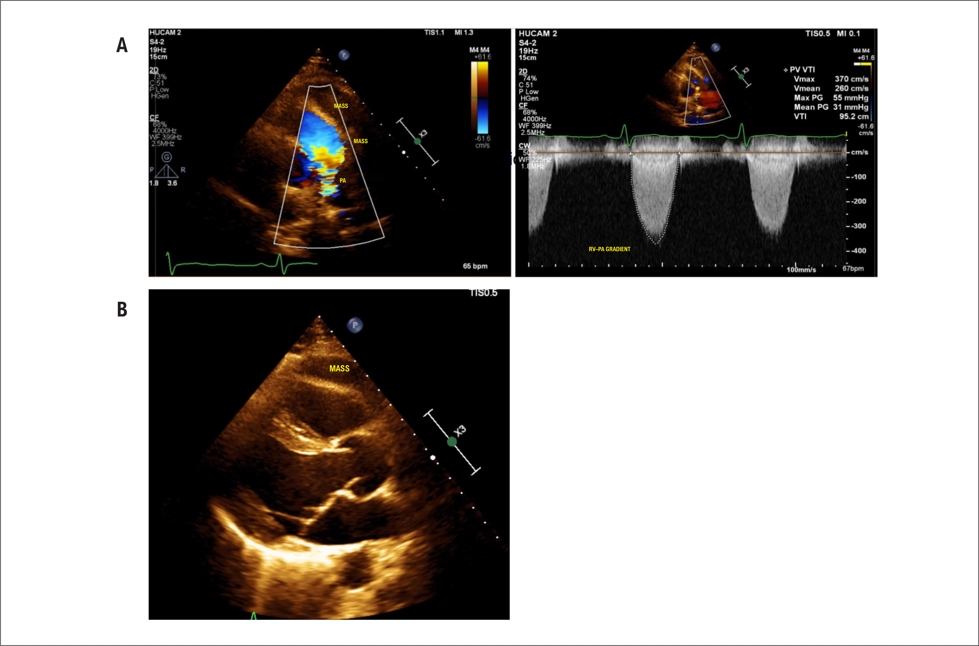
A 33-year-old male patient, without prior comorbidities, presented with cough, dyspnea, and the appearance of a nodular lesion in the anterior cervical region, which progressively enlarged, accompanied by hyperemia and a 15-kg weight loss over 1 year. Physical examination revealed a palpable, painless, and immobile mass approximately 4.5 cm above the suprasternal notch. Cardiac auscultation identified a systolic ejection murmur (3+/6+), audible in all areas and radiating to the suprasternal notch. Chest computed tomography angiography showed a mediastinal mass measuring 11.5 × 9.5 cm, compressing the pulmonary arterial trunk, supra-aortic arterial trunks, superior vena cava, left brachiocephalic vein, and right brachiocephalic vein, with signs suggestive of tumor thrombosis (Figure 1). Transthoracic echocardiography showed right ventricular enlargement and thickening with impaired contractile function, turbulent flow in the pulmonary artery trunk, and bifurcation of the pulmonary arteries, likely due to extrinsic compression or invasion by a mass originating from the anterior mediastinum. The peak right ventricle to pulmonary artery gradient was 79 mmHg, with a peak velocity of 4.4 m/s, and the pulmonary valve remained intact (Figure 2).
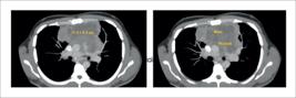
A) Parasternal short-axis view, showing the mass causing pulmonary stenosis. B) Parasternal long-axis view, showing the presence of a mass compressing the right ventricle. PA: pulmonary artery; RV: right ventricle.
Biopsy of the cervical mass identified an undifferentiated malignant neoplasm, and immunohistochemistry confirmed diffuse large B-cell non-Hodgkin lymphoma.
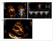
Chemotherapy was initiated, with clinical follow-up and transthoracic echocardiography every 3 months, showing progressive reduction of the mediastinal mass (Figure 3).1 After 9 months of treatment, the patient exhibited complete normalization of right ventricular function and gradients (Figure 4).

A) Mediastinal mass and pulmonary valve gradients showing reduction. B) Parasternal long-axis view, showing reduction of the mass in the mediastinum. PA: pulmonary artery; RV: right ventricle.
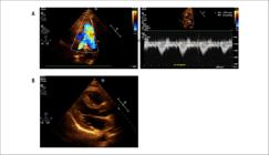
A) Maximum pulmonary valve gradient of 12 mmHg. B) Parasternal long-axis view, showing absence of the mass after treatment. PA: pulmonary artery; RV: right ventricle.
At the end of treatment, the patient developed severe febrile neutropenia, which was difficult to manage, and ultimately died of probable septic shock.
Discussion
Mediastinal lymphomas can involve large vessels, with significant hemodynamic obstructions capable of generating murmurs or symptoms, depending on the location of maximal tumor growth. In cases with cardiac involvement, the most common symptoms are chest pain, dyspnea, and cough, with an audible murmur observed in 81% of patients. Acquired pulmonary arterial stenosis is rare and strongly associated with mediastinal tumors, frequently Hodgkin lymphoma, with uncertain prognosis.2 Chronic obstructions may increase pressures in the right chambers and cause tricuspid regurgitation, ventricular dysfunction, and, in cases with patent foramen ovale, a right-to-left shunt with cyanosis and increased risk of paradoxical embolism.3 Transthoracic echocardiography plays a key role in defining the etiology of pulmonary stenosis.3 In the reported case, the mediastinal mass and its lateral compressive effect were clearly visible in short-axis view, with significant acceleration of flow in the pulmonary artery and normal valve opening, suggesting an external cause of the flow turbulence. Moreover, normalization of pulmonary artery flow and right ventricular function occurred after mass reduction with chemotherapy.
Conclusion
Early identification of this pathology, combined with timely initiation of therapy, can directly influence prognosis.4 Reduction of the mediastinal mass through chemotherapy, radiation therapy, or surgery can relieve compression of the pulmonary artery, leading to normalization of blood flow and improvement in right heart chamber function.5
-
Sources of Funding
There were no external funding sources for this study.
-
Study Association
This article is part of the habilitation thesis of Carolynne Ferreira Machado; Patrick Ventorim Costa, Ana Carolina Main Lucas; Laira Bernabe Mota; Karllayno Camatta Milleri; Fabrício Thebit Bortolon; and Fernando Luiz Torres Gomes at the Cassiano Antônio de Moraes University Hospital (HUCAM/UFES).
-
Ethics Approval and Consent to Participate
This study was approved by the Ethics Committee of the Hospital Universitário Cassiano Antônio de Moraes - HUCAM/UFES under the protocol number 88256725.1.0000.5071. All the procedures in this study were in accordance with the 1975 Helsinki Declaration, updated in 2013. Informed consent was obtained from all participants included in the study.
-
Use of Artificial Intelligence
The authors did not use any artificial intelligence tools in the development of this work.
Availability of Research Data
The underlying content of the research text is contained within the manuscript.
References
-
1 Robinson T, Lynch J, Grech E. Non-Hodgkin's Lymphoma Causing Extrinsic Pulmonary Artery Compression. Eur J Echocardiogr. 2008;9(4):577-8. doi: 10.1093/ejechocard/jen069.
» https://doi.org/10.1093/ejechocard/jen069 -
2 Marshall ME, Trump DL. Acquired Extrinsic Pulmonic Stenosis Caused by Mediastinal Tumors. Cancer. 1982;49(7):1496-9. doi: 10.1002/1097-0142 (19820401)49:7<1496::aid-cncr2820490731>3.0.co;2-3.
» https://doi.org/10.1002/1097-0142 -
3 Voss TH, Arantes FBB. Acquired Pulmonary Stenosis in an Adolescent with Lymphoma. Arq Bras Cardiol: Imagem Cardiovasc. 2022;35(3):eabc294. doi: 10.47593/2675-312X/20223503eabc294.
» https://doi.org/10.47593/2675-312X/20223503eabc294 - 4 Ozer N, Deveci OS, Kaya EB, Demircin M. Mediastinal Lymphoma Causing Extrinsic Pulmonary Stenosis. Turk Kardiyol Dern Ars. 2009;37(6):421-4.
-
5 Pugliatti P, Donato R, Grimaldi P, Nunnari F, Gregorio C, Zito C, et al. Extrinsic Pulmonary Stenosis in Primary Mediastinal B-Cellular Lymphoma. J Clin Ultrasound. 2015;43(1):68-70. doi: 10.1002/jcu.22133.
» https://doi.org/10.1002/jcu.22133
Edited by
-
Editor responsible for the review:
Andrea Vilela