Recurrent Intracardiac Masses in an Orthotopic Heart Transplant Recipient
Keywords
Heart Transplant; Left atrial; Heart Atria; Cardiac MRI; X-Ray Computed Tomography
Palavras-chave
Transplante de Coração; Átrios do Coração; Imageamento por Ressonância Magnética; Tomografia Computadorizada por Raios X
Keywords
Heart Transplant; Left atrial; Heart Atria; Cardiac MRI; X-Ray Computed Tomography
Palavras-chave
Transplante de Coração; Átrios do Coração; Imageamento por Ressonância Magnética; Tomografia Computadorizada por Raios X
Introduction
As long-term survival improves among heart transplant recipients, rare post-transplant complications, including intracardiac masses, are being increasingly recognized.1
Severe left atrial (LA) dilation and atrial arrhythmias may further contribute to blood stasis and thrombus formation in transplant recipients. LA thrombi in these patients may mimic neoplastic masses and represent important diagnostic and therapeutic challenges.2 Contributing factors include atrial dilation, arrhythmias, foreign material, and the components of Virchow's triad, namely abnormal blood flow, endothelial injury, and hypercoagulability.3,4
We report a unique case of a heart transplant recipient who developed recurrent LA thrombi over a 15-year period, requiring surgical resection, long-term anticoagulation, and complex therapeutic decision-making due to bleeding complications.
Case report
A 73-year-old man with a history of orthotopic heart transplantation (OHT) with bicaval anastomosis, performed in 2002 at an outside academic institution, established cardiovascular care at our institution in 2009. During outpatient follow-up, an incidental LA mass was identified on transthoracic echocardiography (TTE). The mass measured 5.5 × 5.1 × 4.3 cm and was located along the posterolateral wall of the LA. The patient was asymptomatic at the time of diagnosis. His medical history was significant for sick sinus syndrome requiring dual-chamber pacemaker implantation, nonobstructive coronary allograft vasculopathy, and monoclonal gammopathy of undetermined significance.
Annual TTE surveillance over the following 7 years demonstrated progressive enlargement of the mass, reaching a maximum size of 7.9 × 6.2 cm. Cardiac computed tomography (CCT) (Figure 1) confirmed the presence of two large LA masses. The first mass originated from the posterolateral wall, with partial calcification and extension through the atrial wall. The second mass arose from the roof of the LA.
CCT demonstrating two LA masses. The larger mass (blue arrow) measured 8 × 6 × 5 cm and exhibited smooth borders with layered calcification. The smaller mass (orange arrow) measured 6 × 5 × 3 cm and was attached to the superior and medial aspects of the left atrium.
After 7 years of imaging surveillance, the patient underwent redo sternotomy with surgical resection of both masses. Histopathological analysis of the first mass demonstrated fibrinopurulent debris with focal dystrophic calcifications, whereas the second mass was confirmed to be thrombotic material. Grocott methenamine silver, periodic acid-Schiff, and Gram staining were all negative. An old epicardial defibrillator pad was also identified, along with an associated thrombus within the pericardial space, which was surgically excised.
The patient was admitted with hypertensive urgency 1 year later. On presentation, blood pressure was 185/112 mmHg, heart rate was 88 beats/min, SpO2 was 98% on room air, and body temperature was normal. Electrocardiography demonstrated normal sinus rhythm with right bundle branch block. During hospitalization, repeat TTE demonstrated recurrence of a mass in the posterolateral LA measuring 3.7 × 4.1 cm. The lesion appeared homogeneous and broadly attached to the atrial wall, findings considered consistent with thrombus formation. Anticoagulation with warfarin was initiated, resulting in complete resolution of the mass 1 year later.
After 4 years of anticoagulation therapy, the patient developed persistent atrial flutter, which was initially managed medically. An additional 2 years later, he underwent electrical cardioversion. Shortly thereafter, he was hospitalized because of a large spontaneous right retroperitoneal hemorrhage and subsequently underwent right renal artery embolization, followed by discontinuation of warfarin therapy. His clinical course was further complicated by acute kidney injury requiring permanent dialysis.
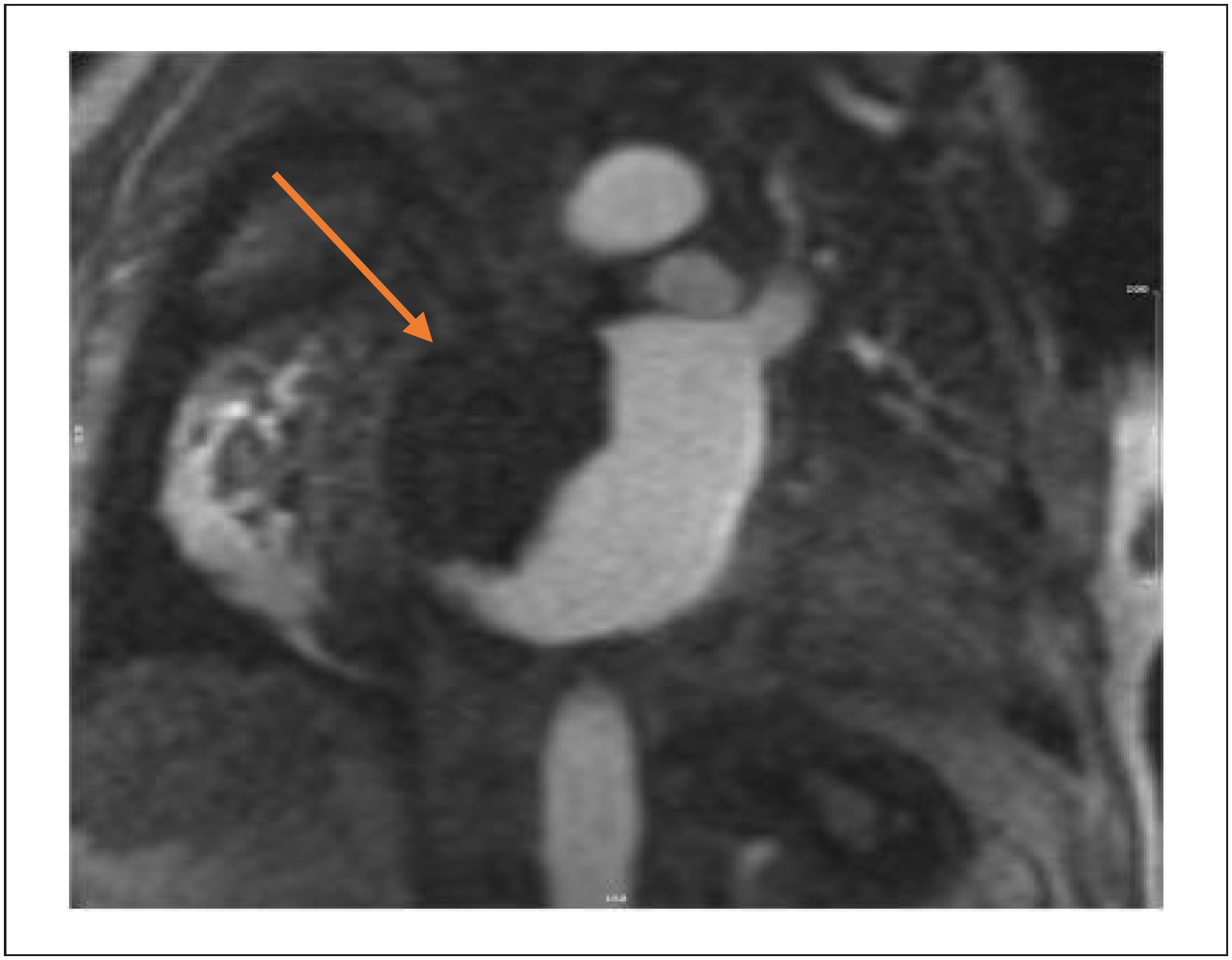
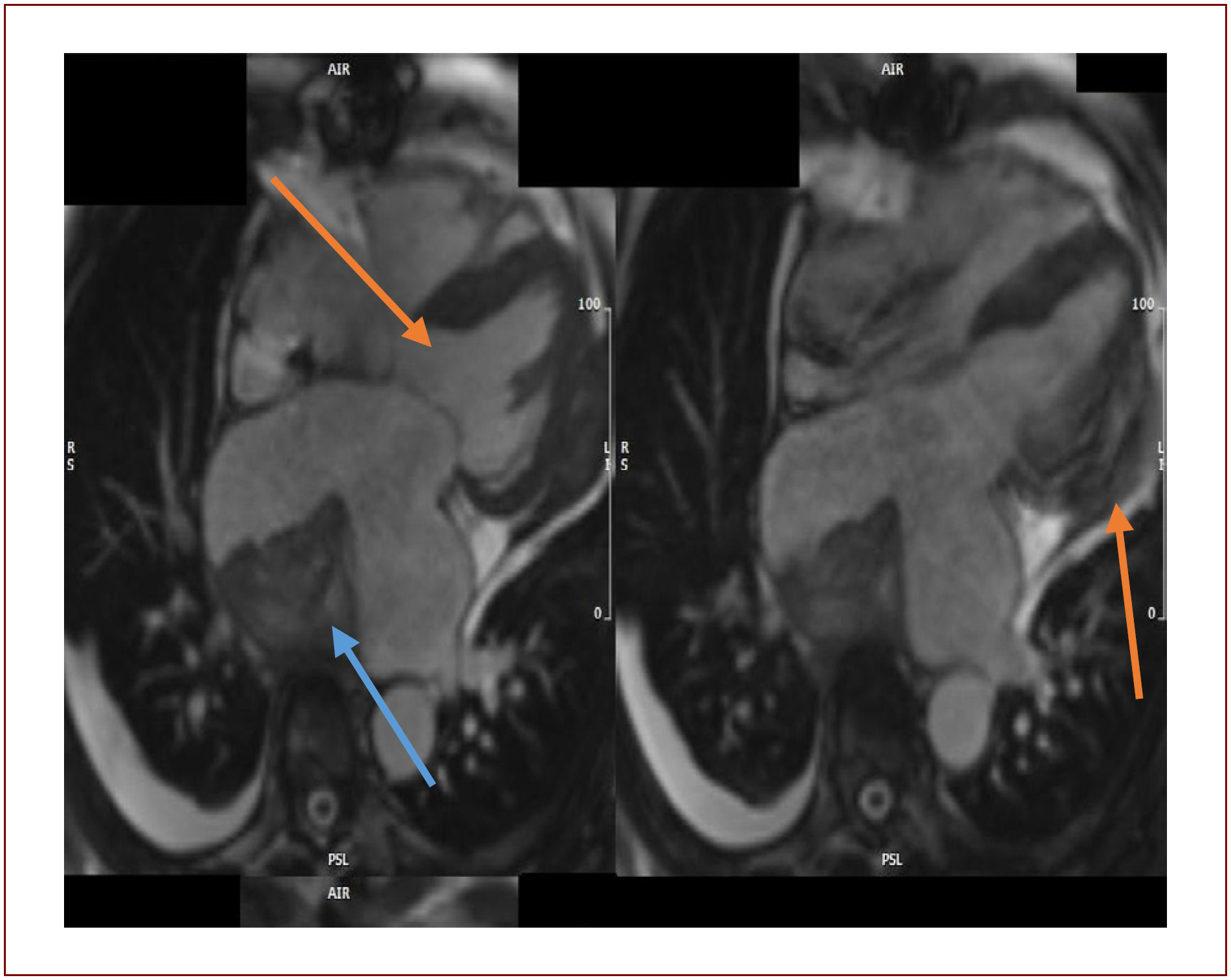
After hospital discharge, routine TTE demonstrated recurrence of a mass along the roof of the LA measuring 7.3 × 6.3 cm. Cardiac magnetic resonance (CMR) was subsequently performed (Figures 2 and 3), revealing severe LA dilation and a large heterogeneous broad-based mass attached to the LA roof, measuring 7.9 × 7.1 × 6.1 cm. A second mass was identified within the pericardial space adjacent to the lateral wall, measuring 4.0 × 1.2 × 3.0 cm. No enhancement was observed on first-pass perfusion or late gadolinium enhancement (LGE) imaging (Figure 4), findings that favored thrombi rather than neoplastic lesions. Consequently, warfarin therapy was resumed.
CMR first-pass perfusion imaging (two-chamber view) demonstrating a hypointense mass attached to the roof of the left atrium with a broad-based attachment and no contrast perfusion. The mass measured 7.9 × 7.1 × 6.1 cm.

CMR cine steady-state free precession sequence (four-chamber view) demonstrating a mass within the pericardial space (orange arrow) adjacent to the lateral wall of the left ventricle, measuring 6.3 × 2.0 cm, along with a large broad-based LA mass (blue arrow) measuring 7.9 × 7.1 × 6.1 cm and exhibiting heterogeneous signal intensity.

High-T1 LGE CMR (three-chamber view) demonstrating a hypointense LA mass with an etched appearance.
Following another 8 months, the patient experienced progressive clinical deterioration, including worsening mental status. He ultimately elected hospice care and died several weeks later. Autopsy was declined.
Discussion
Advances in immunosuppressive therapy and surgical techniques have substantially improved long-term survival after heart transplantation.5 Intracardiac masses remain a rare complication following OHT, and when present, they are typically identified within the first 1-2 years after surgery.1 The most common intracardiac masses in this population include organizing thrombi and primary cardiac tumors, particularly myxomas.2 However, differentiating among the various etiologies of intracardiac masses remains a significant diagnostic challenge and often requires a multimodality imaging approach.
The present case illustrates the unusual occurrence of recurrent thrombi in a patient who underwent OHT using the bicaval technique. Serial annual TTE examinations demonstrated progressive enlargement of the LA mass over several years before surgical resection. Additional imaging modalities, including CCT and CMR, played a critical role in anatomical characterization, as TTE alone could not reliably distinguish among thrombus, tumor, or foreign-body-associated lesions.
In this patient, the initial large LA mass containing fibrinopurulent debris and dystrophic calcification may have represented a reactive process related to the retained epicardial defibrillator pad, contributing to stagnant intra-atrial blood flow in accordance with Virchow's triad.3 Denudation of the extracellular matrix may promote conduction abnormalities, fibrosis, and endocardial infiltration, thereby facilitating thrombogenesis.4
The standard biatrial OHT technique, originally popularized by Shumway and colleagues because of its technical simplicity and shorter ischemic times, may result in anatomical and physiological alterations, including enlarged atrial chambers, blood stasis, atrial thrombosis, and valvular regurgitation. Consequently, the bicaval anastomotic technique was developed to better preserve atrial geometry, reduce atrial arrhythmias, and minimize asynchronous contraction between donor and recipient atrial tissue, all of which may contribute to thrombus formation.6 Despite these theoretical advantages, our patient developed recurrent LA and pericardial thrombi even after removal of the retained surgical material and epicardial defibrillator pad.
Only a limited number of cases describing atrial thrombi confirmed by surgical resection and histopathological examination have been reported in patients undergoing bicaval OHT.2,7-9
CMR is particularly valuable for differentiating thrombus from cardiac tumors through tissue characterization. Imaging features favoring thrombus include absence of first-pass perfusion, lack of LGE, low signal intensity on delayed enhancement sequences, and the presence of a layered or "etched" appearance.10-12
Conclusion
We report a rare case of recurrent LA thrombi in an asymptomatic patient who underwent bicaval OHT. The mass was incidentally detected and demonstrated progressive enlargement over a 7-year period. Multimodality imaging, particularly CCT and CMR, was essential for diagnostic assessment, as TTE alone could not reliably differentiate thrombus from neoplasm or foreign-body-associated lesions.
The initial mass may have been associated with a retained epicardial defibrillator pad, which likely contributed to blood stasis and thrombus formation. Despite surgical resection, thrombus recurrence occurred within months, highlighting the persistent thrombotic risk in this population, even in the absence of atrial arrhythmias.
This case emphasizes the importance of multimodality imaging, histopathological confirmation when feasible, individualized anticoagulation strategies, and long-term surveillance in heart transplant recipients with intracardiac masses. Further studies are needed to better define thrombotic risk factors and optimal anticoagulation duration following bicaval OHT.
-
Sources of Funding
There were no external funding sources for this study.
-
Study Association
This study is not associated with any thesis or dissertation work.
-
Ethics Approval and Consent to Participate
This article does not contain any studies with human participants or animals performed by any of the authors.
-
Use of Artificial Intelligence
The authors did not use any artificial intelligence tools in the development of this work.
Availability of Research Data
The underlying content of the research text is contained within the manuscript.
References
-
1 Velleca A, Shullo MA, Dhital K, Azeka E, Colvin M, DePasquale E, et al. The International Society for Heart and Lung Transplantation (ISHLT) Guidelines for the Care of Heart Transplant Recipients. J Heart Lung Transplant. 2023;42(5):e1-e141. doi: 10.1016/j.healun.2022.10.015.
» https://doi.org/10.1016/j.healun.2022.10.015 -
2 Hale A, Vann J, Henderson P, Harrison T, Trehan S. A Case of a Left Atrial Mass in an Orthotopic Heart Transplant Recipient. CASE. 2019;4(1):33-8. doi: 10.1016/j.case.2019.10.011.
» https://doi.org/10.1016/j.case.2019.10.011 -
3 Lowe GD. Virchow's Triad Revisited: Abnormal Flow. Pathophysiol Haemost Thromb. 2003;33(5):455-7. doi: 10.1159/000083845.
» https://doi.org/10.1159/000083845 -
4 Yamashita T. Virchow Triad and Beyond in Atrial Fibrillation. Heart Rhythm. 2016;13(12):2377-8. doi: 10.1016/j.hrthm.2016.09.007.
» https://doi.org/10.1016/j.hrthm.2016.09.007 -
5 Lund LH, Khush KK, Cherikh WS, Goldfarb S, Kucheryavaya AY, Levvey BJ, et al. The Registry of the International Society for Heart and Lung Transplantation: Thirty-fourth Adult Heart Transplantation Report-2017; Focus Theme: Allograft Ischemic Time. J Heart Lung Transplant. 2017;36(10):1037-46. doi: 10.1016/j.healun.2017.07.019.
» https://doi.org/10.1016/j.healun.2017.07.019 -
6 Dell’Aquila AM, Mastrobuoni S, Bastarrika G, Praschker BL, Agüero PA, Castaño S, et al. Bicaval versus Standard Technique in Orthotopic Heart Transplant: Assessment of Atrial Performance at Magnetic Resonance and Transthoracic Echocardiography. Interact Cardiovasc Thorac Surg. 2012;14(4):457-62. doi: 10.1093/icvts/ivr084.
» https://doi.org/10.1093/icvts/ivr084 -
7 Neuman Y, Tolstrup K, Blanche C, Luthringer D, Kobal S, Miyamoto T, et al. Pseudomyxoma Originating from the Interatrial Septum in a Heart Transplant Patient. J Am Soc Echocardiogr. 2005;18(7):e1. doi: 10.1016/j.echo.2004.09.014.
» https://doi.org/10.1016/j.echo.2004.09.014 -
8 Yousefzai R, Trivedi S, Jain R, Cheema OM, Crouch JD, Thohan V, et al. Expecting the Unexpected: Right Atrial Mass in a Transplant Patient. ESC Heart Fail. 2015;2(4):164-7. doi: 10.1002/ehf2.12065.
» https://doi.org/10.1002/ehf2.12065 -
9 Bartus K, Litwinowicz R, Kapelak B, Filip G, Wierzbicki K, Lee RJ. Giant Left Atrium Associated with Massive Thrombus Formation 14 Years after Orthotopic Heart Transplantation. Braz J Cardiovasc Surg. 2020;35(6):1010-2. doi: 10.21470/1678-9741-2018-0390.
» https://doi.org/10.21470/1678-9741-2018-0390 -
10 Motwani M, Kidambi A, Herzog BA, Uddin A, Greenwood JP, Plein S. MR Imaging of Cardiac Tumors and Masses: A Review of Methods and Clinical Applications. Radiology. 2013;268(1):26-43. doi: 10.1148/radiol.13121239.
» https://doi.org/10.1148/radiol.13121239 -
11 Weinsaft JW, Kim HW, Shah DJ, Klem I, Crowley AL, Brosnan R, et al. Detection of Left Ventricular Thrombus by Delayed-Enhancement Cardiovascular Magnetic Resonance Prevalence and Markers in Patients with Systolic Dysfunction. J Am Coll Cardiol. 2008;52(2):148-57. doi: 10.1016/j.jacc.2008.03.041.
» https://doi.org/10.1016/j.jacc.2008.03.041 -
12 Araoz PA, Eklund HE, Welch TJ, Breen JF. CT and MR Imaging of Primary Cardiac Malignancies. Radiographics. 1999;19(6):1421-34. doi: 10.1148/radiographics.19.6.g99no031421.
» https://doi.org/10.1148/radiographics.19.6.g99no031421
Edited by
-
Editor responsible for the review:
Marcelo Tavares