Successful Occlusion of a Giant Subclavian Artery Aneurysm Sac with a Vascular Plug: A Case Report
Abstract
Subclavian artery aneurysms are rare vascular anomalies that carry significant risks, including thromboembolism, ischemia, and rupture. This case report describes a 71-year-old female patient presenting with left arm ischemia, including pain, numbness, and weakness, caused by a left subclavian artery aneurysm complicated by distal thrombosis. Imaging revealed a 65 × 55 mm aneurysm with a 2.5 cm thrombotic occlusion. The aneurysm was treated with an endovascular approach, utilizing a vascular plug deployed via femoral access. This was preceded by failed attempts to access the lesion through the radial approach due to the presence of thrombotic occlusion and complex vascular anatomy. After the procedure, the patient experienced significant symptom relief, and follow-up imaging confirmed thrombosis of the aneurysm sac. This case highlights the effectiveness of endovascular techniques in managing complex subclavian artery aneurysms and emphasizes the importance of individualized treatment strategies and close follow-up.
Keywords
Subclavian Artery; Endovascular Aneurysm Repair; Thrombosis
Resumo
Aneurismas da artéria subclávia são anomalias vasculares raras que apresentam riscos significativos, incluindo tromboembolismo, isquemia e ruptura. O presente relato de caso descreve uma paciente do sexo feminino de 71 anos de idade que apresentou isquemia no braço esquerdo, incluindo dor, dormência e fraqueza, causada por um aneurisma da artéria subclávia esquerda complicado por trombose distal. A imagem revelou um aneurisma de 65 × 55 mm com uma oclusão trombótica de 2,5 cm. O aneurisma foi tratado com uma abordagem endovascular, utilizando um plugue vascular implantado via acesso femoral. Isso foi precedido por tentativas frustradas de acessar a lesão pela abordagem radial devido à presença de oclusão trombótica e anatomia vascular complexa. Após o procedimento, a paciente apresentou alívio significativo dos sintomas, e a imagem de acompanhamento confirmou a trombose do saco aneurismático. Este caso destaca a eficácia das técnicas endovasculares no manejo de aneurismas complexos da artéria subclávia e enfatiza a importância de estratégias de tratamento individualizadas e acompanhamento rigoroso.
Palavras-chave
Artéria Subclávia; Correção Endovascular de Aneurisma; Trombose
Introduction
Subclavian artery aneurysms are rare, accounting for less than 1% of peripheral artery aneurysms.1 They may be associated with trauma, atherosclerosis, thoracic outlet syndrome, or congenital abnormalities.2 While many remain asymptomatic, symptomatic cases can present with ischemic symptoms due to arterial compression, thromboembolism, or aneurysmal rupture.3 These symptoms often include pain, numbness, weakness, or diminished pulses in the affected arm, along with differential blood pressures between the upper limbs. Given the risk of severe complications such as embolization or rupture, early diagnosis and treatment are crucial. Endovascular repair has emerged as the preferred treatment for subclavian artery aneurysms, offering a minimally invasive alternative to open surgery with lower perioperative risk.4 However, challenges may arise, particularly when aneurysms are complicated by thrombotic occlusion or extensive collateralization. This report presents a 71-year-old woman with symptomatic left arm ischemia caused by a subclavian artery aneurysm with distal thrombosis, successfully treated with endovascular intervention.
Case report
A 71-year-old woman with hypertension presented with pain, numbness, and weakness in her left arm, which had worsened over the past few months. Her heart rate was 80 beats per minute, regular, with no murmurs. Electrocardiogram showed sinus rhythm and incomplete right bundle branch block. Transthoracic echocardiography revealed a left ventricular ejection fraction of 60%, interventricular septal thickness of 12 mm, stage 1 diastolic dysfunction, and mild aortic regurgitation. Physical examination revealed a weak right radial pulse. Blood pressure was 130/70 mmHg in the right arm and 105/65 mmHg in the left arm. A posterior-anterior chest X-ray showed a well-circumscribed round mass in the left hemithorax ( Figure 1 ). Subsequent computed tomographic angiography (CTA) of the thoracic aorta and upper extremities revealed a 65 × 55 mm aneurysm of the left subclavian artery, located just distal to the origin of the vertebral artery. The occluded segment, measuring approximately 2.5 cm, exhibited non-calcified thrombotic material filling the lumen, without evidence of significant extrinsic compression or surrounding soft tissue abnormalities. The mechanism of occlusion appeared to be thrombus formation within the aneurysmal sac, likely due to stasis of blood flow. The aneurysm's etiology was suspected to be degenerative in nature, potentially related to chronic hypertension and atherosclerotic changes, as no history of trauma or congenital abnormality was reported. Collateral vessels reconstructed the left axillary artery, and both the left vertebral artery and the left internal mammary artery (LIMA) demonstrated normal flow ( Figure 2 ). Endovascular repair was planned using stent-graft placement. Access was obtained via the right femoral artery and the left radial artery. However, despite advanced guidewires, the aneurysm could not be reached from the radial approach. An Amplatzer Vascular Plug II (AVP II; Abbott, Plymouth, MN, USA), measuring 10 mm in diameter, was deployed in the left subclavian artery near the aneurysm via the femoral route. The device was delivered using a 6 French guiding catheter, which provided sufficient support for precise positioning of the vascular plug, while preserving access to the left vertebral artery and LIMA. Post-procedural angiography confirmed cessation of aneurysmal flow with preserved flow in the left vertebral artery and LIMA ( Figure 3 ). Following the procedure, the patient was initiated on dual antiplatelet therapy (aspirin 100 mg daily and clopidogrel 75 mg daily) to prevent thromboembolic complications associated with the vascular plug and to maintain patency of critical collateral vessels. The antithrombotic strategy was selected based on the patient's risk profile and the nature of the intervention. At a 1-month follow-up, the patient reported significant improvement in her left arm symptoms, and repeat CTA demonstrated complete thrombosis of the aneurysm sac with cessation of flow ( Figure 4 ). Continued follow-up at 3 and 6 months, including clinical evaluations and imaging, was scheduled to monitor for complications such as migration of the vascular plug, residual flow, or recurrence of ischemic symptoms.
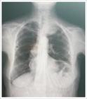
Posterior-anterior chest X-ray showing a well-circumscribed round mass in the left hemithorax.
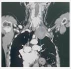
Computed tomography angiography of the thoracic aorta and upper extremities shows a left subclavian artery aneurysm distal to the origin of the vertebral artery.
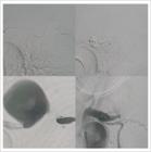
A Destination sheath and vertebral catheter were positioned in the proximal subclavian artery via the femoral artery, while a right Judkins catheter was advanced into the distal subclavian artery through the radial approach (top left). Despite using stiff guidewires, access to the aneurysmal region from the distal end was unsuccessful (bottom left). A 10 mm vascular plug was placed proximal to the aneurysm sac through the vertebral catheter via the femoral route (top right). Post-procedural cine angiography confirmed preserved flow in both the left vertebral artery and the LIMA, with near-complete occlusion of flow within the aneurysm sac (bottom right).

Computed tomography angiography performed 1 month after vascular plug placement in the subclavian artery revealed complete cessation of flow within the aneurysm sac.
Discussion
Subclavian artery aneurysms, although rare, carry significant risks, including embolization, thrombosis, and rupture.5 In this case, the patient presented with upper extremity ischemia, which is typical of symptomatic subclavian aneurysms, manifesting as pain, numbness, weakness, and differential blood pressure. These clinical findings were pivotal in diagnosing a left subclavian artery aneurysm with thrombotic occlusion. Endovascular repair is increasingly preferred due to its minimally invasive nature, reduced morbidity, and faster recovery compared to open surgery.6 However, this case illustrates the technical difficulties that can arise, particularly in navigating complex vascular anatomy complicated by thrombosis and collateralization. In this case, despite initial challenges accessing the aneurysm via the radial route, a vascular plug was successfully deployed via the femoral approach, achieving cessation of aneurysmal flow while preserving perfusion in critical vessels such as the left vertebral artery and LIMA. The patient's favorable outcome, with significant symptom improvement and imaging-confirmed thrombosis of the aneurysm, underscores the efficacy of endovascular management. This case also highlights the importance of thorough imaging assessment and individualized intervention strategies for complex vascular lesions.7 While the outcome in this case was successful, it serves as a reminder of the potential complications of subclavian artery aneurysms and the need for timely diagnosis and intervention to prevent more severe consequences, such as rupture or embolization. Continued follow-up is essential to monitor for recurrence or complications related to the vascular plug.
Conclusion
This case demonstrates the successful endovascular treatment of a rare subclavian artery aneurysm complicated by thrombosis and presenting with upper extremity ischemia. Despite procedural challenges, the deployment of a vascular plug effectively halted aneurysmal flow while preserving critical vessel patency. Early diagnosis, tailored intervention, and ongoing follow-up are crucial to achieving optimal outcomes in such complex cases.
-
Sources of Funding
There were no external funding sources for this study.
-
Study Association
This study is not associated with any thesis or dissertation work.
-
Ethics Approval and Consent to Participate
This article does not contain any studies with human participants or animals performed by any of the authors.
References
-
1 Counts S, Zeeshan A, Elefteriades J. Giant Subclavian Artery Aneurysm. Int J Angiol. 2016;25(2):128-30. doi: 10.1055/s-0034-1372245.
» https://doi.org/10.1055/s-0034-1372245 -
2 Maślanka K, Zielinska N, Karauda P, Balcerzak A, Georgiev G, Borowski A, et al. Congenital, Acquired, and Trauma-Related Risk Factors for Thoracic Outlet Syndrome-Review of the Literature. J Clin Med. 2023;12(21):6811. doi: 10.3390/jcm12216811.
» https://doi.org/10.3390/jcm12216811 -
3 Dougherty MJ, Calligaro KD, Savarese RP, Laurentis DA. Atherosclerotic Aneurysm of the Intrathoracic Subclavian Artery: A Case Report and Review of the Literature. J Vasc Surg. 1995;21(3):521-9. doi: 10.1016/s0741-5214(95)70297-0.
» https://doi.org/10.1016/s0741-5214(95)70297-0 -
4 Zha B, Chen Z, Ou-Yang H. Endovascular Repair for Giant Right Subclavian Artery Aneurysm with Gore Viabahn and Pull-Through Procedure: A Case Report. Front Surg. 2022;9:939818. doi: 10.3389/fsurg.2022.939818.
» https://doi.org/10.3389/fsurg.2022.939818 -
5 Banning SK, Ur R, Malleis J, Hamlat CA, Byers PH, Shalhub S. Extrathoracic Subclavian Artery Aneurysm in a Patient with Suspected Genetic Arteriopathy. J Vasc Surg Cases Innov Tech. 2020;7(1):46-50. doi: 10.1016/j.jvscit.2020.11.008.
» https://doi.org/10.1016/j.jvscit.2020.11.008 -
6 Daye D, Walker TG. Complications of Endovascular Aneurysm Repair of the Thoracic and Abdominal Aorta: Evaluation and Management. Cardiovasc Diagn Ther. 2018;8(Suppl 1):S138-S156. doi: 10.21037/cdt.2017.09.17.
» https://doi.org/10.21037/cdt.2017.09.17 -
7 Christie A, Roditi G. Vascular Imaging: The Evolving Role of the Multidisciplinary Team Meeting in Peripheral Vascular Disease. Semin Intervent Radiol. 2014;31(4):320-9. doi: 10.1055/s-0034-1393968.
» https://doi.org/10.1055/s-0034-1393968
Edited by
-
Editor responsible for the review:
Tiago Magalhães