My Approach to Echocardiographic Assessment of Left Ventricular Filling Pressures: From Ambiguity to Precision With New Guidelines
Abstract
The echocardiographic estimation of left ventricular (LV) filling pressures is a cornerstone in the assessment of heart failure, particularly in patients with heart failure with preserved ejection fraction (HFpEF). The 2016 American Society of Echocardiography/European Association of cardiovascular Imaging algorithm standardized this evaluation using key variables, but a substantial proportion of cases remained indeterminate in clinical practice. The recent update of the American guideline reorganized the approach into a hierarchical framework, incorporating age-related adjustments and formalizing the role of left atrial strain (LAS) as a tie-breaking parameter, while also providing a more detailed characterization of special clinical scenarios. In parallel, the 2024 British Society of Echocardiography guideline emphasizes the pathophysiological interpretation of ventricular filling, complementing the operational framework proposed by the American document. Multicenter studies with invasive validation support these updates, establishing LAS as a robust marker of increased filling pressures. In this article, we present My Approach to echocardiographic assessment of LV filling pressures, based on an initial morphofunctional evaluation, followed by structured screening (Step 1) and further refinement (Step 2) incorporating LAS and additional parameters. We also provide a comparison between guidelines, discuss common pitfalls and algorithm limitations, and include case-based videos for practical application.
Keywords
Left Ventricular Function; Echocardiography; Heart Failure
Resumo
A estimativa ecocardiográfica das pressões de enchimento do ventrículo esquerdo (VE) é um pilar na avaliação da insuficiência cardíaca, particularmente em pacientes com insuficiência cardíaca com fração de ejeção preservada (ICFEp). O algoritmo da American Society of Echocardiography (ASE)/European Association of Cardiovascular Imaging (EACVI) de 2016 padronizou essa avaliação utilizando variáveis-chave, mas uma proporção substancial de casos permaneceu indeterminada na prática clínica. A atualização recente da diretriz americana reorganizou a abordagem em um modelo hierárquico, incorporando ajustes relacionados à idade e formalizando o papel do strain atrial esquerdo (SAE) como parâmetro de desempate, além de fornecer uma caracterização mais detalhada de cenários clínicos especiais. Em paralelo, a diretriz de 2024 da British Society of Echocardiography enfatiza a interpretação fisiopatológica do enchimento ventricular, complementando o modelo operacional proposto pelo documento americano. Estudos multicêntricos com validação invasiva sustentam essas atualizações, estabelecendo o SAE como um marcador robusto de pressões de enchimento elevadas. Neste artigo, apresentamos Como Eu Faço a avaliação ecocardiográfica das pressões de enchimento do VE, baseada em uma avaliação morfofuncional inicial, seguida por uma triagem estruturada (Etapa 1) e refinamento adicional (Etapa 2), incorporando o SAE e parâmetros adicionais. Também apresentamos uma comparação entre diretrizes, discutimos armadilhas comuns e limitações dos algoritmos, e incluímos vídeos baseados em casos para aplicação prática.
Palavras-chave
Função Ventricular Esquerda; Ecocardiografia; Insuficiência Cardíaca
Introduction
The echocardiographic assessment of left ventricular (LV) filling pressures has evolved from a mitral Doppler-centered approach to an integrated model incorporating both morphological and functional parameters, supported by increasingly structured algorithms. The 2016 American Society of Echocardiography (ASE)/European Association of cardiovascular Imaging (EACVI) guideline for assessing LV diastolic function by echocardiography marked a pivotal step in this transition by proposing a simplified framework based on four core variables;1 however, a substantial proportion of examinations remained indeterminate with respect to filling pressure estimation. This limits diagnostic accuracy, particularly in conditions such as heart failure with preserved ejection fraction (HFpEF).
In addition, specific clinical conditions (e.g., atrial fibrillation [AF], pulmonary hypertension, and valvular heart disease) have continued to pose challenges to the applicability and interpretation of these parameters.2
Recent updates have advanced this field along two complementary directions. The 2024 British Society of Echocardiography guideline for assessing LV diastolic function3 reinforces the pathophysiological basis of ventricular filling and formalizes left atrial strain (LAS) as a refinement tool. In parallel, the ASE 2025 update reorganizes the decision-making process, incorporates age-related adjustments, and integrates atrial strain in a similar manner,4 supported by multicenter studies with invasive validation (Table 1).5
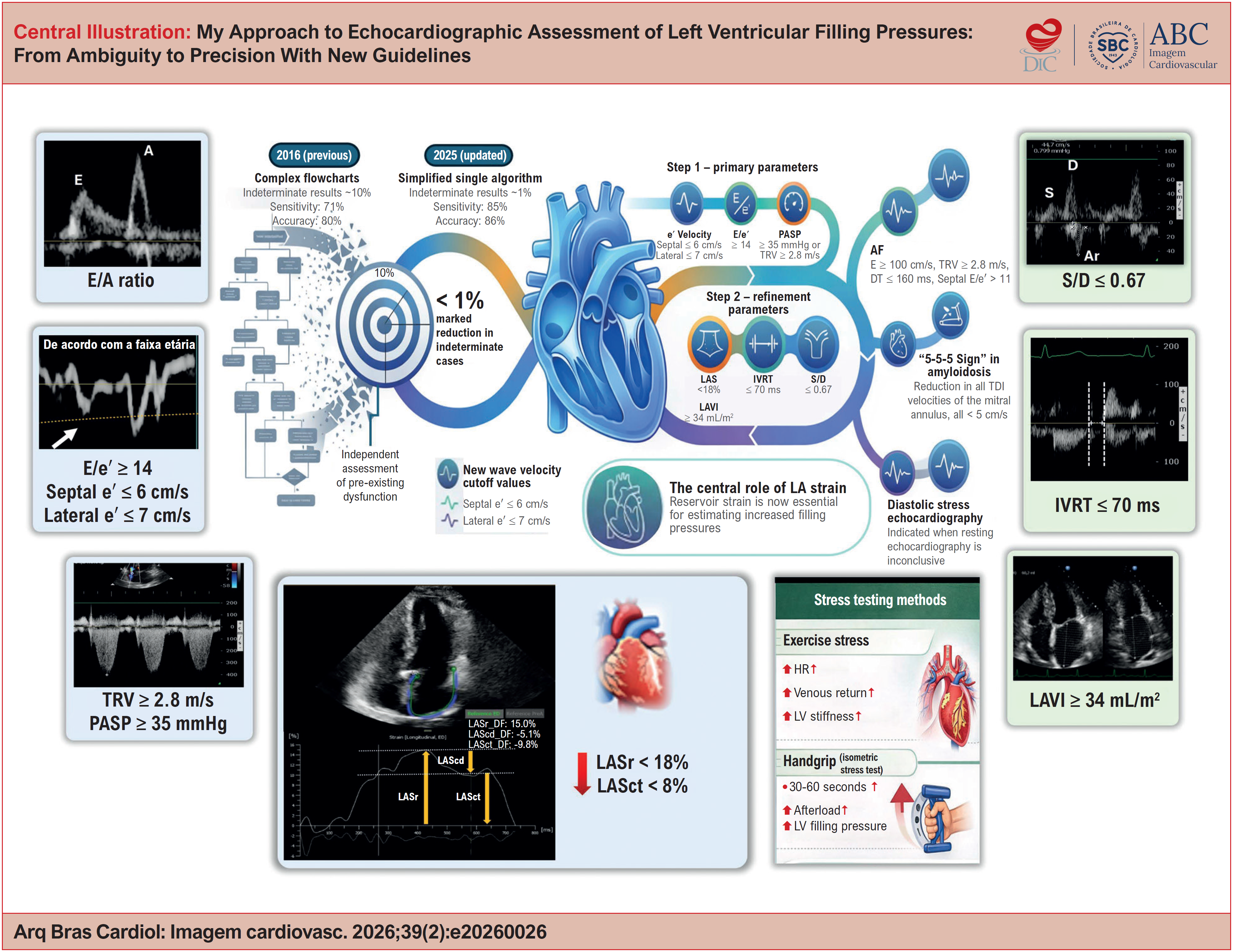
This article translates these advances into daily clinical practice through My Approach to echocardiographic assessment of LV filling pressures, an approach designed to be practical, reproducible, and clinically meaningful while preserving a strong physiological foundation (Central Illustration).
Integration and hierarchical application of parameters and techniques for the evaluation of LV filling pressures. The strategy begins with morphofunctional assessment and structured screening, followed by targeted refinement in indeterminate or discordant cases (Step 2), in which LAS and complementary parameters play a decisive role in the final diagnostic classification. AF: atrial fibrillation; DT: deceleration time; ED: end-diastole; HR: heart rate; IVRT: isovolumetric relaxation time; LAS: left atrial strain; LAScd: left atrial conduit strain; LASct: left atrial contraction strain; LASr: left atrial reservoir strain; LAVI: left atrial volume index; LV: left ventricular; PASP: pulmonary artery systolic pressure; TDI: tissue Doppler imaging; TRV: tricuspid regurgitation velocity.
Before the algorithm: the integrated view of the echocardiographer
The assessment of diastolic function should always begin with clinical contextualization (e.g., age, symptoms, cardiac rhythm, valvular heart disease, and hemodynamic status) because the algorithm addresses a specific clinical question and should not be applied indiscriminately.3,4
Before applying any flowchart, a two-dimensional morphofunctional assessment allows the echocardiographer to rapidly integrate patterns of hypertrophy, LA size, ventricular geometry, mitral annular motion, signs of pulmonary hypertension, and the overall visual impression of ventricular compliance. This approach enables the clinician to position the patient within a probable phenotype: low, intermediate, or high likelihood of increased filling pressures.
This initial impression does not replace the algorithm; rather, it prevents both the mechanical application of numerical thresholds and conclusions drawn without objective criteria. It serves as a conceptual framework that guides interpretation and may be refined or corrected by the structured model.
Strategy overview: two steps, one clinical question
The assessment should be structured to answer a simple and clinically meaningful question: Is there consistent evidence of increased LV filling pressures? Table 2 summarizes the parameters used for this evaluation.
Step 1: initial screening
Updated guidelines emphasize that aging is associated with a physiological decline in myocardial relaxation. As a result, values considered abnormal in younger individuals may be expected in older people. Therefore, an isolated reduction in e′, particularly in older people, should not be automatically interpreted as indicative of increased filling pressures. Age-adjusted reference values are essential to support this interpretation.4
-
e’ velocity: relaxation with age adjustment
Both septal and lateral e′ velocities should be routinely assessed. A decreased e′ suggests impaired relaxation but does not necessarily indicate increased filling pressures, particularly in older individuals, in whom lower values are common and may occur in the presence of normal pressures.3 Thus, e′ is highly informative for characterizing relaxation but must be interpreted in conjunction with other parameters.
New/current concept: the 2025 guideline refines normal reference thresholds and acknowledges that septal e′ ≤ 6 cm/s or lateral e′ ≤ 7 cm/s is frequently observed in individuals aged > 60-70 years and, in isolation, does not define increased filling pressures.4
-
E/e′ ratio: useful but not definitive
The E/e′ ratio remains a cornerstone parameter due to its practicality; however, it should be interpreted as supportive evidence rather than a standalone determinant.3,4
New/current concept: an average E/e′ ≥ 14 supports the presence of increased filling pressures, whereas lower values make this less likely. However, an intermediate "gray zone" (particularly 8-14) is common and, according to the 2025 guideline, requires further refinement using additional parameters. Limitations must also be recognized in the presence of significant mitral valve disease, irregular rhythms without adequate beat averaging, and ventricles with preserved ejection fraction.5,6
-
Tricuspid regurgitation velocity (TRV): a link to pulmonary hemodynamics
When adequately measured, TRV provides an indirect estimate of pulmonary pressure and may support the presence of increased LV filling pressures (post-capillary), provided that primary pulmonary disease (pre-capillary) is excluded. The cutoff of ≥ 2.8 m/s remains consistent across recent guidelines.3
New/current concept: the ASE update also considers an estimated pulmonary artery systolic pressure (PASP) ≥ 35 mmHg as suggestive of increased filling pressures, provided that right atrial pressure estimation based on inferior vena cava parameters is technically reliable.4
Step 2: refinement — where the recent updates have truly shifted practice
Step 2 focuses on the assessment of LA/LV remodeling markers and indicators of increased filling pressures.
-
E/A ratio and deceleration time (DT): the mitral pattern still matters
The E/A ratio remains a central physiological marker of transmitral filling, guiding the distinction between impaired relaxation and decreased compliance.1 An E/A ratio ≤ 0.8 suggests impaired relaxation (common with aging), whereas E/A ≥ 2.0 combined with DT < 160 ms (particularly in patients with decreased LVEF) indicates a restrictive filling pattern and increased pressures.
The main limitation is pseudonormalization (E/A 0.8-2.0 in the presence of increased pressures), which underscores that E/A should never be interpreted in isolation.
New/current concept: the 2016 guideline emphasized its role in grading diastolic dysfunction, and the 2025 update preserves its physiological relevance while prioritizing a more objective and reproducible decision-making framework.4
-
Left atrial volume index (LAVI): a marker of chronic exposure rather than current pressure
LAVI > 34 mL/m2 is a well-established marker of chronic LA exposure to increased filling pressures, with important diagnostic and prognostic value in HF, AF, valvular heart disease, and cardiomyopathies.7-9
New/current concept: in the 2025 update, LAVI is no longer a central parameter but assumes a supportive role since it reflects chronic remodeling rather than current hemodynamic status. Therefore, it should be interpreted alongside markers that are less influenced by transient changes.
-
Left atrial strain (LAS) (reservoir and contractile): the key contemporary tie-breaker
Recent studies have demonstrated a strong correlation between LAS and invasive measures of filling pressure, establishing left atrial reservoir strain (LASr) as a marker of increased pressures and left atrial contractile strain (LASct) as a tool to exclude them.10,11
New/current concept: atrial strain represents the most relevant practical innovation in current guidelines since it captures both LA function and hemodynamic history. LASr < 18% (particularly < 16%) suggests increased filling pressures by reflecting decreased atrial compliance, whereas LASct > 14% in patients with preserved EF effectively excludes increased pressures, even in the presence of borderline E/e′ values.4,10
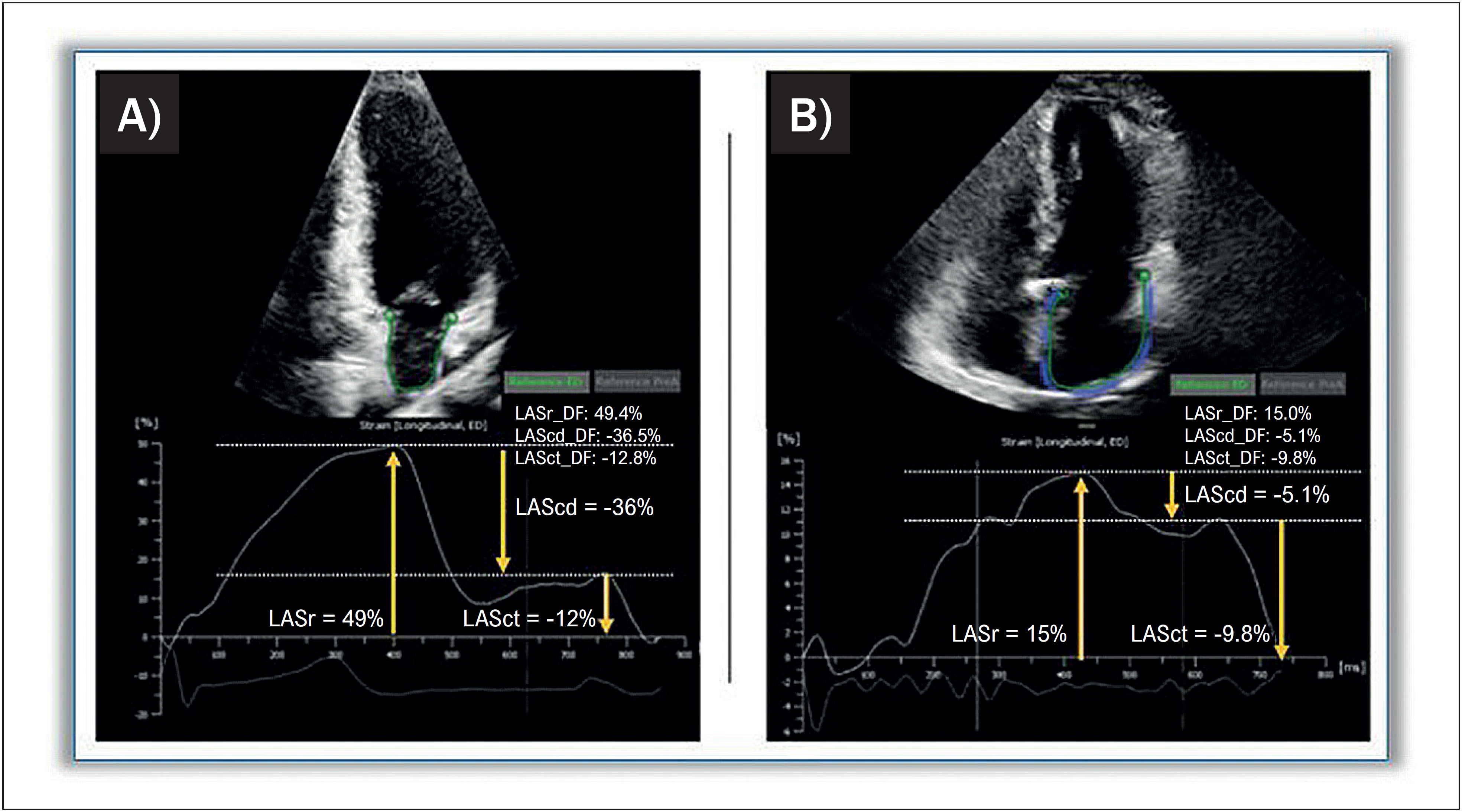
Technical acquisition: LAS should be measured in apical four- and two-chamber views, with the R-R interval defining the cardiac cycle. Adequate frame rates (> 60 fps), appropriate depth, and optimized image acquisition are essential. Speckle-tracking analysis should exclude pulmonary veins and the LA appendage. The average of both views should be reported (Figure 1, Video 1).
-
Isovolumetric relaxation time (IVRT): useful in discordant or challenging scenarios
IVRT corresponds to the interval between aortic valve closure and mitral valve opening, reflecting early active ventricular relaxation.12
New/current concept: initially described as an auxiliary parameter in the 2009 guidelines and maintained as a complementary measure in 2016, IVRT has regained relevance as a refinement tool in discordant cases. Although not part of the primary decision-making core, a shortened IVRT (≤ 70 ms) suggests increased filling pressures, particularly when associated with a restrictive filling pattern or decreased atrial strain. It is especially useful when tissue Doppler measurements are unreliable, such as in AF or mitral annular calcification.4
-
Pulmonary venous flow (S/D and Ar-A): when additional confirmation is needed
Pulmonary venous flow assessment can be technically challenging but provides valuable information when adequately acquired. A diastolic predominance (S/D ≤ 0.67) supports increased filling pressures. However, patients with preserved LVEF may exhibit S/D > 0.67 despite increased pressures, requiring confirmation with additional parameters.
An Ar-A duration difference > 30 ms may be useful in selected conditions, such as hypertrophic cardiomyopathy and mitral regurgitation (MR).3,4,13
-
Additional supplementary parameters
When primary and refinement parameters are unavailable or unreliable, additional measures may support clinical interpretation. These include: peak diastolic pulmonary regurgitation velocity ≥ 2 m/s; pulmonary artery diastolic pressure ≥ 16 mmHg; mitral inflow L-wave velocity ≥ 50 cm/s; Ar-A duration > 30 ms; ≥ 50% reduction in mitral E/A during the Valsalva maneuver; E/Vp ≥ 2.5; A-wave transit time ≤ 45 ms; and IVRT/TE e′ < 2.
In addition, an LV mass index > 95 g/m2 in women or > 115 g/m2 in men may indicate structural remodeling consistent with diastolic dysfunction.3,4
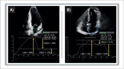
Role of LAS in refining the assessment of LV filling pressure. Assessment of LA deformation using speckle-tracking echocardiography. A) LASr, LAScd, and LASct measurements in a normal subject, with values of 49%, −36%, and −12%, respectively; B) corresponding strain curves from a patient with increased filling pressures, showing values of 15%, −5.1%, and −9.8%, respectively. LA: left atrium; LAS: LA strain; LAScd: LA conduit strain; LASct: LA contractile strain; LASr: LA reservoir strain; LV: left ventricle.
Video 1
HFpEF with inconclusive resting assessment, clarified by LAS. In: http://abcimaging.org/supplementary-material/2026/3902/ABCImag-2026-0026_AR_Video_1.mp4
Interpretation and integration of parameters (algorithm-based approach)
If all primary parameters assessed in Step 1 (e′, TRV, and E/e′) are within normal limits, LV filling pressures are considered normal. Conversely, if all three parameters are abnormal, increased filling pressures are present.
When e′ is decreased (based on age-adjusted reference values) and the E/A ratio is ≤ 0.8, this pattern is consistent with grade I diastolic dysfunction and normal filling pressures.
Diagnostic uncertainty arises in intermediate or discordant scenarios, including cases in which only e′ is decreased with E/A > 0.8, isolated increase of TRV/PASP or E/e′, or when any two primary variables are abnormal. In these situations, refinement using Step 2 parameters becomes essential.
These include LASr, IVRT, S/D, LAVI, and additional supplementary parameters. If one or more of these refinement markers are abnormal, increased filling pressures are confirmed. An E/A ratio < 2 supports the classification of grade II diastolic dysfunction, whereas E/A ≥ 2 indicates grade III diastolic dysfunction.4
Figure 2 shows the application of the algorithm.
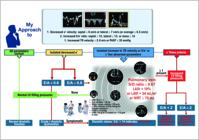
Decision algorithm for estimating LV filling pressures. Practical flowchart based on the 2025 ASE guideline. Step 1 relies on core parameters of myocardial relaxation and filling pressure assessment. In cases of discordance (e.g., a single abnormal parameter or borderline values), Step 2 is applied, prioritizing LASr and IVRT. Integration of these findings allows both grading of diastolic dysfunction and definitive classification of LV filling pressure status. ASE: American Society of Echocardiography; IVRT: isovolumetric relaxation time; LASr: left atrial reservoir strain; LAVI: left atrial volume index; LV: left ventricle; LVMI: LV mass index; PADP: pulmonary artery diastolic pressure; PASP: pulmonary artery systolic pressure; TR: tricuspid regurgitation.
Special situations
The 2025 guideline reinforces that a "one-size-fits-all" approach is not applicable. Specific clinical scenarios require adaptation of both acquisition and interpretation strategies (Table 3).
-
AF
Beat-to-beat variability increases the risk of measurement error; therefore, averaging multiple cardiac cycles is essential. Ideally, measurements should be obtained at a controlled heart rate < 100 bpm. Patients with decreased variability in mitral inflow tend to have increased filling pressures (Figure 3).3,14
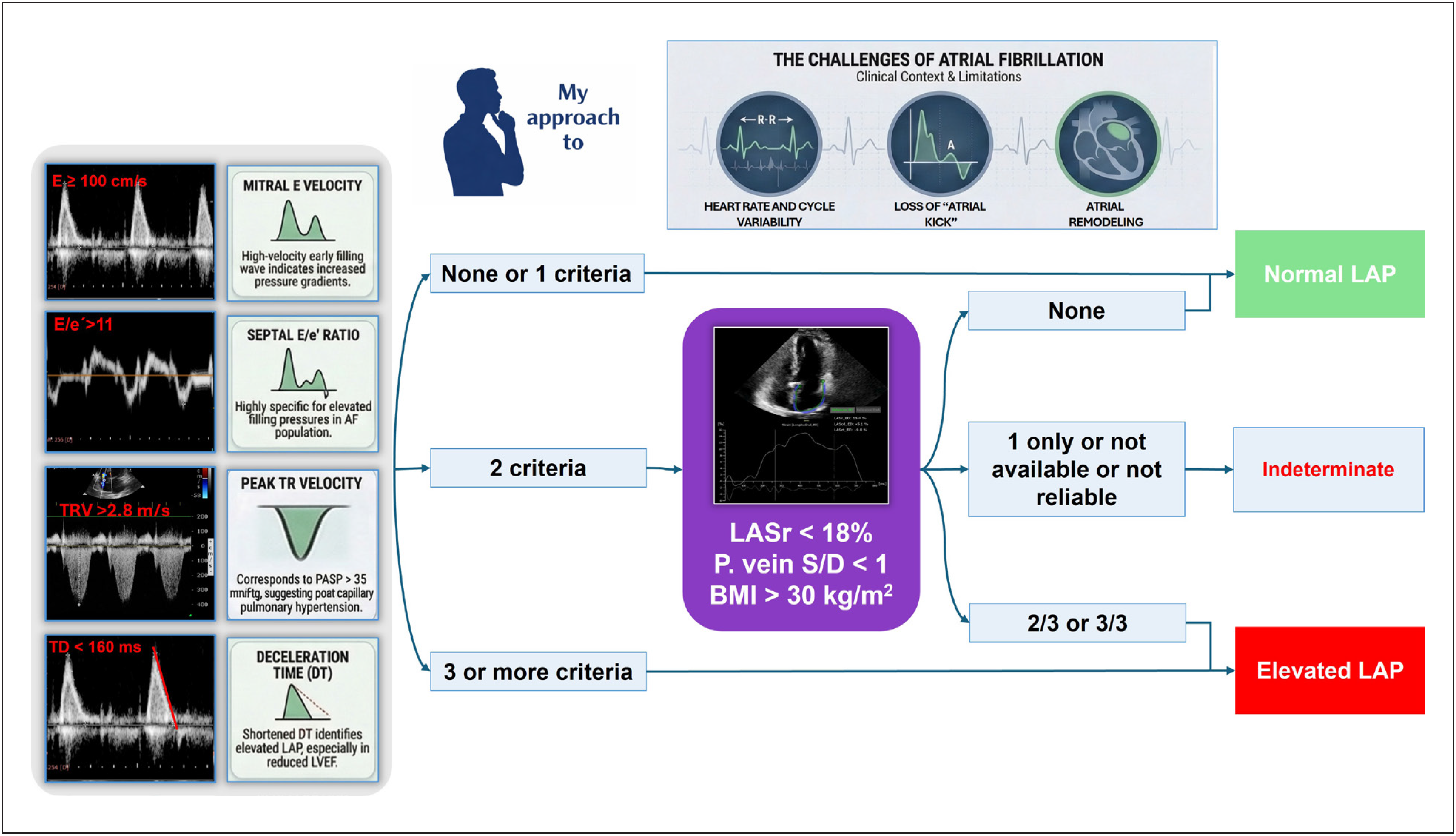
My Approach to AF: the assessment follows a two-step framework. In Step 1, the following parameters are considered: E ≥ 100 cm/s, septal E/e′ ≥ 11, TRV > 2.8 m/s or PASP > 35 mmHg, and DT ≤ 160 ms.
If none or only one parameter is abnormal, filling pressures are considered normal. If ≥ three parameters are abnormal, filling pressures are increased. If two parameters are abnormal, refinement is required using Step 2 markers, including LASr < 18% and S/D < 1. BMI > 30 kg/m2 further supports the diagnosis of HFpEF.
An average of 5-10 cardiac cycles should be used. If none of the parameters are abnormal, filling pressures are normal. If two of the three refinement parameters are abnormal, increased filling pressures are present. If only one parameter is abnormal or data are unavailable, the result should be considered indeterminate.
Caution: LASct is not present in AF; however, LASr remains informative. Very low values (< 16%) indicate decreased atrial compliance and increased filling pressures (Video 2).
-
Mitral valve disease
In mitral stenosis, the E/e′ ratio should not be used. In significant MR, the E-wave may be increased due to volume overload rather than increased filling pressures.3,4,15
My Approach to mitral valve disease: in MR, greater emphasis should be placed on pulmonary venous flow patterns and IVRT. LAS should be interpreted cautiously, as regurgitant volume may artificially increase LASr.
-
Cardiac amyloidosis
In this setting, isolated numerical values may fail to capture the underlying pathophysiology; the two-dimensional phenotype and overall functional pattern are key determinants. The presence of increased LV wall thickness associated with an "apical sparing" pattern on longitudinal strain should prompt evaluation for a restrictive diastolic filling pattern.3,16
My Approach to cardiac amyloidosis: a characteristic dissociation is often observed, with markedly decreased e′ velocities (septal and lateral < 5 cm/s) in contrast to a high mitral E-wave and shortened DT. This classic restrictive pattern strongly supports the presence of increased filling pressures, often obviating the need for complex algorithmic assessment.
-
Pulmonary hypertension
The E/e′ ratio, particularly the septal measurement, may be misleading in pre-capillary pulmonary hypertension. In such cases, greater emphasis should be placed on the lateral E/e′ and on LAS, especially when distinguishing pre- from post-capillary mechanisms in borderline scenarios3,4,17 (Video 3).
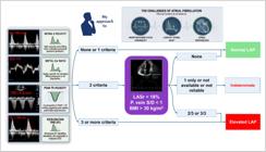
Algorithm for estimating mean LA in AF. Initial assessment is based on four parameters: mitral E velocity ≥ 100 cm/s, septal E/e′ > 11, TR velocity > 2.8 m/s (or PASP > 35 mmHg), and DT ≤ 160 ms. The presence of none or only one abnormal parameter suggests normal LAP. When two parameters are abnormal, additional markers, including LASr < 18%, S/D < 1, and BMI > 30 kg/m2, are used to refine classification as normal, increased, or indeterminate LAP. BMI: body mass index; DT: deceleration time; LA: left atrium; LAP: LA pressure; LASr: LA reservoir strain; LV: left ventricle; LVEF: LV ejection fraction; PASP: pulmonary artery systolic pressure; TR: tricuspid regurgitation; AF: atrial fibrillation.
Video 2
AF: importance of beat averaging and the use of IVRT and LAS for refinement. In: http://abcimaging.org/supplementary-material/2026/3902/ABCImag-2026-0026_AR_Video_2.mp4
Video 3
Pre-capillary pulmonary hypertension with borderline parameters and preserved LAS. In: http://abcimaging.org/supplementary-material/2026/3902/ABCImag-2026-0026_AR_Video_3.mp4
The role of diastolic stress testing and invasive hemodynamic assessment
In patients with exertional dyspnea (New York Heart Association classes II and III) and a resting echocardiography that is normal or indeterminate, even after incorporation of LAS, evaluation should not be discontinued.3,4,18
Diastolic stress echocardiography using a supine bicycle or treadmill is recommended.3,4 Maneuvers that increase LV preload, such as passive leg raising, may also help unmask increased filling pressures in patients with decreased ventricular compliance. These approaches may serve as alternatives when formal exercise testing is unavailable, although a negative result does not exclude clinically significant diastolic dysfunction.3
Some studies have also proposed handgrip stress to increase afterload.19 In selected cases, stress echocardiography may be combined with simultaneous invasive hemodynamic assessment to confirm dynamic increases in pulmonary capillary pressure, thereby supporting the diagnosis of HFpEF when noninvasive findings are inconclusive.
My Approach to stress testing: I assess changes in the E/e′ ratio and TRV at peak exercise. An increase in average E/e′ > 14 or TRV > 2.8 m/s (or > 3.2 m/s in some studies to improve specificity) during exertion indicates dynamic increase of filling pressures and supports the diagnosis of HFpEF not evident at rest (Table 4; Video 4; Video 5).
Video 4
Passive leg raising demonstrating increased filling pressures in a patient with exertional dyspnea. In: http://abcimaging.org/supplementary-material/2026/3902/ABCImag-2026-0026_AR_Video_4.mp4
Video 5
Diastolic stress testing with handgrip confirming increased filling pressures in unexplained dyspnea. In: http://abcimaging.org/supplementary-material/2026/3902/ABCImag-2026-0026_AR_Video_5.mp4
Lung ultrasound (LUS) and venous excess ultrasound
LUS and the venous excess ultrasound (VExUS) score have emerged as complementary tools for the assessment of congestion. LUS identifies B-lines as markers of interstitial edema, whereas VExUS integrates inferior vena cava assessment with Doppler interrogation of intra-abdominal veins to characterize systemic venous congestion.
Although these methods do not replace diastolic function analysis, they expand bedside hemodynamic evaluation and may reinforce the suspicion of increased filling pressures in complex clinical scenarios.4
Artificial intelligence (AI) in the assessment of LV diastolic function
AI has emerged as a promising tool in the assessment of diastolic dysfunction and HFpEF, particularly due to its ability to integrate multiple echocardiographic and clinical variables into predictive models that outperform isolated parameters.
Machine learning algorithms can identify subtle phenotypic patterns, reduce the rate of indeterminate cases, and improve the estimation of filling pressures. Although still undergoing broad validation, AI is expected to function primarily as a decision-support tool, refining traditional algorithms without replacing the clinical judgment of the echocardiographer.4,20
What should be included in the report?
The echocardiographic report of diastolic function should address a clear clinical question rather than simply reproduce an algorithm. Classification as grade I, II, III, or indeterminate is insufficient on its own; it is essential to explicitly state whether there is consistent evidence of increased filling pressures and to describe the reasoning underlying this conclusion.
The echocardiographer should integrate available parameters and clearly articulate the interpretative logic, presenting key data alongside a direct and accountable conclusion. A high-quality report is one that informs clinical management: technical rigor has value only when it translates into clarity and actionable insight.
Conclusion
The assessment of LV filling pressures has evolved from a rigid, algorithm-driven exercise to an integrated physiological interpretation. The 2025 ASE update provides greater flexibility, allowing adaptation of the assessment according to age and comorbidities.
Rather than representing a purely mechanical application of predefined criteria, this evaluation should be understood as the structured integration of physiological data in support of clinical decision-making. We measure velocities and deformation, but our ultimate goal is to understand the hemodynamic mechanisms underlying symptoms.
When performed with technical rigor and contextualized interpretation, echocardiography not only estimates filling pressures but also elucidates underlying mechanisms. This ability to translate quantitative data into clinically meaningful insight underpins its central role in contemporary cardiology practice.
-
Sources of Funding
There were no external funding sources for this study.
-
Study Association
This study is not associated with any thesis or dissertation work.
-
Ethics Approval and Consent to Participate
This article does not contain any studies with human participants or animals performed by any of the authors.
-
Use of Artificial Intelligence
During the preparation of this work, the author(s) used ChatGPT to improve the readability and language quality of the manuscript. After using this tool/service, the author(s) reviewed and edited the content as needed and take full responsibility for the content of the published article.
Availability of Research Data
The underlying content of the research text is contained within the manuscript.
References
-
1 Nagueh SF, Smiseth OA, Appleton CP, Byrd BF 3rd, Dokainish H, Edvardsen T, et al. Recommendations for the Evaluation of Left Ventricular Diastolic Function by Echocardiography: An Update from the American Society of Echocardiography and the European Association of Cardiovascular Imaging. J Am Soc Echocardiogr. 2016;29(4):277-314. doi: 10.1016/j.echo.2016.01.011.
» https://doi.org/10.1016/j.echo.2016.01.011 -
2 van de Bovenkamp AA, Enait V, de Man FS, Oosterveer FTP, Bogaard HJ, Vonk Noordegraaf A, et al. Validation of the 2016 ASE/EACVI Guideline for Diastolic Dysfunction in Patients with Unexplained Dyspnea and a Preserved Left Ventricular Ejection Fraction. J Am Heart Assoc. 2021;10(18):e021165. doi: 10.1161/JAHA.121.021165.
» https://doi.org/10.1161/JAHA.121.021165 -
3 Robinson S, Ring L, Oxborough D, Harkness A, Bennett S, Rana B, et al. The Assessment of Left Ventricular Diastolic Function: Guidance and Recommendations from the British Society of Echocardiography. Echo Res Pract. 2024;11(1):16. doi: 10.1186/s44156-024-00051-2.
» https://doi.org/10.1186/s44156-024-00051-2 -
4 Nagueh SF, Sanborn DY, Oh JK, Anderson B, Billick K, Derumeaux G, et al. Recommendations for the Evaluation of Left Ventricular Diastolic Function by Echocardiography and for Heart Failure with Preserved Ejection Fraction Diagnosis: An Update from the American Society of Echocardiography. J Am Soc Echocardiogr. 2025;38(7):537-69. doi: 10.1016/j.echo.2025.03.011.
» https://doi.org/10.1016/j.echo.2025.03.011 -
5 Lababidi H, Rahi W, Smiseth OA, Billick K, Inoue K, Khan FH, et al. New Algorithm for Estimating Left Ventricular Filling Pressure by Echocardiography. Circulation. 2025;152(7):424-35. doi: 10.1161/CIRCULATIONAHA.125.074974.
» https://doi.org/10.1161/CIRCULATIONAHA.125.074974 -
6 Nagueh SF, Middleton KJ, Kopelen HA, Zoghbi WA, Quiñones MA. Doppler Tissue Imaging: A Noninvasive Technique for Evaluation of Left Ventricular Relaxation and Estimation of Filling Pressures. J Am Coll Cardiol. 1997;30(6):1527-33. doi: 10.1016/s0735-1097(97)00344-6.
» https://doi.org/10.1016/s0735-1097(97)00344-6 -
7 Nedios S, Koutalas E, Sommer P, Arya A, Rolf S, Husser D, et al. Asymmetrical Left Atrial Remodelling in Atrial Fibrillation: Relation with Diastolic Dysfunction and Long-Term Ablation Outcomes. Europace. 2017;19(9):1463-9. doi: 10.1093/europace/euw225.
» https://doi.org/10.1093/europace/euw225 -
8 Ahmeti A, Bytyçi FS, Bielecka-Dabrowa A, Bytyçi I, Henein MY. Prognostic Value of Left Atrial Volume Index in Acute Coronary Syndrome: A Systematic Review and Meta-Analysis. Clin Physiol Funct Imaging. 2021;41(2):128-35. doi: 10.1111/cpf.12689.
» https://doi.org/10.1111/cpf.12689 -
9 Cameli M, Lisi M, Giacomin E, Caputo M, Navarri R, Malandrino A, et al. Chronic Mitral Regurgitation: Left Atrial Deformation Analysis by Two-Dimensional Speckle Tracking Echocardiography. Echocardiography. 2011;28(3):327-34. doi: 10.1111/j.1540-8175.2010.01329.x.
» https://doi.org/10.1111/j.1540-8175.2010.01329.x -
10 Inoue K, Khan FH, Remme EW, Ohte N, García-Izquierdo E, Chetrit M, et al. Determinants of Left Atrial Reservoir and Pump Strain and Use of Atrial Strain for Evaluation of Left Ventricular Filling Pressure. Eur Heart J Cardiovasc Imaging. 2021;23(1):61-70. doi: 10.1093/ehjci/jeaa415.
» https://doi.org/10.1093/ehjci/jeaa415 -
11 Lundberg A, Johnson J, Hage C, Bäck M, Merkely B, Venkateshvaran A, et al. Left Atrial Strain Improves Estimation of Filling Pressures in Heart Failure: A Simultaneous Echocardiographic and Invasive Haemodynamic Study. Clin Res Cardiol. 2019;108(6):703-15. doi: 10.1007/s00392-018-1399-8.
» https://doi.org/10.1007/s00392-018-1399-8 -
12 Appleton CP, Hatle LK, Popp RL. Relation of Transmitral Flow Velocity Patterns to Left Ventricular Diastolic Function: New Insights from a Combined Hemodynamic and Doppler Echocardiographic Study. J Am Coll Cardiol. 1988;12(2):426-40. doi: 10.1016/0735-1097(88)90416-0.
» https://doi.org/10.1016/0735-1097(88)90416-0 -
13 Kuecherer HF, Muhiudeen IA, Kusumoto FM, Lee E, Moulinier LE, Cahalan MK, et al. Estimation of Mean Left Atrial Pressure from Transesophageal Pulsed Doppler Echocardiography of Pulmonary Venous Flow. Circulation. 1990;82(4):1127-39. doi: 10.1161/01.cir.82.4.1127.
» https://doi.org/10.1161/01.cir.82.4.1127 -
14 Khan FH, Zhao D, Ha JW, Nagueh SF, Voigt JU, Klein AL, et al. Evaluation of Left Ventricular Filling Pressure by Echocardiography in Patients with Atrial Fibrillation. Echo Res Pract. 2024;11(1):14. doi: 10.1186/s44156-024-00048-x.
» https://doi.org/10.1186/s44156-024-00048-x -
15 Diwan A, McCulloch M, Lawrie GM, Reardon MJ, Nagueh SF. Doppler Estimation of Left Ventricular Filling Pressures in Patients with Mitral Valve Disease. Circulation. 2005;111(24):3281-9. doi: 10.1161/CIRCULATIONAHA.104.508812.
» https://doi.org/10.1161/CIRCULATIONAHA.104.508812 -
16 Pagourelias ED, Mirea O, Duchenne J, Van Cleemput J, Delforge M, Bogaert J, et al. Echo Parameters for Differential Diagnosis in Cardiac Amyloidosis: A Head-to-Head Comparison of Deformation and Nondeformation Parameters. Circ Cardiovasc Imaging. 2017;10(3):e005588. doi: 10.1161/CIRCIMAGING.116.005588.
» https://doi.org/10.1161/CIRCIMAGING.116.005588 -
17 Inoue K, Andersen OS, Remme EW, Khan FH, Andreassen AK, Skulstad H, et al. Echocardiographic Evaluation of Left Ventricular Filling Pressure in Patients with Pulmonary Hypertension. JACC Cardiovasc Imaging. 2024;17(5):566-7. doi: 10.1016/j.jcmg.2023.12.004.
» https://doi.org/10.1016/j.jcmg.2023.12.004 -
18 Borlaug BA, Nishimura RA, Sorajja P, Lam CS, Redfield MM. Exercise Hemodynamics Enhance Diagnosis of Early Heart Failure with Preserved Ejection Fraction. Circ Heart Fail. 2010;3(5):588-95. doi: 10.1161/CIRCHEARTFAILURE.109.930701.
» https://doi.org/10.1161/CIRCHEARTFAILURE.109.930701 -
19 Samuel TJ, Beaudry R, Haykowsky MJ, Sarma S, Nelson MD. Diastolic Stress Testing: Similarities and Differences between Isometric Handgrip and Cycle Echocardiography. J Appl Physiol. 2018;125(2):529-35. doi: 10.1152/japplphysiol.00304.2018.
» https://doi.org/10.1152/japplphysiol.00304.2018 -
20 Akerman AP, Porumb M, Scott CG, Beqiri A, Chartsias A, Ryu AJ, et al. Automated Echocardiographic Detection of Heart Failure with Preserved Ejection Fraction Using Artificial Intelligence. JACC Adv. 2023;2(6):100452. doi: 10.1016/j.jacadv.2023.100452.
» https://doi.org/10.1016/j.jacadv.2023.100452
Edited by
-
Editor responsible for the review:
Marcelo Tavares