Impact of Isometric Exercise on Left Ventricular Mechanics Assessed by Global Longitudinal Strain and Myocardial Work in Healthy Adults
Abstract
Background: Traditional volumetric parameters have limitations in detecting subtle left ventricular (LV) systolic dysfunction. Global longitudinal strain (GLS) and myocardial work (MW) allow a more sensitive assessment of ventricular mechanics.
Objectives: To evaluate changes in GLS and MW indices during isometric handgrip exercise compared with resting conditions.
Methods: A total of 30 healthy individuals (29.3 ± 6.1 years; 50% male) were included in the sample. Echocardiography was performed at rest and during handgrip exercise (30%-40% of maximal strength). GLS, LV ejection fraction (LVEF), and MW indices were assessed: i) global work index (GWI), ii) global constructive work (GCW), iii) global wasted work (GWW), and iv) global work efficiency (GWE). Comparisons were performed using paired tests. Statistical significance was set at p < 0.05.
Results: Handgrip exercise increased both systolic blood pressure (115 ± 16 vs 133 ± 18 mmHg; p < 0.0001) and diastolic blood pressure (69 ± 9 vs 79 ± 13 mmHg; p = 0.0002), without significant changes in LVEF (64.8% vs 64.4%; p = 0.62). A decrease in GLS was observed (20.38% ± 2.57% vs 19.60% ± 2.52%; p = 0.028), along with increases in GWI (+244 mmHg%; p = 0.0002), GCW (+313 mmHg%; p < 0.0001), and GWW (+52 mmHg%; p = 0.0008) as well as a decrease in GWE (94.8% ± 1.8% vs 93.6% ± 2.5%; p = 0.022).
Conclusions: Handgrip exercise induces measurable ventricular mechanical changes in healthy individuals, reflecting a physiological response to acute pressure overload.
Keywords:
Exercise; Echocardiography; Left Ventricular Dysfunction
Resumo
Fundamento: Parâmetros volumétricos tradicionais apresentam limitações na detecção de disfunção sistólica sutil do ventrículo esquerdo (VE). O strain longitudinal global (SLG) e o trabalho miocárdico (TM) permitem uma avaliação mais sensível da mecânica ventricular.
Objetivos: Avaliar as alterações do SLG e dos índices de TM durante o exercício isométrico de preensão manual, em comparação às condições de repouso.
Métodos: Ao todo, 30 indivíduos saudáveis (29,3 ± 6,1 anos; 50% do sexo masculino) foram incluídos na amostra. A ecocardiografia foi realizada em repouso e durante o exercício de preensão manual (30%-40% da força máxima). Foram avaliados o SLG, a fração de ejeção do VE (FEVE) e os índices de TM: i) índice de trabalho global (ITG), ii) trabalho construtivo global (TCG), iii) trabalho desperdiçado global (TDG) e iv) eficiência global do trabalho (EGT). As comparações foram realizadas por testes pareados, considerando-se significância estatística quando p < 0,05.
Resultados: O exercício de preensão manual promoveu aumento da pressão arterial (PA) sistólica (115 ± 16 vs 133 ± 18 mmHg; p < 0,0001) e diastólica (69 ± 9 vs 79 ± 13 mmHg; p = 0,0002), sem alteração significativa da FEVE (64,8% vs 64,4%; p = 0,62). Observou-se redução do SLG (20,38% ± 2,57% vs 19,60% ± 2,52%; p = 0,028), aumento do ITG (+244 mmHg%; p = 0,0002), do TCG (+313 mmHg%; p < 0,0001) e do TDG (+52 mmHg%; p = 0,0008), além de redução da EGT (94,8% ± 1,8% vs 93,6% ± 2,5%; p = 0,022).
Conclusões: O exercício de preensão manual induz alterações mecânicas ventriculares mensuráveis em indivíduos saudáveis, refletindo resposta fisiológica à sobrecarga pressórica aguda.
Palavras-chave:
Exercício Físico; Ecocardiografia; Disfunção Ventricular Esquerda
Introduction
Assessment of left ventricular (LV) systolic function is central to contemporary echocardiography. Although LV ejection fraction (LVEF) is widely used, its dependence on ventricular geometry and loading conditions limits the detection of subclinical myocardial dysfunction.1 In this context, more sensitive techniques, such as speckle-tracking echocardiography (STE) and global longitudinal strain (GLS), have expanded the ability to assess myocardial mechanical performance.
GLS, which is obtained by STE, quantifies shortening of LV subendocardial fibers and provides a sensitive measure of myocardial contractility.2 GLS is an early marker of ventricular dysfunction with established prognostic value and is often altered before changes in LVEF become apparent.3 However, its sensitivity to variations in loading conditions, including preload and afterload, limits its isolated interpretation, which has justified the development of methods capable of integrating myocardial deformation into the hemodynamic context.
Myocardial work (MW) integrates myocardial deformation with the noninvasively estimated systolic pressure gradient through pressure-strain curves, allowing a more comprehensive assessment of LV mechanics under different loading conditions.4 These indices show good correlation with invasive measures of ventricular performance and lower afterload dependence compared with strain alone, thereby expanding their clinical applicability.4-7
Isometric handgrip exercise is a simple, safe, and reproducible method for inducing cardiovascular stress, promoting an acute increase in systolic blood pressure (BP) and afterload.6,9 Classic studies demonstrated that individuals with preserved ventricular reserve increase systolic work, whereas patients with ventricular dysfunction exhibit adverse hemodynamic responses, including increased end-diastolic pressure and decreased MW efficiency.9,10 More recent echocardiographic protocols have confirmed that handgrip exercise reproduces controlled hemodynamic stress and enables sensitive assessment of ventricular mechanical adaptations, including increased global work index (GWI) and a slight decrease in global work efficiency (GWE).7,11
This study evaluated, in healthy young adults, changes in GLS and MW indices during isometric handgrip exercise compared with resting conditions, aiming to characterize the physiological LV response to acute pressure overload.
Methods
Study design and population
This was a prospective, cross-sectional study that included healthy young adults (18-40 years) undergoing echocardiographic evaluation at rest and during isometric handgrip exercise. Individuals with preserved myocardial function and no clinical comorbidities were included. Participants with significant structural heart disease, arrhythmias, limiting musculoskeletal disorders, or contraindications to stress echocardiography were excluded.
Sample size calculation was based on normative MW values described by Olsen et al.,5 using GWI as the primary outcome and considering a paired design (rest vs handgrip exercise). A conservative increase of 150 mmHg% in GWI was adopted, with an estimated standard deviation of differences of 240 mmHg%, based on data from Cebrowska et al.12 Considering a significance level of 5% and statistical power of 80%, the formula for paired mean comparisons indicated a minimum requirement of 21 individuals. To increase the precision of estimates and analytical robustness, 30 participants were included in the final sample.
Echocardiographic acquisition and analysis
Transthoracic echocardiography was performed using the Vivid™ E95 Ultrasound System (GE Vingmed Ultrasound AS, Horten, Norway), equipped with a 3.5-MHz MS5 sector transducer. Standard 2D images were acquired over three cardiac cycles, synchronized to the QRS complex, and stored in digital format for offline analysis using EchoPAC™ software (version 206; GE Vingmed Ultrasound AS, Horten, Norway), according to the recommendations of the American Society of Echocardiography.13
LVEF was obtained using the biplane Simpson method. Diastolic function was assessed according to current guidelines. GLS was analyzed using STE with standard apical views.
MW assessment
MW indices (i.e., GWI, global constructive work [GCW], global wasted work [GWW], and GWE) were automatically calculated from pressure-strain curves. For this purpose, brachial BP measured at the time of the examination was used.
Handgrip exercise protocol
The handgrip protocol consisted of sustained isometric contraction at 40% of maximal voluntary strength, previously determined by dynamometry. The effort was maintained for 2-3 minutes, with echocardiographic image acquisition performed between the second and third minutes.
Statistical analysis
Comparisons between resting and handgrip conditions were performed using paired tests according to data distribution. Statistical significance was set at p < 0.05. Effect size was calculated to estimate the magnitude of the observed differences.
Ethical considerations
The study was approved by the local human research ethics committee, and all participants provided written informed consent.
Results
A total of 30 healthy individuals were evaluated, 50% of whom were male, with a mean age of 29.3 ± 6.1 years, and all completed the isometric handgrip exercise protocol. Overall clinical characteristics demonstrated mean values consistent with the studied age range. During handgrip exercise, significant increases were observed in systolic BP (115 mmHg vs 133 mmHg; p < 0.0001), diastolic BP (69 mmHg vs 79 mmHg; p = 0.0002), and heart rate (72 bpm vs 81 bpm; p < 0.0001), characterizing the typical hemodynamic response to isometric effort (Table 1).
Structural echocardiographic measurements demonstrated ventricular dimensions and LV mass within normal ranges, without relevant morphological abnormalities. Global systolic function remained preserved throughout the protocol, with no changes in LVEF between rest and stress conditions (64.8% vs 64.4%; p = 0.6163). Diastolic function parameters also remained stable, with a mild decrease in lateral e’ velocity (16.4 cm/s vs 14.9 cm/s; p = 0.0123), without relevant changes in the functional pattern (Table 2).
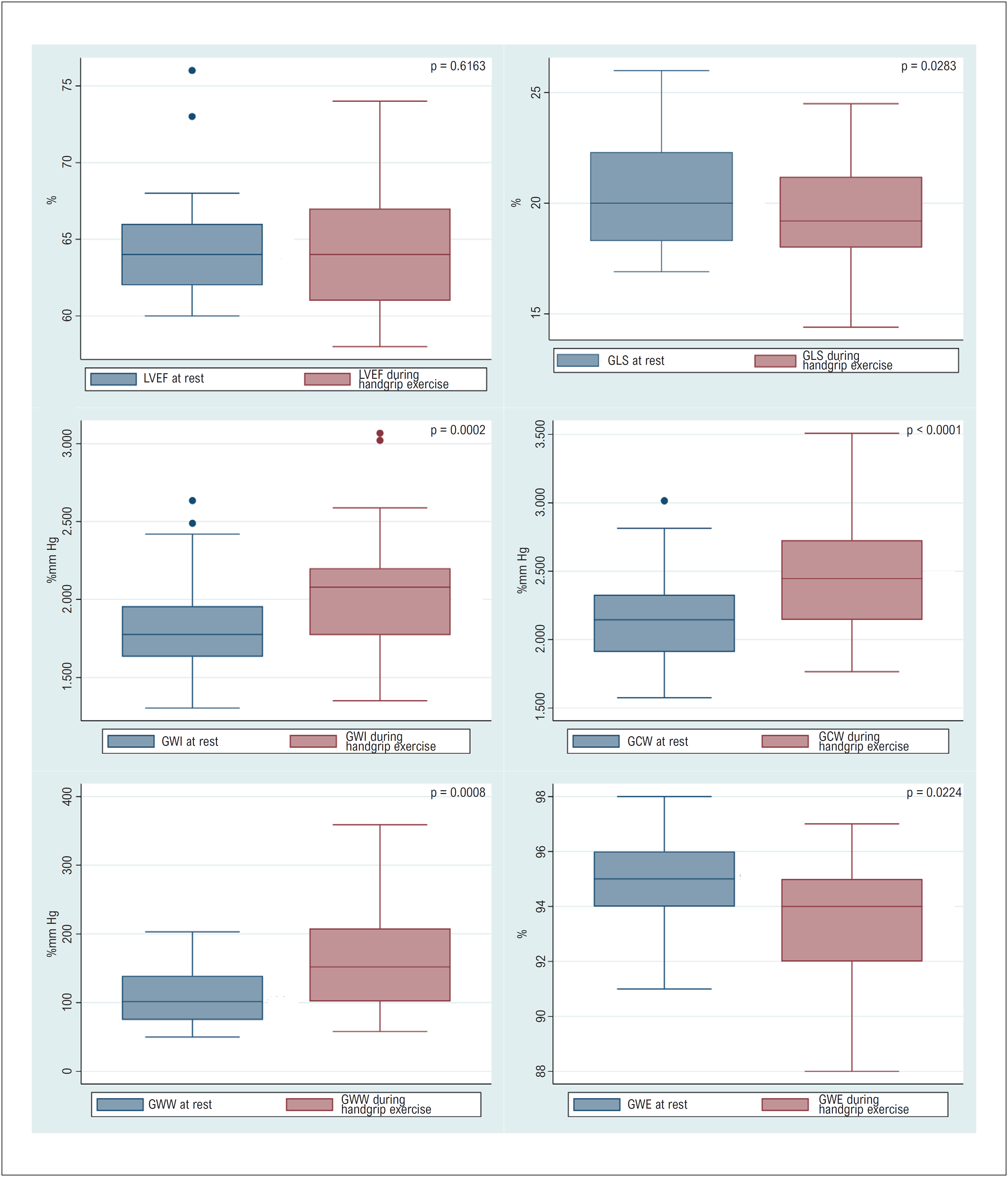
In the GLS analysis, a slight absolute decrease was observed during handgrip exercise (20.3% vs 19.6%; p = 0.0283), with a small effect size (Cohen's d = 0.42). The decrease was more evident in basal segments, also with a small effect size (d = 0.47), whereas mid and apical segments showed minimal variations and very small effect sizes (d = 0.11 and 0.16, respectively), without statistical significance (Table 3).
Regarding MW, there was a significant increase in GWI (1,810 mmHg% vs 2,054 mmHg%; p = 0.0002), with a moderate effect size (d = 0.77), and in GCW (2,172 mmHg% vs 2,486 mmHg%; p < 0.0001), which showed a large effect size (d = 1.05), representing the greatest magnitude among the evaluated parameters. GWW also increased, with a moderate effect size (d = 0.68). GWE showed a slight decrease, with a small effect size (d = 0.44).
Segmental MW analysis demonstrated increases in basal segments (1,719 mmHg% vs 1,959 mmHg%; p = 0.0003) and mid segments (1,655 mmHg% vs 2,047 mmHg%; p = 0.0001), with moderate effect sizes (d = 0.81 and 0.72, respectively), whereas apical segments showed no significant variation. These results are presented in detail in Table 3.
Figure 1 graphically summarizes the distribution of the main evaluated parameters. LVEF remained stable, GLS showed a slight decrease, and consistent increases were observed in GWI, GCW, and GWW, accompanied by a mild decrease in GWE.
Variation in ventricular function, GLS, and MW parameters between rest and handgrip exercise. GCW: global constructive work; GLS: global longitudinal strain; GWE: global work efficiency; GWI: global work index; GWW: global wasted work; LVEF: left ventricular ejection fraction.
Discussion
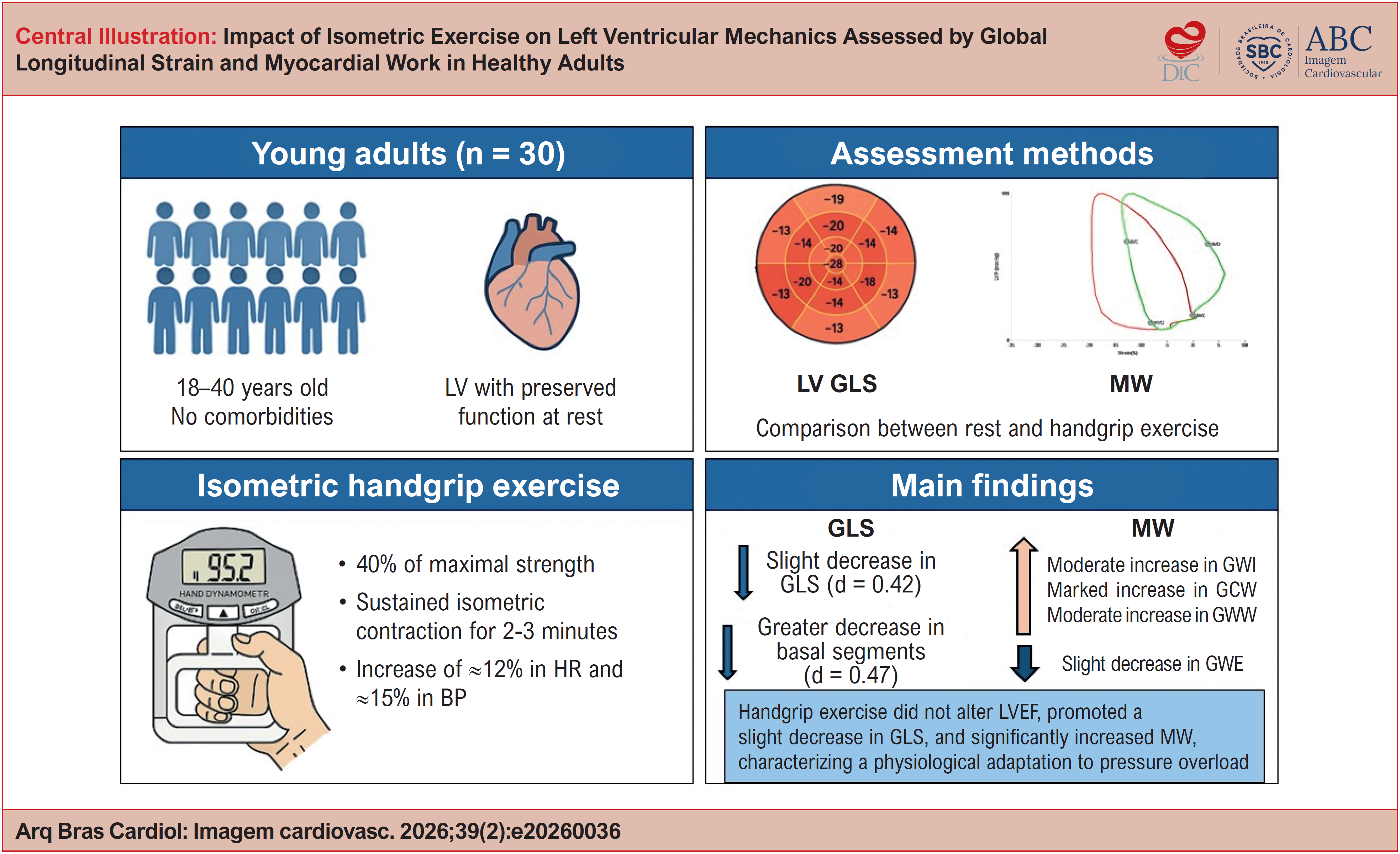
The present study contributes by demonstrating the integrated LV response to isometric handgrip stress in healthy individuals through the combination of GLS and MW indices. Our findings show that acute afterload increase promotes a consistent rise in BP, preservation of LVEF, a slight decrease in GLS, and increased MW indices as well as increased GWW and a mild decrease in GWE (Central Illustration). These results expand the understanding of the physiological myocardial adaptation to pressure stress and reinforce the value of a multiparametric approach for identifying changes not detectable by LVEF alone.
The consistent increase in systolic and diastolic BP during handgrip exercise confirms the role of this maneuver as a reproducible hemodynamic stressor, in agreement with the classic findings of Helfant et al.8 and Kivowitz et al.,9 who described the physiological mechanisms underlying the pressor response to isometric effort. These authors demonstrated that increased sympathetic tone and peripheral vascular resistance are the main determinants of BP increase, whereas HR shows only a modest increase, a pattern also observed in the present investigation. More recent studies, such as that by Samuel et al.,14 further support the usefulness of handgrip exercise as a practical, accessible alternative to more complex dynamic stress protocols, especially in the assessment of subtle changes in ventricular performance.
The stability of LVEF both at rest and during stress highlights the limitation of volumetric parameters in detecting subtle contractile changes induced by loading variations, corroborating observations by Thomas et al.15 and Clemmensen et al.16 They demonstrated that LVEF may remain unchanged despite relevant modifications in systolic mechanics, which underscores the need for more sensitive tools such as GLS and MW.
The slight decrease in GLS during handgrip exercise represents an expected physiological finding. This behavior, described by Flachskampf and Chandrashekar,4 reflects the sensitivity of strain to afterload changes. The greater decrease in basal segments reinforces the regional heterogeneity of the mechanical response, as suggested by Thomas et al.15 These regions exhibit higher wall stress and depend more directly on longitudinal shortening, making them more susceptible to acute pressure overload. The relative stability of mid and apical segments suggests preservation of overall contractile reserve in healthy individuals.
MW indices provided relevant complementary information. The significant increase in GWI and GCW during handgrip exercise is consistent with the physiological increase in mechanical energy required to overcome the greater systolic load. Studies by Zhu et al.2 and Caminiti et al.7 demonstrated similar behavior both in healthy individuals and in populations with hypertension or coronary artery disease, reinforcing the sensitivity of the pressure-strain model in quantifying contractile adjustments in response to acute stimuli.
The increase in GWW represents another physiologically consistent finding. In scenarios of acute afterload increase, as described by Russell et al.17 and summarized by Flachskampf and Chandrashekar,4 part of the energy generated by the myocardium is expected not to be converted into useful work because of temporal dyssynchrony between tension generation and effective fiber shortening. This mechanism contributes to the slight decrease in GWE, which nevertheless remained within the physiological range. The observed values are consistent with normative limits previously described by Olsen et al.,5 reinforcing the validity of these findings in a healthy population.
Segmental LV analysis revealed an additional aspect of the physiological adaptation to isometric stress. During handgrip exercise, a decrease in GLS was observed in basal segments, accompanied by increased MW in these same regions, a pattern also observed in mid segments. This dissociation between lower deformation and greater MW suggests a physiological adjustment to acute afterload increase, in which reduced longitudinal shortening is compensated by greater mechanical energy generation to maintain overall performance. Taken together, the observed pattern remained aligned with the normal phenotype described by Grandperrin et al.,18 which underscores that the regional response to stress represents physiological contractile adaptation rather than subclinical dysfunction.
This study has limitations that should be considered. The exclusive inclusion of healthy young adults and the relatively small sample size limit the generalizability of the results to clinical populations. In addition, echocardiographic acquisition during isometric effort may introduce technical variability in image quality. On the other hand, the prospective and standardized design, the homogeneous sample without comorbidities, which allowed physiological characterization with reduced external interference, and the integrated evaluation of GLS and MW are important strengths, increasing sensitivity for detecting subtle changes in ventricular mechanics. The inclusion of effect size analysis also adds interpretative value by allowing assessment of the practical relevance of the observed differences.
Taken together, these findings reinforce the value of handgrip exercise as a practical, safe, and reproducible submaximal stress tool. The integrated GLS and MW approach proved capable of detecting acute physiological modifications not identifiable by traditional methods such as LVEF and may be particularly relevant in contexts requiring assessment of contractile reserve or identification of subclinical dysfunction.
In addition to characterizing the physiological LV response to isometric stress, this study contributes to consolidating the role of GLS and MW as central tools in the contemporary assessment of ventricular mechanics, highlighting handgrip exercise as a valuable strategy for both physiological studies and clinical applications.
Conclusions
Isometric handgrip exercise induced measurable hemodynamic and mechanical changes in healthy individuals, characterized by stable LVEF, a slight decrease in GLS, and significant increases in GWI and GCW as well as increased GWW and a mild decrease in GWE. These findings reflect physiological contractile adaptation to acute pressure overload and reinforce handgrip exercise as a simple, safe, reproducible tool for assessing ventricular mechanics beyond traditional volumetric parameters.
-
Sources of Funding
There were no external funding sources for this study.
-
Study Association
This study is not associated with any thesis or dissertation work.
-
Ethics approval and consent to participate
This study was approved by the Ethics Committee of the Hospital Carlos Macieira under the protocol number 7.784.405. All the procedures in this study were in accordance with the 1975 Helsinki Declaration, updated in 2013. Informed consent was obtained from all participants included in the study.
-
Use of Artificial Intelligence
The authors did not use any artificial intelligence tools in the development of this work.
Data Availability Statement
The underlying content of the research text is contained within the manuscript.
References
-
1 Stanton T, Leano R, Marwick TH. Prediction of All-Cause Mortality from Global Longitudinal Speckle Strain: Comparison with Ejection Fraction and Wall Motion Scoring. Circ Cardiovasc Imaging. 2009;2(5):356-64. doi: 10.1161/CIRCIMAGING.109.862334.
» https://doi.org/10.1161/CIRCIMAGING.109.862334 -
2 Zhu H, Guo Y, Wang X, Yang C, Li Y, Meng X, et al. Myocardial Work by Speckle Tracking Echocardiography Accurately Assesses Left Ventricular Function of Coronary Artery Disease Patients. Front Cardiovasc Med. 2021;8:727389. doi: 10.3389/fcvm.2021.727389.
» https://doi.org/10.3389/fcvm.2021.727389 -
3 Oikonomou EK, Kokkinidis DG, Kampaktsis PN, Amir EA, Marwick TH, Gupta D, et al. Assessment of Prognostic Value of Left Ventricular Global Longitudinal Strain for Early Prediction of Chemotherapy-Induced Cardiotoxicity: A Systematic Review and Meta-Analysis. JAMA Cardiol. 2019;4(10):1007-18. doi: 10.1001/jamacardio.2019.2952.
» https://doi.org/10.1001/jamacardio.2019.2952 -
4 Flachskampf FA, Chandrashekar Y. Myocardial Work and Work Index: Related but Different for Clinical Usage. JACC Cardiovasc Imaging. 2022;15(8):1521-3. doi: 10.1016/j.jcmg.2022.07.001.
» https://doi.org/10.1016/j.jcmg.2022.07.001 -
5 Olsen FJ, Skaarup KG, Lassen MCH, Johansen ND, Sengeløv M, Jensen GB, et al. Normal Values for Myocardial Work Indices Derived from Pressure-Strain Loop Analyses: From the CCHS. Circ Cardiovasc Imaging. 2022;15(5):e013712. doi: 10.1161/CIRCIMAGING.121.013712.
» https://doi.org/10.1161/CIRCIMAGING.121.013712 -
6 Olsen FJ, Bjerregaard CL, Skaarup KG, Lassen MCH, Johansen ND, Modin D, et al. Impact of Echocardiographic Analyses of Valvular Event Timing on Myocardial Work Indices. Eur Heart J Cardiovasc Imaging. 2023;24(3):314-23. doi: 10.1093/ehjci/jeac171.
» https://doi.org/10.1093/ehjci/jeac171 -
7 Caminiti G, Volterrani M, Iellamo F, Marazzi G, D’Antoni V, Calandri C, et al. Acute Changes in Myocardial Work during Isometric Exercise in Hypertensive Patients with Ischemic Heart Disease: A Case-Control Study. J Clin Med. 2024;13(19):5955. doi: 10.3390/jcm13195955.
» https://doi.org/10.3390/jcm13195955 -
8 Helfant RH, De Villa MA, Meister SG. Effect of Sustained Isometric Handgrip Exercise on Left Ventricular Performance. Circulation. 1971;44(6):982-93. doi: 10.1161/01.cir.44.6.982.
» https://doi.org/10.1161/01.cir.44.6.982 -
9 Kivowitz C, Parmley WW, Donoso R, Marcus H, Ganz W, Swan HJ. Effects of Isometric Exercise on Cardiac Performance. The Grip Test. Circulation. 1971;44(6):994-1002. doi: 10.1161/01.cir.44.6.994.
» https://doi.org/10.1161/01.cir.44.6.994 -
10 Fisher ML, Nutter DO, Jacobs W, Schlant RC. Haemodynamic Responses to Isometric Exercise (Handgrip) in Patients with Heart Disease. Br Heart J. 1973;35(4):422-32. doi: 10.1136/hrt.35.4.422.
» https://doi.org/10.1136/hrt.35.4.422 -
11 Samuel TJ, Beaudry R, Haykowsky MJ, Sarma S, Park S, Dombrowsky T, et al. Isometric Handgrip Echocardiography: A Noninvasive Stress Test to Assess Left Ventricular Diastolic Function. Clin Cardiol. 2017;40(12):1247-55. doi: 10.1002/clc.22818.
» https://doi.org/10.1002/clc.22818 -
12 Cebrowska K, Mińczykowski A, Krauze T, Guzik P, Szczepanik A, Wykrętowicz A. The Pressure-Strain Work Indices in Response to Isometric Handgrip Exercise. Kardiol Pol. 2021;79(4):455-7. doi: 10.33963/KP.15912.
» https://doi.org/10.33963/KP.15912 -
13 Lang RM, Badano LP, Mor-Avi V, Afilalo J, Armstrong A, Ernande L, et al. Recommendations for Cardiac Chamber Quantification by Echocardiography in Adults: An Update from the American Society of Echocardiography and the European Association of Cardiovascular Imaging. J Am Soc Echocardiogr. 2015;28(1):1-39.e14. doi: 10.1016/j.echo.2014.10.003.
» https://doi.org/10.1016/j.echo.2014.10.003 -
14 Samuel TJ, Haykowsky MJF, Sarma S, Nelson MD. Diastolic Stress Testing: Have You Considered Isometric Handgrip Echocardiography? JACC Cardiovasc Imaging. 2019;12(10):2095-7. doi: 10.1016/j.jcmg.2019.07.023.
» https://doi.org/10.1016/j.jcmg.2019.07.023 -
15 Thomas JD, Edvardsen T, Abraham T, Appadurai V, Badano L, Banchs J, et al. Clinical Applications of Strain Echocardiography: A Clinical Consensus Statement from the American Society of Echocardiography Developed in Collaboration with the European Association of Cardiovascular Imaging of the European Society of Cardiology. J Am Soc Echocardiogr. 2025;38(11):985-1020. doi: 10.1016/j.echo.2025.07.007.
» https://doi.org/10.1016/j.echo.2025.07.007 -
16 Clemmensen TS, Eiskjær H, Mikkelsen F, Granstam SO, Flachskampf FA, Sørensen J, et al. Left Ventricular Pressure-Strain-Derived Myocardial Work at Rest and during Exercise in Patients with Cardiac Amyloidosis. J Am Soc Echocardiogr. 2020;33(5):573-82. doi: 10.1016/j.echo.2019.11.018.
» https://doi.org/10.1016/j.echo.2019.11.018 -
17 Russell K, Eriksen M, Aaberge L, Wilhelmsen N, Skulstad H, Gjesdal O, et al. Assessment of Wasted Myocardial Work: A Novel Method to Quantify Energy Loss Due to Uncoordinated Left Ventricular Contractions. Am J Physiol Heart Circ Physiol. 2013;305(7):H996-1003. doi: 10.1152/ajpheart.00191.2013.
» https://doi.org/10.1152/ajpheart.00191.2013 -
18 Grandperrin A, Schnell F, Donal E, Galli E, Hedon C, Cazorla O, et al. Specific Alterations of Regional Myocardial Work in Strength-Trained Athletes Using Anabolic Androgenic Steroids Compared to Athletes with Genetic Hypertrophic Cardiomyopathy. J Sport Health Sci. 2023;12(4):477-85. doi: 10.1016/j.jshs.2022.07.004.
» https://doi.org/10.1016/j.jshs.2022.07.004
Edited by
-
Editor responsible for the review:
Marcelo Tavares