 Indeterminate cell histiocytosis with ETV3::NCOA2 fusion detected by optical genome mapping
Dear Editor,
Following observation of a generalized eruption in a young patient with a unique histopathology, the term Indeter-minate Cell Histiocytosis (ICH) was described and coined by Wood GS et al. in 1985.1 It is a rare cutaneous proliferative disorder characterized by a proliferation of CD1a + and CD207/langerinmononuclear phagocytic cells, lacking Birbeck granules on electron microscopy. It is currently included in Group L of the 2016 revised clas-sification of histiocytosis, catalogued as ‘‘indeterminate dendritic cell histiocytosis’’ and as ‘‘indeterminate den-dritic cell tumor’’ in the 2022 WHO classification of dendritic cell neoplasms.2 This classification has been reaffirmed in the 5th edition (2024) of the WHO Classification of Haematolymphoid Tumours.3 Recent advances have shown that a subset of patients carries the ETV3::NCOA2 fusion gene, generally detected by Fluorescence In Situ Hybridiza-tion (FISH) or Next-Generation Sequencing (NGS).4-6 In this paper, we report a case of a patient with florid ICH in which the ETV3::NCOA2 fusion was identified for the first time using Optical Genome Mapping (OGM), achieving a favorable outcome after treatment with cladribine.
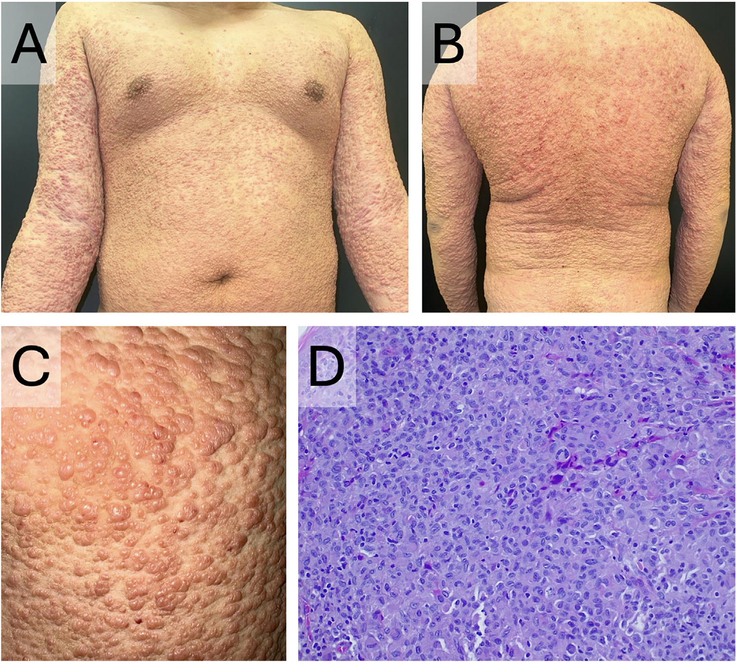
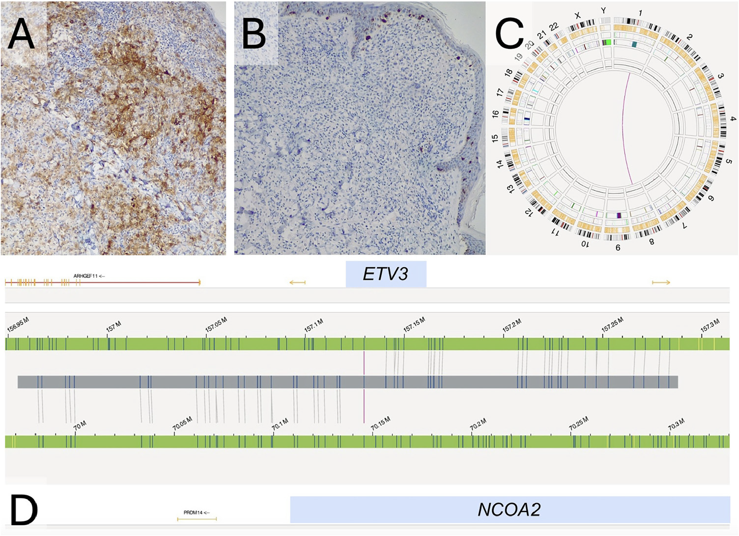
A 17-year-old male presented with rapidly progress-ing cutaneous lesions over the last 6-months. Physical examination revealed numerous flesh-colored papules with a generalized, cobblestone-like distribution, respect-ing the acrofacial regions (Fig. 1A-C). There were no lymphadenopathies, mucosal involvement, or systemic symptoms. Laboratory tests, including blood count, autoim-munity (ANA, myositis, and sclerosis blots), blood smear, and serologies, were normal. Histopathology (Fig. 1D) showed a dermal infiltrate that dissected the collagen fibers, consist-ing mainly of dense histiocytoid cells, with few lymphocytes and no eosinophils. Immunohistochemistry, the tumor cells were positive for CD1a (Fig. 2A) and S100, but nega-tive for CD207/langerin (Fig. 2B). His clinical presentation and histopathological findings supported the diagnosis of ICH.
Clinical presentation of the lesions and histopathological examination of one of the lesions. (A-B) Generalized flesh-colored papules with a ’cobblestone’ appearance of the skin. (C) Close-up view of the lesions. (D) Dermal infiltrate, predominantly histiocytic, with few lymphocytes, plasma cells and no eosinophils (Hematoxylin & eosin, ×200).
Immunohistochemistry of one of the lesions and optical genomic mapping of the tumor sample. (A) Diffuse positivity for CD1a (CD1a, 10×). (B) Negativity for CD207/langerin (CD207/langerin, 10×). (C and D) Graphical representation of the Optical Genomic Mapping with the interchromosomal translocation t(1;8) (q23.1;q13.3).
As part of the extension study, a positron emission tomography-computed tomography scan and bone marrow biopsy were performed, both of which were normal. A NGS study using the AmpliseqTM Comprehensive Cancer Panel was performed on DNA from the surgical specimen, cov-ering more than 400 genes, including BRAF, KRAS, NRAS, MAP2K1, NTRK1-3, and was negative except for the pres-ence of the variants c.1288C > T p.(Pro430Ser) in the MLLT10 gene, with a Variant Allele Frequency (VAF) of 49.7% and the variant c.49997 G > A p.(Gly1666Asp) in the RNF213 gene, with a VAF of 43.4%, both of them of unknown clinical relevance.
Subsequently, an OGM analysis was performed on a papu-lar specimen, which had been previously snap-frozen in liquid nitrogen at -196 ◦C for 3-minutes and subsequently stored at -80 ◦C. Data analysis was conducted using Bio-nano Access® Software (Bionano Genomics), applying the ‘‘Rare Variant Analysis’’ pipeline with the GRCh38 genome as reference. This analysis (Fig. 2C-D) identified an inter-chromosomal translocation t(1;8)(q23.1;q13.3) with a VAF of 35%, leading to the formation of the ETV3::NCOA2 fusion gene.
The patient was started on prednisone 30 mg/d and methotrexate 15 mg/week. However, due to progression of the lesions after two months, treatment with cladrib-ine 5 mg/m2/day (days 0-5) was started for 4-months.
He experienced a complete response of the lesions with no adverse effects or relapse at 8-month follow-up.
In recent years, advances in genetics and molecular biol-ogy techniques have led to the identification of two subtypes of ICH. The first is characterized by a predominance of mutations in the BRAF, KRAS, and MAP2K1 pathways and is often associated with multisystemic disease and other hematological disorders (especially chronic myelomonocytic leukemia). The second, defined by the presence of the ETV3::NCOA2 fusion gene (detected by FISH, NGS or, in our case, by OGM), generally has a more indolent course and a favorable prognosis.2,5,6
This case highlights the utility of OGM for detecting cryp-tic structural variants that may go unnoticed by conventional techniques, providing valuable diagnostic and prognostic information.7
Numerous treatments have been reported, with surgical excision generally being the first choice for local-ized cutaneous forms, and methotrexate, phototherapy or chemotherapeutic agents in monotherapy (cyclophos-phamide, busulfan, cladribine, vinblastine) for extensive cutaneous forms. Polychemotherapy regimens and/or hematopoietic cell transplantation are typically preferred for progressive cutaneous or multisystem involvement.4,5
Research data availability
Does not apply.
References
- 1 Wood GS, Hu CH, Beckstead JH, Turner RR, Winkelmann RK. The indeterminate cell proliferative disorder: report of a case man-ifesting as an unusual cutaneous histiocytosis. J Dermatol Surg Oncol. 1985;11:1111-9.
- 2 Zanella S, Berti E, Bonometti A, for Associazione Italiana Ricerca Istiocitosi ONLUS. Indeterminate cell histiocytosis: a systematic review of the literature with a comprehensive revision of clinical, histopathological, and molecular features. J Eur Acad Dermatol Venereol. 2023;37:1559-75.
- 3 World Health Organization Classification of Tumours Editorial Board. In: WHO classification of tumours: haematolymphoid tumours part A. 5th ed. Lyon: IARC; 2024.
- 4 Brown RA, Kwong BY, McCalmont TH, Ragsdale B, Ma L, Cheung C, et al. ETV3-NCOA2 in indeterminate cell histiocytosis: clonal translocation supports sui generis. Blood. 2015;126:2344-5.
- 5 Ozkaya N, Melloul Benizri S, Venkataraman G, Karai LJ, Fraitag S, Razanamahery J, et al. Indeterminate DC histiocytosis is dis-tinct from LCH and often associated with other hematopoietic neoplasms. Blood Adv. 2024;8:5796-805.
- 6 Kemps PG, Woei-A-Jin FJSH, Quint KD, van den Bos C, Naeije L, van Laar JAM, et al. Recurrent ETV3::NCOA2 fusions and MAPK pathway mutations in indeterminate dendritic cell histiocytosis. Blood Adv. 2025;9:439-44.
- 7 Moore S, McGowan-Jordan J, Smith AC, Rack K, Koehler U, Stevens-Kroef M, et al. Genome mapping nomenclature. Cyto-genet Genome Res. 2023;163:236-46.
Edited by
-
Editor
Hiram Larangeira de Almeida Jr.