 Radiographic and ultrasonographic findings of secondary synovial osteochondromatosis in dog - case report
ABSTRACT
Secondary synovial osteochondromatosis is a benign condition characterized by the formation of osteocartilaginous nodules within the synovial lining of joints because of chronic joint abnormalities. Imaging modalities, including radiography and ultrasonography, are essential for accurate diagnosis. This report describes a 9-month-old female Labrador Retriever with left pelvic limb lameness persisting for five months following trauma, unresponsive to anti-inflammatory therapy, and presenting with joint swelling and pain during flexion-extension movements. Radiographic examination revealed a circular intra-articular structure with peripheral soft tissue opacity and a central area of bone density. Ultrasonographic evaluation demonstrated a hypoechoic nodule with a hyperechoic center. These findings were consistent with synovial osteochondromatosis. Histopathological analysis of an intra-articular fragment obtained during arthrotomy confirmed the diagnosis. Concurrent cranial cruciate ligament rupture and meniscal injury were surgically treated by tibial plateau leveling osteotomy and meniscectomy. No clinical signs or nodular recurrence were observed during postoperative follow-up.
Keywords:
joint; osteochondroma; radiography; ultrasonography
RESUMO
A osteocondromatose sinovial secundária é uma condição benigna caracterizada pela formação de nódulos osteocartilaginosos no revestimento sinovial das articulações, como resultado de anomalias articulares crônicas. Modalidades de imagem, incluindo radiografia e ultrassonografia, são essenciais para um diagnóstico preciso. Este relato descreve o caso de uma cadela Labrador Retriever de 9 meses com claudicação do membro pélvico esquerdo persistente por cinco meses após trauma, sem resposta à terapia anti-inflamatória e apresentando edema articular e dor durante movimentos de flexão-extensão. O exame radiográfico revelou estrutura intra-articular circular com opacidade do tecido mole periférico e uma área central de densidade óssea. A avaliação ultrassonográfica demonstrou um nódulo hipoecóico com centro hiperecóico. Esses achados foram consistentes com osteocondromatose sinovial. A análise histopatológica do fragmento intra-articular obtido durante a artrotomia confirmou o diagnóstico. A ruptura concomitante do ligamento cruzado cranial e a lesão meniscal foram tratadas cirurgicamente por osteotomia de nivelamento do platô tibial e meniscectomia. Nenhum sinal clínico ou recorrência nodular foi observado durante o acompanhamento pós-operatório.
Palavras-chave:
articulação; osteocondroma; radiografia; ultrassonografia
INTRODUCTION
Synovial osteochondromatosis (SOC) is a benign condition, also referred to as synovial chondrometaplasia or synovial chondromatosis (Szilasi et al., 2022). It is characterized by the development of cartilaginous nodules, known as chondromas, which may undergo ossification and form osteochondromas within the synovial lining of joints (Smith et al., 2012; Tas et al., 2013). In both humans and animals including dogs (Diaz-Bertrana et al., 2010; Aeffner et al., 2012; Smith et al., 2012), cats (Tas et al., 2013; Szilasi et al., 2022), and horses (Seghrouchni et al., 2019) the disease most commonly affects the synovial membrane of joints.
SOC can be classified as either primary or secondary. Primary SOC is a rare condition that typically affects a single joint and develops spontaneously, without any predisposing joint abnormality. In contrast, secondary SOC represents a metaplastic response to chronic irritation associated with preexisting joint pathology and is considered relatively common in humans (Aeffner et al., 2012; Smith et al., 2012; Tas et al., 2013). Secondary SOC may arise from osteoarthritis, osteonecrosis, trauma, osteochondritis dissecans, rheumatoid arthritis, neuropathic osteoarthropathy, or tuberculosis (Aeffner et al., 2012; Tas et al., 2013; Terazaki et al., 2014). However, secondary SOC remains rare in veterinary medicine.
Imaging modalities such as radiography, ultrasonography, arthrography, computed tomography (CT), and magnetic resonance imaging (MRI) are essential for diagnosis, as they provide valuable information regarding joint involvement, lesion extent, and the presence of underlying changes that may promote the formation of osseous fragments thereby assisting in the distinction between primary and secondary forms (Friedenberg et al., 2018).
The imaging appearance varies according to the composition of the intra-articular nodules. When the nodules are cartilaginous (chondromas), they are not radiopaque and therefore may not be visible on radiographs, appearing instead as soft-tissue densities. On ultrasonography, they typically appear as homogeneous hypoechoic nodules. On CT, they exhibit low attenuation, whereas on MRI they appear hyperintense on T2-weighted images and of intermediate intensity on T1-weighted images, similar to the signal of muscle tissue (Friedenberg et al., 2018; Szilasi et al., 2022).
When mineralization occurs, osteochondromas appear as radiopaque nodules on radiographs. On ultrasonography, they appear hyperechoic and produce posterior acoustic shadowing. CT images show high attenuation, while MRI reveals intermediate signal intensity on T1 and high intensity on T2, with focal areas of low signal representing mineralization (Murphey et al., 2007).
In humans, radiographic changes are visible in only about 70% of SOC cases (McKenzie et al., 2008; Tas et al., 2013), as the disease may be evaluated before ossification occurs. Ultrasonography is a dynamic imaging technique that allows assessment of both ossified and non-ossified intra-articular fragments, facilitating early detection. To date, however, no reports have described the use of ultrasonography for SOC diagnosis in veterinary species. Histopathological examination remains the gold standard for definitive diagnosis (Murphey et al., 2007; Diaz-Bertrana et al., 2010; Smith et al., 2012).
Given the scarcity of reports on secondary SOC and the lack of descriptions regarding its ultrasonographic diagnosis in veterinary medicine, the objective of this study was to describe the clinical features and treatment of secondary SOC in dogs, as well as the radiographic and ultrasonographic findings that enabled its diagnosis.
CASE REPORT
A 9-month-old neutered female Labrador Retriever weighing 27.8kg was presented to the Veterinary Hospital of the Federal University of Goiás with a primary complaint of left pelvic limb lameness that had persisted for five months following trauma. The condition was unresponsive to treatment with nonsteroidal anti-inflammatory drugs.
Physical examination revealed swelling of the left stifle joint, accompanied by marked pain during flexion and extension. The tibial compression test was positive, indicating cranial cruciate ligament insufficiency. Examination of the contralateral stifle joint yielded normal findings.
The patient had no clinical history or physical examination findings suggestive of concurrent systemic disease. Complete blood count, serum biochemical analysis (creatinine and alanine aminotransferase), and urinalysis results were within reference ranges for the species.
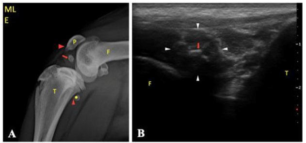
Radiographic evaluation of the left femorotibiopatellar (FTP) joint in mediolateral and craniocaudal projections was performed under outpatient anesthesia using Philips KL.74/20.40 radiographic equipment (55kV, 15mAs), a Bucky table with a floating homogeneous top, and an FCR CAPSULA X digital imaging system. Radiographs revealed a well-defined circular intra-articular structure with a peripheral region of soft-tissue radiopacity (1.19×0.76 cm) and a central core of bone radiopacity (0.76×0.59 cm). Additional findings included misalignment of the FTP joint axis, reduced articulation between the femoral condyles and the tibial plateau, caudodistal displacement of the popliteal sesamoid bone, and enthesophyte formation at the origin and insertion sites of the patellar and popliteal ligaments (Figure 1A).
(A) Mediolateral radiographic image and (B) longitudinal ultrasonographic image (7.5-9 MHz) of the lateral aspect of the femorotibiopatellar joint in a 9-month-old female Labrador Retriever. (A) Intra-articular bone-density structure located cranial to the femoral condyles and caudodistal to the patella (arrow); loss of femorotibiopatellar (FTP) joint axis alignment; caudodistal displacement of the popliteal sesamoid bone (*); and enthesophyte formation at the origin and insertion sites of the patellar and popliteal ligaments (arrowheads). (B) Circular hypoechoic structure (between arrowheads) with a central irregular hyperechoic component (arrow) producing posterior acoustic shadowing. F, femur; P, patella; T, tibia.
To assess soft tissue involvement, ultrasonographic evaluation of the left femorotibiopatellar (FTP) joint was performed using an FT 422 SAEVO system equipped with a linear multifrequency transducer (7.5-14.5 MHz). Longitudinal scans were obtained from the lateral, medial, cranial, and caudal aspects of the joint. Ultrasonography revealed a well-defined, homogeneous, circular hypoechoic structure (1.15×1.00cm) with a hyperechoic center producing posterior acoustic shadowing (0.50 cm; Figure 1B).
The combined radiographic and ultrasonographic findings were consistent with synovial osteochondromatosis (SOC). The patient was subsequently referred for arthrotomy, during which the intra-articular structure identified on imaging was surgically excised. Intraoperatively, a cranial cruciate ligament rupture and a medial meniscal tear were also observed. Accordingly, the patient underwent tibial plateau leveling osteotomy and partial meniscectomy of the caudal pole of the medial meniscus.
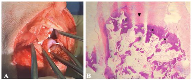
The excised intra-articular fragment was irregularly shaped, brown in color, firm, and elastic in consistency, measuring approximately 1.50×1.00×0.80cm (Figure 2A). Histopathological examination revealed abundant mature fibrous connective tissue interspersed with sparse blood vessels and extensive areas of cartilaginous and osseous metaplasia, findings consistent with a diagnosis of SOC (Figure 2B).
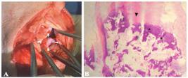
(A) Intraoperative image and (B) histopathological micrograph of the osteocartilaginous fragment excised from the left femorotibiopatellar joint of a 9-month-old female Labrador Retriever. (A) Intra-articular fragment (between arrowheads) observed during arthrotomy. (B) Micrograph showing lamellar bone (asterisk), cartilage (arrowhead), and loose connective tissue (HE stain, 10× magnification).
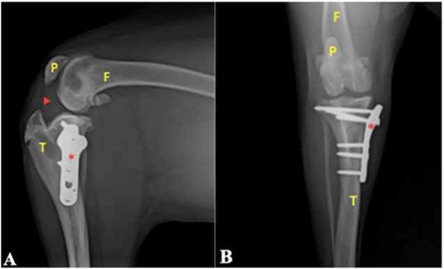
The patient was re-evaluated 75 days after surgery with no evidence of orthopedic abnormalities. A follow-up radiographic examination was performed to assess possible recurrence of the osteocartilaginous nodule (Figure 3). The radiographs demonstrated the presence of the metallic implant, absence of the previously identified osteocartilaginous nodule, proper alignment of the femorotibiopatellar (FTP) joint axis, and persistent enthesophyte formation at the origin and insertion sites of the patellar ligament. The sesamoid bone of the popliteal muscle was not visible.

Postoperative radiographs of the left femorotibiopatellar joint in a 9-month-old female Labrador Retriever. (A) Mediolateral projection showing no abnormalities in the intra-articular region (arrowhead), presence of a metallic implant (*), proper alignment of the femorotibiopatellar (FTP) joint axis, and enthesophyte formation at the origin sites. The insertion of the patellar ligament and the sesamoid bone of the popliteal muscle are not visible. (B) Craniocaudal projection demonstrating bone and joint structures within normal radiographic limits. F, femur; P, patella; T, tibia.
DISCUSSION
In the present case, the patient had a history of previous trauma. During physical examination, the tibial compression test produced cranial displacement of the tibia relative to the femur, indicative of cranial cruciate ligament instability (Hayashi et al., 2004; Czerwik et al., 2019). Based on these findings, radiographic evaluation was performed to assist in surgical planning. The radiographs demonstrated loss of alignment of the femorotibiopatellar (FTP) joint axis and reduced contact between the femoral condyles and the tibial plateau, findings consistent with the clinical examination.
Additionally, a circular bone-density structure was identified in the intra-articular region, surrounded by soft-tissue radiopacity. These findings supported a diagnosis of SOC. Terazaki et al. (2014) reported that nodules may calcify in irregular patterns or concentric layers, consistent with the observations in the present case.
To assess soft-tissue involvement, ultrasonographic examination was performed, revealing a homogeneous hypoechoic structure with a central nucleus producing posterior acoustic shadowing, indicative of mineralization findings consistent with descriptions in the literature (Terazaki et al., 2014; Maghear et al., 2018). A hypoechoic halo surrounding the mineralized area suggested adjacent soft-tissue reaction (Kim et al., 2014; Terazaki et al., 2014). Although ultrasonography has not previously been reported as a diagnostic tool for SOC in veterinary medicine, the findings observed here were consistent with those described in human cases (Maghear et al., 2018; Czerwik et al., 2019).
The primary diagnostic suspicion in this case was SOC, resulting from chronic irritation associated with predisposing joint disease (Aeffner et al., 2012; Smith et al., 2012; Tas et al., 2013). In this patient, the underlying condition was a ruptured cranial cruciate ligament. In addition to a history of trauma and concomitant joint disease confirmed radiographically, only a single osteocartilaginous nodule was identified, a feature that McKenzie et al. (2008) consider characteristic of secondary SOC. In contrast, primary SOC typically presents with multiple nodules of uniform size, whereas secondary SOC is associated with fewer nodules of variable dimensions. Although this disease is well documented in humans, no published reports in dogs are currently available (Terazaki et al., 2014; Ji et al., 2015; Wang et al., 2016).
Histopathological evaluation revealed no evidence of malignancy, and the diagnosis of synovial SOC was established based on the imaging findings and the patient’s clinical history (Aeffner et al., 2012; Kim et al., 2014). The absence of nuclear atypia further supported the diagnosis of secondary SOC (Murphey et al., 2007; Czerwik et al., 2019).
The combined interpretation of radiographic, ultrasonographic, surgical, and histopathological findings underscored the importance of multimodal diagnostic assessment in the evaluation of this disease. Comparison of the fragment’s size and location as described on radiographic (Fig. 1A) and ultrasonographic (Fig. 1B) images with those observed intraoperatively (Figure 2A) demonstrated close agreement, although imaging slightly underestimated the actual dimensions.
In humans, magnetic resonance imaging and computed tomography are recommended for diagnostic confirmation (Nasri et al., 2023). These advanced imaging techniques were not employed in the present case, as the radiographic and ultrasonographic findings provided sufficient diagnostic information for clinical decision-making, allowing definitive management and closure of the case.
CONCLUSION
Based on the radiographic and ultrasonographic evaluations of the FTP joint, together with histopathological analysis of the osteocartilaginous nodule, a diagnosis of SOC was established in this dog. These findings demonstrate that the combined use of radiography, ultrasonography, and histopathology is highly effective for diagnosing synovial SOC in canine patients. In addition to enabling an accurate diagnosis, these complementary diagnostic tools provided valuable information on lesion extent, involvement of adjacent structures, and contributed to guiding treatment planning and prognosis.
REFERENCES
- AEFFNER, F.; WEEREN, R.; MORRISON, S. et al Synovial osteochondromatosis with malignant transformation to chondrosarcoma in a dog. Vet. Pathol, v.49, p.1036-1039, 2012.
- CZERWIK, A.; OLSZEWSKA, A.; STARZOMSKA, B. et al Multiple cartilaginous exostoses in a Swiss Mountain dog causing thoracolumbar compressive myelopathy. Acta Vet. Scand, v.61, p.32-40, 2019.
- DIAZ-BERTRANA, C.; DURALL, I.; RIAL, J.M. Extra- and intra-articular synovial chondromatosis and malignant transformation to chondrosarcoma. Vet. Comp. Orthop. Traumatol., v.23, p.277-283, 2010.
- FRIEDENBERG, S. G., VANSTEENKISTE, D., YOST, O et al A de novo mutation in the EXT2 gene associated with osteochondromatosis in a litter of American Staffordshire Terriers. J. Vet. Intern. Med, v.32, p.986-992, 2018.
- HAYASHI, K.; MANLEY, P.A.; MUIR, P. Cranial cruciate ligament pathophysiology in dogs with cruciate disease: a review. J. Am. Anim. Hosp. Assoc., v.40, p.385-390. 2004.
- JI, J.H.; SHAFI, M.; JEONG, D.S. Secondary synovial chondromatosis of the shoulder. Knee Surg. Sports Traumatol. Arthrosc., v.23, p.2624-2627, 2015.
- KIM, T.K.; LEE, D.H.; PARK, J.H. et al Synovial osteochondromatosis in the subacromial bursa mimicking calcific tendinitis: sonographic diagnosis. J. Clin. Ultrasound, v.42, p.237-240, 2014.
- MAGHEAR, L.; SERBAN, O.; PAPP, I. et al Multimodal ultrasonographic evaluation in a case with unossified primary synovial osteochondromatosis. Med. Ultrason, v.20, p.527-530, 2018.
- MCKENZIE, G.; RABY, N.; RITCHIE, D. A pictorial review of primary synovial osteochondromatosis. Eur. Radiol., v.18, p.2662-2669, 2008.
- MURPHEY, M.D.; VIDAL, J.A.; FANBURG-SMITH, J.D.; GAJEWAKI, D.A. Imaging of synovial chondromatosis with radiologic-pathologic correlation. Radiographics, v.27, p.1465-1488, 2007.
- NASRI, S.; BELHARTI, A.; KENNOUDI, N. et al Primary synovial osteochondromatosis of the ankle: a case report and review of the literature. Radiol. Case Rep, v.18, p.701-703, 2023.
- SEGHROUCHNI, M.; BOLLO, E.; PIRO, M. et al Osteochondroma of the first phalanx in Tbourida horses. Front. Vet. Sci, v.5, p.328-330, 2019.
- SMITH, T.J.; BALTZER, W.I.; LOHR, C. Primary synovial osteochondromatosis of the stifle in an English Mastiff. Vet. Comp. Orthop. Traumatol, v.25, p.160-166, 2012.
- SZILASI, A.; KOLTAI, Z.; DÉNES, L. et al In situ hybridization of feline leukemia virus in a case of osteochondromatosis. Vet. Sci, v.9, p.59-65, 2022.
- TAS, O.; COCK, H.; LEMMENS, P. Synovial osteochondromatosis and sclerosing osteosarcoma in a cat. Vet. Comp. Orthop. Traumatol, v.26, p.160-164, 2013.
- TERAZAKI, C.R.T.; TRIPPIA, C.R.; TRIPPIA, C.H.; CABOCLO, M.S.F.S. Synovial chondromatosis of the shoulder: imaging findings. Radiol. Bras., v.47, p.38-42, 2014.
- ANG, Y.; LI, L.; CHEN, M.; YANG, C. Osteochondroma with secondary synovial chondromatosis in the temporomandibular joint. Br. J. Oral Maxillofac. Surg., v.54, p.454-456, 2016.
The research data are available within the article itself.