
 Cutaneous cryptococcosis with atypical location in a cat - case report
ABSTRACT
Cryptococcosis is a rare systemic mycosis among pathogenic fungi, yet it is recognized for its clinical importance in felines. This paper reports on a clinical case of cutaneous cryptococcosis in a domestic cat (Felis catus), presenting a single lesion on the abdominal wall, without ulceration, an uncommon presentation in veterinary literature. The 7-year-old cat had normal laboratory parameters, which contrast with most reports of systemic fungal infections. Diagnosis was based on cytology obtained through fine-needle aspiration (FNA), stained with Panoptic stain and complemented by the India ink technique, highlighting the polysaccharide capsule of Cryptococcus spp. Antifungal therapy with itraconazole and fluconazole, both administered orally, was initiated. Despite early treatment, the cat died of cardiorespiratory arrest on the fourth day of hospitalization. This report emphasizes the importance of early diagnosis, even in atypical clinical presentations, and highlights cytology with specific staining as an accessible and effective diagnostic tool, as well as alerting to the possibility of fulminant disease progression even in the absence of evident clinical and laboratory changes.
Keywords:
encapsulated yeasts; cytology; India ink staining; systemic mycosis
RESUMO
A criptococose é uma micose sistêmica rara entre os fungos patogênicos, mas, ainda assim, reconhecida por sua importância clínica em felinos. Suas formas clínicas mais comuns envolvem acometimento rinonasal e cutâneo e, em casos mais graves, comprometimento do sistema nervoso central. Este trabalho relata um caso clínico de criptococose cutânea em felino doméstico (Felis catus), com lesão única na parede abdominal, sem ulceração, apresentação considerada incomum na literatura. O animal, de sete anos, apresentou parâmetros laboratoriais normais, contrastando com a maioria dos relatos de infecções fúngicas sistêmicas. O diagnóstico foi realizado por citologia obtida via punção aspirativa por agulha fina (PAAF), com coloração de Panótico, complementada pela técnica com tinta da China, evidenciando a cápsula polissacarídica de Cryptococcus spp. Instituiu-se terapia antifúngica com itraconazol e fluconazol, ambos por via oral. Apesar do início precoce do tratamento, o animal evoluiu para óbito por parada cardiorrespiratória no quarto dia de internação. O relato destaca a importância do diagnóstico precoce, mesmo em apresentações clínicas atípicas, e reforça a citologia com coloração específica como ferramenta diagnóstica eficaz, além de alertar para a possibilidade de evolução fulminante da doença, mesmo na ausência de alterações clínicas e laboratoriais evidentes.
Palavras-chave:
leveduras encapsuladas; citologia; coloração com nanquim; micose sistêmica
INTRODUCTION
Cryptococcosis is a rare systemic mycosis among pathogenic fungi, yet it is recognized for its clinical importance in cats (Larsson et al., 2003; Caswell and Williams, 2007). It is caused by the encapsulated yeasts Cryptococcus neoformans and C. gatti, which, although uncommon in companion animals, are clinically significant due to the severity of systemic conditions and their zoonotic potential (Zhao et al., 2022; Pedroso and Cândido, 2006). These fungi are found in pigeon droppings, tree trunks and decomposing organic matter, where they can remain viable for years (Reolon et al., 2004; Nelson and Couto, 2015). Infection occurs mainly through inhalation of spores, with possible haematogenous spread to the central nervous system, skin and other organs (Hosho et al., 2003). Infection in the respiratory tract of cats causes clinical signs such as serous or bloody nasal discharge, nasal deformity and occlusion, rhinitis, sneezing, sinusitis, and oro-nasal lesions with crusts or ulcers, as well as respiratory noises (Betty et al., 2004; Norsworthy et al., 2004). Pereira and Coutinho (2023) and Ettinger et al. (2017) describe the appearance of lesions with a characteristic ‘clown nose’ appearance. When the central nervous system is affected, the symptoms to be observed include convulsions, paresis, ataxia, depression, and blindness (Ker, 2003).
Considering the increase in antifungal resistance, the scarcity of effective therapies, and high mortality in immunosuppressed individuals, the World Health Organization (WHO) has classified C. neoformans as a critical priority pathogen and C. gatti as a medium priority pathogen (Fungal…, 2022). The diagnosis can be confirmed by cytology, microbiological culture, histopathology, or molecular techniques (Hnilica and Medleau, 2012; Medleau et al., 2003; Trindade et al., 2008). Despite this, many cases are only identified in post-mortem findings (Rondelli et al., 2010), and treatment requires prolonged use of systemic antifungals (Malik et al., 2013). Although renal forms are the most common in cats (Caswell and Williams, 2007; Malik et al., 2002), cutaneous manifestations may also occur, especially by haematogenous dissemination or direct inoculation (Malik et al., 2013). However, involvement in atypical regions, especially the abdominal cavity, is considered rare, with few cases described in cats in the literature (Souza et al., 2023).
This study aims to report a clinical case of feline cryptococcosis with atypical cutaneous presentation in the abdominal region and discuss the laboratory findings, emphasising the importance of cytology as a diagnostic tool, in addition to the therapeutic challenges involved.
Although the rhinonasal form is more frequently reported in cats (Malik et al., 2002; Caswell and Williams, 2007), cutaneous manifestations may also occur, especially in cases of haematogenous dissemination or direct inoculation (Malik et al., 2013). However, the occurrence of subcutaneous lesions located in unusual anatomical regions, such as the abdominal wall, is considered uncommon in the veterinary literature, with few reports available in cats (Souza et al., 2023). Thus, this study aims to report a clinical case with atypical cutaneous presentation and discuss the laboratory findings, emphasizing the importance of cytology as a diagnostic tool and the therapeutic challenges involved.
CASE REPORT
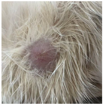
A female, spayed, mixed-breed cat weighing 3.0kg and aged 7 years was admitted to the Veterinary Hospital of Universidade Brasil - UB presenting a non-ulcerated, rounded, mobile nodule in the lateral region of the abdomen measuring approximately 3 cm in diameter (Fig. 1).
The first consultation took place on 18 August 2024, when the guardian reported that the lump had appeared 15 days earlier. The animal had been anorexic for two days and was presenting with polydipsia, polyuria and normal defecation, with no gastrointestinal, neurological, locomotor or cardiovascular changes. On physical examination, the physiological parameters (heart and respiratory rate, rectal temperature, capillary perfusion time, and mucosal coloration) were within normal limits for the species, and no changes were found on pulmonary auscultation. Based on the history and clinical signs, cutaneous neoplasia was suspected.

Non-ulcerated, rounded, mobile nodule located in the lateral region of the abdomen, approximately 3 cm in diameter.
Additional tests were requested, such as a complete blood count and serum biochemistry of renal and hepatic enzymes. The Erythrogram showed mild normocytic normochromic anaemia. The other results were within normal limits for the species. A cytopathological examination was performed on material obtained by fine needle aspiration (FNA) of the nodule. The slides were initially stained using the panotic method for general cell evaluation. In addition, India ink staining was used to highlight the characteristic polysaccharide capsule of Cryptococcus spp., facilitating the identification of the etiological agent by visualizing the clear halo around the fungal cells, confirming the presence of the pathogen.
RESULTS AND DISCUSSION
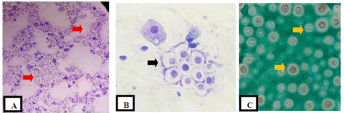
In the cytological evaluation, numerous yeast-like structures were observed. In some fields, the yeasts had large, clear capsules with narrow bases (Fig. 2).

Microorganisms of the genus Cryptococcus are observed, characterized by medium to large yeasts with a thick, clear and well-defined capsule. In image A (10x objective), there are numerous yeasts scattered across the bottom of the slide, some grouped together (red arrows), with a clear halo compatible with the mucopolysaccharide capsule. In image B (40x objective), encapsulated yeasts are identified, located both in clusters (black arrows) and inside macrophages, exhibiting budding with a narrow base. In image C (40x objective), yeast-like structures stained in intense green are noted, with an evident translucent capsule, a characteristic enhanced by the specific staining technique (yellow arrows).
After diagnosis, oral antifungal therapy with itraconazole and fluconazole was instituted. Itraconazole was administered at a dose of 10mg/kg once daily (SID) for 6 months. Fluconazole was used as adjunctive therapy at a dose of 5mg/kg SID, according to clinical protocol. On 21 August 2024, the animal suffered cardiorespiratory arrest and died.
The nodule was located in the abdominal region of the cat, an area considered unusual for cutaneous manifestations of cryptococcosis. The literature describes that cutaneous forms generally occur in regions such as the nasal plane, face, ears, and paws, which are more susceptible to direct inoculation or hematogenous dissemination (Malik et al., 2002; Malik et al., 2013). In the present case, the lesion presented as a firm subcutaneous nodule with no signs of ulceration, which also differs from the most common clinical pattern of cutaneous cryptococcosis, which usually presents with ulceration, scabs, exudate, or fistulas (Caswell and Williams, 2007).
Given this, the importance of early diagnosis, especially through cytological evaluation, is emphasized. Cytology of the nodule was performed by fine needle aspiration (FNA), followed by rapid panoptic staining and India ink staining. The yeasts observed had a diameter between 4 and 15 µm, a rounded shape, thick wall, and evident mucopolysaccharide capsule, with narrow base budding. These characteristics are compatible with Cryptococcus spp., especially when associated with positivity in India ink staining.
Differentiation from other yeast-like fungi, such as Histoplasma capsulatum-which is smaller (2-4µm), intracellular, and has no obvious capsule-and Blastomyces dermatitidis, which has broad-based budding, is essential for accurate diagnosis. Recognition of these typical structures in cytological smears is a quick and effective screening tool, especially in nodular skin presentations.
The use of India ink staining is a rapid, low-cost diagnostic technique that is widely recognized in the literature for cases of cryptococcosis (Malik et al., 2013; Pennisi et al., 2013).
The complete blood count revealed mild normocytic normochromic anemia, evidenced by a hematocrit of 23% (reference range: 24-45%), mean corpuscular volume of 55fL (reference range: 39-55fL), and mean corpuscular hemoglobin concentration of 36g/dL (reference: 30-36g/dL). The white blood cell count showed a total leukocyte count of 9,400/mm³ (reference: 5,500-19,500/mm³), with no significant changes in differential distribution. The platelet count was 375,000/mm³ (reference: 230,000-680,000/mm³), and total plasma protein was 6.8g/dL (reference: 4.5-7.8g/dL). In serum biochemistry, creatinine (1.3mg/dL; reference: 0.8-1.8mg/dL) and ALT (60U/L; reference: up to 90 U/L) values remained within physiological limits. However, this does not exclude the potential severity of the infection, since cryptococcosis can present systemic progression even with apparently normal laboratory tests, especially in the early stages or in localized presentations (Caswell and Williams, 2007; Malik et al., 2013). This finding highlights the importance of not underestimating fungal infection even in the absence of obvious clinical changes.
The patient was an adult female cat, aged 7 years. The literature reports that feline cryptococcosis most frequently affects young cats and young adults, between 2 and 5 years of age, with occurrence in older animals being less common (Malik et al., 2002; Caswell and Williams, 2007), which contributes to the atypical nature of the present case.
Based on the presumptive diagnosis, an antifungal protocol was instituted with oral itraconazole and fluconazole. The regimen used was itraconazole at 10mg/kg once daily (SID) and fluconazole at 5mg/kg SID, with 50mg tablets adapted to the patient's body weight (3kg). Current studies indicate that fluconazole is often better tolerated, with good tissue penetration, especially in infections affecting the central nervous system (European Advisory Board on Cat Diseases, 2024).
Considering the need for early therapeutic intervention, cytological diagnosis plays an essential role in cases of nodular skin presentation. This type of lesion can mimic neoplasms, abscesses or other infectious dermatoses, making immediate clinical diagnosis difficult. In this context, aspiration cytology, especially when associated with India ink staining, represents an accessible, rapid, and effective screening tool for the early recognition of fungal infection (Malik et al., 2013; Pennisi et al., 2013). The cytological finding of yeasts with mucopolysaccharide capsules and narrow-base budding is essential for differentiation from other yeast-like fungi. Thus, even in the face of initial laboratory tests within normal limits, well-founded clinical suspicion and targeted cytological investigation are essential for early diagnosis and the establishment of timely therapeutic conduct.
Despite early treatment, the animal's condition deteriorated and it died of cardiorespiratory arrest on the fourth day of hospitalization. No previous laboratory changes were observed that would justify organ failure, suggesting a possible acute and fulminant progression, as already described in other cases of disseminated cryptococcosis or initially misdiagnosed (Souza et al., 2023). This highlights the aggressive potential of the disease, even in apparently localized presentations, and the importance of early recognition and rigorous therapeutic intervention.
CONCLUSION
This report describes an atypical case of cutaneous cryptococcosis in a cat, with a nodular lesion located in the abdominal region, without signs of ulceration and with laboratory parameters within reference ranges. The diagnosis was confirmed by cytology with Panoptic staining and complemented with India ink, revealing the characteristic capsule of Cryptococcus spp. Despite early initiation of antifungal treatment with itraconazole and fluconazole, the animal died within a few days, demonstrating the aggressive potential of the infection, even in apparently localized presentations. This case highlights the importance of considering cryptococcosis as a differential diagnosis in nodular skin lesions, even when classic signs such as ulceration or laboratory abnormalities are absent and reinforce the essential role of cytology as a rapid and effective tool for the early diagnosis of the disease.
REFERENCES
- BETTY, D.; FRANCESCHI, C.; MARTINS, M.C. Criptococose felina: revisão de literatura. Clín. Vet., v.54, p.42-46, 2004.
- CASWELL, J.L.; WILLIAMS, K.J. Cryptococcus spp. In: MAXIE, M.G. (Ed.). Jubb, Kennedy, and Palmer’s pathology of domestic animals 5.ed. Philadelphia: Saunders Elsevier, 2007. p.157-161.
- ETTINGER, S.J.; FELDEMAN, E.C.; CÔTÉ, E. Textbook of veterinary internal medicine 8.ed. St Louis: Elsevier, 2017. p.1502-1504.
-
EUROPEAN Advisory board on cat diseases (ABCD). ABCD guidelines: Cryptococcosis in cats. 2024. Available in: https://www.abcdcatsvets.org/. Accessed in: 6 Jul. 2025.
» https://www.abcdcatsvets.org - HNILICA, K.A.; MEDLEAU, L. Small animal dermatology: a color atlas and therapeutic guide. 3.ed. St Louis: Elsevier, 2012. p.270-272.
- HOSHO, K.; HARTMANN, K.; LLORETT, A. et al Cryptococcus neoformans infection in a cat: cytologic diagnosis and treatment. J. Vet. Med. Sci., v.65, p.1367-1369, 2003.
- KER, J.E. Neurological manifestations of systemic fungal infections in cats. J. Feline Med. Surg, v.5, p.161-165, 2003.
- LARSSON, C.E.; OTSUKA, M.; MICHALANY, N.S. et al Criptococose em felinos domésticos: revisão e relato de casos. Braz. J. Vet. Res. Anim. Sci, v.40, p.269-274, 2003.
- MALIK, R.; WIGNEY, D.I.; MUIR, D.B. et al Cryptococcosis in cats: clinical and mycological features, diagnosis and therapeutic recommendations. Aust. Vet. J., v.80, p.749-758, 2002.
- MALIK, R.; KROKENBERG, D.J.; FYFE, J. et al Cutaneous cryptococcosis in cats: clinical presentation and treatment outcomes. Vet Dermatol., v.24, p.235-240, 2013.
- MEDLEAU, L.; MARKS, M.A.; BROWN, J.; BORGES, W.L. Diagnosis of cryptococcosis in cats: clinical, cytological and histopathological aspects. Vet. Dermatol, v.14, p.193-198, 2003.
- NELSON, R.W.; COUTO, C.G. Small animal internal medicine 5.ed. St Louis: Mosby Elsevier, 2015. p.720-722.
- NORSWORTHY, G.D.; GRACE, S.F.; CRYSTAL, M.A. The feline patient 4.ed. Ames: Wiley-Blackwell, 2004. p.231-233.
- PEDROSO, P.M.O.; CÂNDIDO, L.M. Cryptococcosis in domestic animals: clinical and pathological features. Vet. Pathol, v.43, p.651-658, 2006.
- PENNISI, M.G.; HARTMANN, K.; FERRER, L. et al Cryptococcus neoformans and Cryptococcus gattii infections in cats and dogs: ABCD guidelines for diagnosis and management. J. Feline Med. Surg, v.15, p.611-618, 2013.
- PEREIRA, M.C.L.; COUTINHO, M.B. Nasal deformities in feline cryptococcosis: clinical features and differential diagnoses. Braz. J. Vet. Med, v.45, p.e202345, 2023.
- REOLON, L.; MEIRELES, M.C.A.; CLEFF, M.B. et al Fungal flora in urban pigeon droppings and implications for public health. Rev. Soc. Bras. Med. Trop, v.37, p.424-428, 2004.
- RONDELLI, M.I.; NOGUEIRA, A.F.S.; ANAI, L.A. et al Cryptococcosis in cats: post mortem findings and histopathology. Mycopathologia, v.169, p.249-253, 2010.
- SOUZA, T.M.; BIANCH, M.V.; PAVARINI, S.R. et al Abdominal cutaneous cryptococcosis in cats: case report and review of the literature. Braz. J. Vet. Pathol, v.16, p.102-107, 2023.
- TRINDADE, E.; ROCHA, L.H.B.; ANDRADE, M.C.R.; ALMOSNY, N.R.P. Diagnóstico rápido de criptococose em felinos através da técnica de tinta da china. Rev. Bras. Med. Vet., v.30, p.221-224, 2008.
- FUNGAL priority pathogens list to guide research, development and public health action. Geneva: WHO, 2022.
- ZHAO, Y.; SOHN, J.H.; WARNER, J.R. et al Global status of cryptococcosis in veterinary species. Mycoses, v.65, p.617-625, 2022.
The data supporting the findings of this study are available within the article.

 Source: Personal archive.
 Source: Author's personal archive (2025).