 Molecular prevalence and risk factors analysis of Theileria annulata in cattle at Jeddah, Saudi Arabia
ABSTRACT
Theileria annulata is a tick-borne heamoprotozoan parasite responsible for tropical theileriosis in bovine populations, which causes substantial economic losses to the livestock sector. This study aimed to determine the prevalence of T. annulata infection in cattle in Jeddah, Saudi Arabia, and its associated risk factors using microscopic examination of Giemsa-stained blood smears (GSBS), PCR targeting the 18S rRNA and cytochrome b (cyt b) gene, and sequencing analysis. Material and Methods: A total of 200 blood samples were collected randomly from cattle between December 2022 and May 2023. Results: Results revealed that 53 samples (26.5%) were positive for Theileria piroplasms using GSBS and increased to 71 samples (35.5%) using PCR. The risk of T. annulata infection was reported to be non-significantly higher among young, female, and imported cattle. The study revealed that 53 samples (26.5%) were positive for Theileria piroplasms using GSBS, while the PCR technique showed 71 samples (35.5%). The level of kappa agreement between GSBS and PCR was slight (0.135). Further, GSBS showed 51% accuracy, 18.31% sensitivity, and 68.99% specificity when compared to PCR results. Analysis of the cyt b sequence of Saudi isolates of T. annulata showed 99.27-100% identity, and with 18S rRNA sequences the identity was > 98%. Conclusion: The high infection of T. annulata in slaughtered cattle in Jeddah requires the implementation of adopted programs to reduce the burden of theileriosis on the Saudi economy. PCR is considered a better test for conducting periodic examinations and control programs for bovine theileriosis.
Keywords:
Cattle; Polymerase chain reaction; T. annulata; Theileriosis
RESUMO
Theileria annulata é um parasita hemoprotista transmitido por carrapatos responsável pela teileríase tropical em populações bovinas, causando perdas econômicas substanciais ao setor pecuário. Este estudo teve como objetivo determinar a prevalência da infecção por T. annulata em bovinos em Jeddah, Arábia Saudita, e seus fatores de risco associados, utilizando exame microscópico de esfregaços de sangue corados com Giemsa (GSBS), PCR direcionado ao gene 18S rRNA e citocromo b (cyt b) e análise de sequenciamento. Material e métodos: Um total de 200 amostras de sangue foram coletadas aleatoriamente de bovinos entre dezembro de 2022 e maio de 2023. Resultados: Os resultados revelaram que 53 amostras (26,5%) foram positivas para piroplasmas Theileria usando GSBS e aumentaram para 71 amostras (35,5%) usando PCR. O risco de infecção por T. annulata foi relatado como não significativamente maior entre bovinos jovens, fêmeas e importados. O estudo revelou que 53 amostras (26,5%) foram positivas para piroplasmas Theileria usando GSBS, enquanto a técnica de PCR mostrou 71 amostras (35,5%). O nível de concordância kappa entre GSBS e PCR foi leve (0,135). Além disso, o GSBS apresentou 51% de precisão, 18,31% de sensibilidade e 68,99% de especificidade quando comparado aos resultados da PCR. A análise da sequência cyt b de isolados sauditas de T. annulata mostrou 99,27-100% de identidade, e com sequências de rRNA 18S a identidade foi >98%. A alta infecção por T. annulata em bovinos abatidos em Jeddah requer a implementação de programas adotados para reduzir o impacto da teileríase na economia saudita. A PCR é considerada um teste melhor para a realização de exames periódicos e programas de controle da teileríase bovina.
Palavras-chave:
bovinos; reação em cadeia da polimerase; T. annulata; Teileríase
INTRODUCTION
Bovine theileriosis is a tick-borne protozoal disease caused by Theileria annulata and transmitted by ixodid ticks of the genus Hyalomma (Mohammed-Ahmed et al., 2018). This disease causes significant financial losses to farmers due to mortality, morbidity, reduced milk production, and high costs of acaricides, treatment, and vaccination (Perera et al., 2014). Although T. annulata causes productivity losses in exotic cattle breeds and their crosses, naive indigenous cattle, particularly calves and adults under endemic instability, are also affected (Kasaija et al., 2021). The clinical signs of theileriosis are variable and may include fever, mild nasal and ocular discharge, anorexia, salivation, enlargement of superficial lymph nodes, respiratory distress, acute anemia, jaundice, and death due to asphyxia (Osman and Al-Gaabary, 2007). The most common pathological lesions include enlargement of the lymph nodes and spleen, pulmonary emphysema, and subcutaneous and intramuscular hemorrhages.
Accurate and conclusive laboratory testing is essential for confirming Theileria infection. Although theileriosis is commonly diagnosed by microscopic examination of Giemsa-stained peripheral blood smears or by detecting macro schizonts in the lymph node biopsy smears (Perera et al., 2014; Hayati et al., 2020), this method is not sufficiently sensitive to detect infection in carrier animals (Pienaar et al., 2020). Furthermore, serological diagnosis using IFA and ELISA may yield false positives due to cross reactivity or inaccurate specific immune response (Billiouw et al., 2005). Blood and biochemical profiles, such as bilirubin, cholesterol, albumin, also total proteins, can be useful within diagnosing several diseases (Alnahari et al., 2025). However, since its introduction, PCR has demonstrated greater sensitivity, specificity, and accuracy for detecting T. annulata in infected animal hosts, even in cases of low parasitemia (Bilgic et al., 2010).
To date, several studies have been carried out in different regions of Saudi Arabia to identify and diagnose theileriosis in cattle. In Taif, Riyadh, and Al-Ahsa, studies (Ghafar and Amer, 2019; Alanazi et al., 2019) reported that more than 5% of cattle were infected with T. annulata by PCR. In contrast, studies conducted in Qassim and Jizan (Omer et al., 2002; Al-Khalifa et al., 2009) reported a prevalence of 15.4-25% based on the examination of blood smears. However, there is currently no available data on theileriosis in cattle in Jeddah, except for one study that reported T. ovis in sheep and goats from Jeddah using PCR targeting the 18S rRNA gene (Metwally et al., 2021). Therefore, the objectives of the present study were to estimate the prevalence and associated risk factors of theileriosis in cattle in Jeddah, to evaluate the diagnostic performance of Giemsa-stained blood smears compared to PCR, and to genetically characterize T. annulata isolates by targeting the 18S rRNA and cytochrome b (cyt b) genes.
ETHICAL ASPECTS
The research was submitted to the Ethics Committee on Animal Use of the University of Jeddah, Saudi Arabia, and approved under the number (UJ-232430040).
MATERIALS AND METHODS
From December 2022 to May 2023, a total of 200 blood samples were randomly collected from cattle from the central slaughterhouse in Jeddah city, located in the western region of Saudi Arabia (21° 32′ 36″ N, 39° 10′ 22″ E). Data including sex, breed, and age (categorized into three groups according to reference 4) were recorded. Blood samples (5 mL) were aseptically collected from the jugular vein using a vacuum tube with ethylenediamine tetra acetic acid (EDTA) and transported in an ice box to the Department of the College of Science at the University of Jeddah Laboratory for preparation of thin blood smears and preserved at −20°C for molecular analysis (Selim et al., 2022).
Briefly, a small drop of fresh blood was placed on the first third of a clean glass microscopic slide, then spread with another slide and left to air dry. The blood film was fixed in methanol alcohol for 2 min and then stained with Giemsa stain for 30 min and examined under an oil immersion lens at x100 (Lempereur et al., 2017).
DNA extraction was performed using the Qiagen DNA Purification Kit (Promega, USA), as per the manufacturer’s instructions, and stored at - 20(C until future use. For the amplification of targeted genes of Theileria spp., both on genus and species-specific levels. The PCR was performed as per previously described PCR assays. The amplification of the 18S rRNA gene (778 bp) was performed using previously described primers: F: 5'-GAAACGGCTACCACATCT-'3, R: 5'-AGTTTCCCCGTGTTGAGT-'3 (Cao et al., 2013). The cycling conditions were a cycle at 95(C for 5 min and 35 cycles of 94(C for 1 min, 60(C for 1 min, and 72(C for 90 s, and a final extension for 10 min at 72(C. The amplification of the cyt b gene (312 bp) was performed using previously described primers; F: 5(-ACT TTG GCC GTA ATG TTA AAC-3( and R: 5(-CTC TGG ACC AAC TGT TTG G-3( (Bilgiç et al., 2013). The cycling conditions of a cycle at 94(C for 5 min followed by 35 cycles of 94(C for 30 s, 57(C for 30 s and 72(C for 30 s and a final extension at 72(C for 10 min. Positive and negative (PCR-grade water) controls were included for each run. PCR product (10μL) was electrophoresed on a 2% agarose gel containing 5μL/ml ethidium bromide in Tris-acetate-EDTA buffer at 110 V and visualized under UV light.
The PCR products of the Cyt b (312 bp) and 18S rRNA (778 bp) genes were purified by the QIAquick PCR purification kit (QIAGEN) and subjected to sequencing by using an automated DNA sequencer (ABI 3730XL, Solgent Co. Ltd., South Korea). DNA BaserV3 software was used to read the sequences. Sequences identified were aligned with the previously published T. annulata sequences in GenBank by the Basic Local Alignment Search Tool (BLASTn) (http://www.ncbi.nlm.nih.gov/BLAST), and analyzed by the MegAlign software (DNASTAR, Madison, WI, USA) using the Clustal W method. Divergence estimation within the Cyt b and 18S rRNA genes sequenced in this study was calculated in MEGA X by comparing with the other available Cyt b and 18S rRNA sequences of T. annulata from the GenBank. Divergence is calculated by MEGA X.
For the phylogenetic tree, the analysis was performed by using the Maximum Likelihood method (ML) based on the Tamura-Nei model (Tamura and Nei, 1993). The percentage of trees in which the associated taxa clustered together is shown next to the branches. The initial tree for the heuristic search was obtained by applying Neighbor-Join and BioNJ algorithms to a matrix of pairwise distances estimated using the Tamura-Nei model and then selecting the topology with superior log likelihood value. The tree is drawn to scale, with branch lengths measured in the number of substitutions per site. Codon positions included were 1st+2nd+3rd+noncoding. In the bootstrap analysis, 1000 replications were used. All positions containing gaps and missing data were eliminated. Evolutionary analyses were conducted in MEGAX (Tamura et al., 2021).
The SPSS software (SPSS for Windows Version 22, SPSS Inc., Chicago, USA) was used to analyze the data. The category variables were examined using Chi-square test. Statistical significance was defined as a P-value < 0.05.
RESULTS
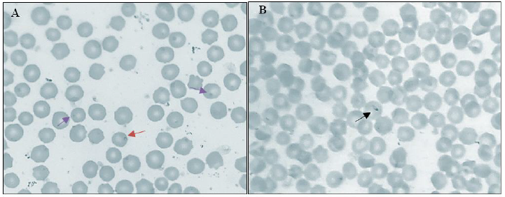
Out of 200 samples, 53 (26.5%) samples were found positive in GSBS, showing characteristic shaped piroplasm. Microscopic examination of stained thin blood smears revealed T. annulata (Fig. 1). Theileria piroplasms were observed mostly as signet rings or dots (Fig. 1A); some were detected in pyriform shape inside the red blood cells (Fig. 1B). As seen in Tables 1 and 2, the prevalence rate of T. annulata piroplasms in imported breed of cattle was higher (26.8%, 95% CI: 0.19-0.35) than in local ones (25.97%, 95% CI: 0.18-0.26). Regarding the age factor, a high prevalence rate was recorded among cattle ≤ 1 year old (27.7%, 95% CI: 0.18-0.39) compared to cattle >1 year old. Furthermore, 33.3% (95% CI: 0.06-0.79) prevalence was reported among females compared to 26.4% (95% CI: 0.21-0.33) prevalence in males. All differences were statistically non-significant (Tables 1 and 2).
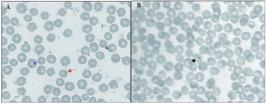
Blood smears demonstrating intracellular piroplasm of T. annulata (X 100). The presence of (A) signet ring (red arrow) and dot (blue arrow), (B) pyriform shaped (black arrow)
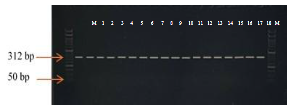
The PCR assay detected 71 samples (35.5%) positive for T. annulata. As shown in Table 1 and Fig. 2, prevalence increased non-significantly among cattle > 1 year of age (37.8%, 95% CI: 0.30 - 0.46, P=0.2), females (66.7%, 95% CI: 0.20 - 0.94, P=0.3), and imported cattle (39.8%, 95% CI: 0.32 - 0.49, P=0.11). The PCR included 57 samples previously tested negative by Giemsa staining (Table 3).
A comparison of the diagnostic characteristics between GSBS and PCR in the examination of 200 blood samples collected showed that the PCR detected most of the samples and the GSBS detected the least. All percentages of positivity for each test are seen in Table 1. The PCR's ability to identify more blood samples increased the number of T. annulata-positive blood samples to 71, compared to 53 positive samples using GSBS. Not all samples positive with GSBS were also positive with PCR (Table 3). Therefore, 57 GSBS-negative blood samples were PCR positive. According to the negative and positive results, the Kappa agreement between the GSBS and PCR revealed slight agreement (0.135; 95% CI: 0.261 -0.008; Table 3). In addition to the results of kappa, GSBS showed 51.00% accuracy, 18.31% sensitivity, and 68.99% specificity in the detection of T. annulata in cattle when compared to PCR results as a gold standard test (Table 4).
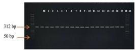
Detection of T. annulata DNA in native cattle. Lane M (50 bp DNA ladder). Lane 1 (positive control of T. annulata). Lane 2 to 18 (positive samples)
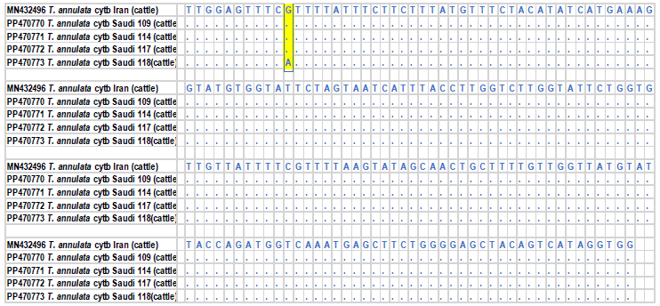
The partial Cyt b gene sequence (312 bp) was sequenced. The sequences of the four Saudi isolates were aligned with the related T. annulata Cyt b gene sequences in the GenBank database, and the alignment of the identified sequences with retrieved Cyt b sequences showed 99.27-100% identity. The BLASTn search of the identified nucleotide sequences with the reference T. annulata Cyt b gene (MN492496) showed 100% identity among the three sequences (PP470770-2) and with the four-sequence (PP470773) showed 99.9% identity with a substitution at one nucleotide (Fig. 3). Pairwise genetic distance of the four sequences with the reference sequence revealed one nucleotide substitution, indicating a very little genetic distance of 0.01. These results suggest a little genetic diversity between Saudi isolates based on the Cyt b gene.
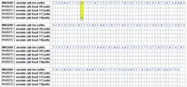
Nucleotide sequence alignment of Saudi T. annulata cyt b gene variants (this study, GenBank Accession No. PP70770-3) with reference T. annulata isolate from Iran (GenBank Accession No. MN432496). Identical bases (dots) and sites of variation (yellow box)
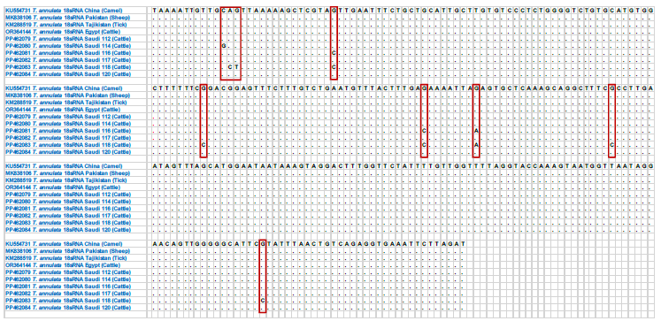
The 778 bp nucleotide sequences corresponding to the partial 18S rRNA gene sequence were amplified and sequenced. The sequences of the six Saudi isolates were compared with the other related 18S rRNA gene sequences in the GenBank database and the alignment of the identified sequences with reference T. annulata 18S rRNA sequences showed > 98 % identity. Six sequences were deposited in the GenBank database under the accession numbers PP462079, PP462080, PP462081, PP462082, PP462083, and PP462084. Alignment of the six sequenced T. annulata isolates with the reference T. annulata 18SrRNA gene (KU554731, KM288519, OR364144, and MK838106) showed that three sequences (PP462079, PP462082, and PP462084) were 100% identical. But lower identity (97.95-99.99%) was estimated when aligned with the other three sequences, PP462080, PP462081, and PP462083, which showed 1, 3, and 8 nucleotide substitutions, respectively, with a genetic distance of 0.01 - 0.02 (Fig. 4).
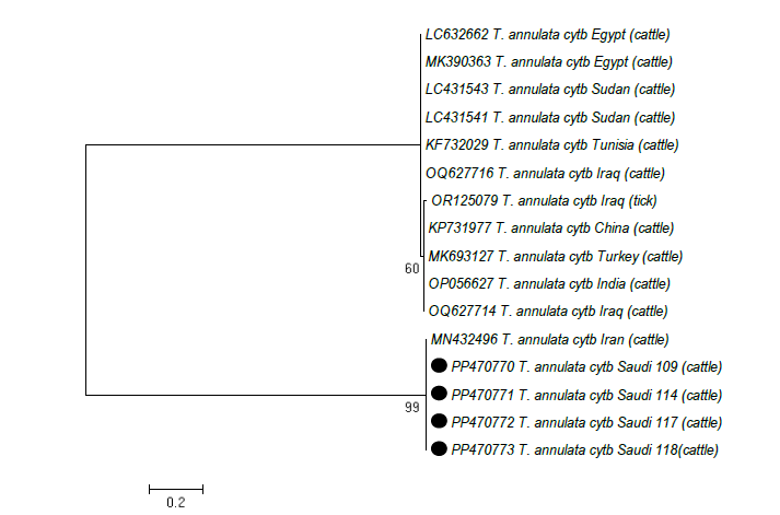
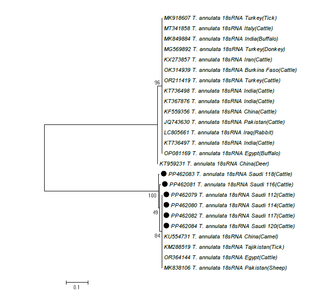
A phylogenetic tree based on the partial Cyt b sequences (312 bp) of Saudi isolates and sequences from Egypt, Sudan, Iran, Turkey, Iraq, China, Tunisia, and India was generated using the Maximum Likelihood method. The tree is divided into two clades. The studied isolates grouped into a single clade and were closely related to the Iranian T. annulata isolate with 99% nodal support but distinct from other known T. annulata from Egypt, Sudan, Turkey, Iraq, China, Tunisia, and India, and the estimated pairwise distance ranged between 0.48 and 0.51 (Fig. 5). Further, phylogenetic tree based on 18S rRNA gene sequences classified the obtained isolates into three subtypes (ST). ST1 included one sequence (PP462083) with eight nucleotide substitutions. ST2 included one sequence (PP462081) with three nucleotide substitutions. ST3 included four sequences (PP462079, PP462082, and PP462084) with one nucleotide substitution. These isolates were closely related to T. annulata from Egypt, China, Tajikistan, and Pakistan but distinct from other known T. annulata from other countries (Fig. 6).
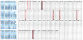
Nucleotide sequence alignment of Saudi T. annulata 18S rRNA gene variants (this study, GenBank Accession No. PP462079-83) with reference T. annulata isolate from China (GenBank Accession No. KU554731), Pakistan (MK838106), Tajikistan (KM288519), and Egypt (OR364144). Identical bases (dots) and sites of variation (red boxes)
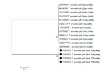
Molecular phylogenetic analysis of the partial cyt b gene sequence of T. annulata by the maximum likelihood method
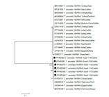
Molecular phylogenetic analysis of the partial 18S rRNA gene sequence of T. annulata by the maximum likelihood method
DISCUSSION
Animal husbandry plays an important role in the economy. However, several haemoprotozoan diseases constrain livestock production in many developing countries and are responsible for high rates of morbidity and mortality, leading to reduced production of meat, milk, and other livestock by-products (Khan et al., 2004). Bovine tropical theileriosis (T. annulata infection) is of great economic importance to many countries where millions of cattle are at risk of exposure to this disease. The present study aimed to estimate the prevalence of T. annulata infection in cattle at the main slaughterhouse in Jeddah city. The findings confirmed that theileriosis is endemic among cattle in Jeddah, western Saudi Arabia, as the infection was detected in most slaughtered cattle. Furthermore, the infection showed no significant association with sex, origin, or age, suggesting that all cattle were equally exposed to the pathogen.
The overall prevalence of T. annulata infection was 26.5% by GSBS and 35.5% by PCR. Previous studies in Saudi Arabia have reported varying prevalence rates, ranging from 1-5% in Taif, Riyadh, and Al-Ahsa by PCR testing (Ghafar and Amer, 2019; Alanazi et al., 2019), and 15.4 - 25% in Qassim and Jazan by blood smear testing (Omer et al., 2002; Al-Khalifa et al., 2009). To the best of our knowledge, this is the first report confirming the presence of T. annulata in cattle in Jeddah, KSA. In contrast, the prevalence obtained in this study was higher than the 28.5% reported in cattle from Pakistan (Parveen et al., 2021) and the 18.2% reported in China (Guo et al., 2018). Such variation in prevalence rates may be attributed to differences in sample size, animal species, diagnostic techniques, cattle population density, and the level of farmers’ awareness (Elhaig and Wahdan, 2023).
Although PCR enhanced the detection of Theileria in GSBS-negative blood samples, we found a slight kappa agreement (0.135, 95% CI = 0.261 - 0.008) between GSBS and PCR results. Furthermore, GSBS demonstrated 51% accuracy, 18.31% sensitivity, and 68.99% specificity when compared to PCR results. The results agree with a previous study conducted in Pakistan, which reported that PCR was more sensitive than microscopic examination for detecting T. annulata in cattle with moderate kappa agreement (κ = 0.6) between the microscopic examination and PCR (Ullah et al., 2021). Furthermore, globally, PCR is now used to diagnose Theileria infection, especially to identify carrier animals (Bilgic et al., 2010; Shayan and Rahbari, 2005) and microscopic examination was reported to have lower sensitivity (Azizi et al., 2008; Noaman, 2014) due to the destruction of the piroplasmic form due to RBC hemolysis, thick smear development, poor staining, lack of microscopic skill, and low parasitemia (Hoghooghi et al., 2011; Nayel et al., 2012). Our results and previous ones indicate that PCR is better for the diagnosis of theileriosis.
In the current survey, the risk of T. annulata infection non-significantly increased by 1.3 times in cattle > 1 year compared to cattle ≤ 1 year. A previous report found that T. annulata infection was higher in adult cattle (1-3 years) than in young cattle aged < 1 year (Abaker et al., 2017). Another study has found an increased infection rate of T. annulata in adult cattle (≥ 6 years) compared to young cattle (< 2 years), with a significant association between the infection and cattle age (Sallemi et al., 2018). Although there was no significant difference between age groups and T. annulata infection, this emphasized the influence of increasing age factors in this study, possibly due to older cattle being frequently re-infected, ticks being less attractive to calves, or the persistence of colostral antibodies (Gharbi et al., 2014).
Although the sex difference was not statistically significant, females had a higher prevalence of T. annulata (risk ratio = 1.9) than males, which could be attributed to sampling bias. This is endorsed by findings from a comparable study in Tunisia (Sallemi et al., 2018). The tendency of females to examine more positively compared to males is consistent with a previous study in Pakistan (Ullah et al., 2021). This could be due to female cattle having more hormonal fluctuations, a weak or disturbed immune response during pregnancy or lactation, and the presence of more ticks on their bodies, all of which make females more susceptible to T. annulata infection (Tuli et al., 2015; Parveen et al., 2021).
Regarding the origin of cattle, the prevalence of T. annulata was high in imported cattle, with a risk ratio about 1.9 times higher (P > 0.05) than that in local cattle. A similar result was documented in Pakistan (Ullah et al., 2021), which may reflect reduced tick infestation in local breeds because of an elevated level of tick resistance developed during generations of long-term exposure to vector ticks (Tuli et al., 2015; Glass, 2001). Additionally, the native breed would become more resistant to stresses that could make them more susceptible to infection after becoming acclimated to the local environment (Durrani et al., 2010; Parveen et al., 2021). In contrast to our results, a report from Sudan found a higher prevalence of T. annulata in local breeds compared to crossbred cattle using GSBS, IFAT, and PCR. This may be due to the exotic breeds being intensively managed including the use of insecticides to control ticks, good nutrition, hygiene, and careful veterinary care (Abaker et al., 2017).
Four and six sequences herein this study were partially sequenced using the Cyt b and 18S rRNA gene, respectively, and recognized as T. annulate. The phylogenetic tree based on the 18S rRNA was useful to infer the association between the collected Saudi isolates with a high bootstrap value. The Saudi sequences were classified into three subtypes, and all subtypes clustered with T. annulata strains from Egypt, China, Tajikistan, and Pakistan. Further, forming a separate clade on the evolutionary tree (Kundave et al., 2014; George et al., 2015). The analysis of 18S rRNA of T. annulata showed genetic diversity within seven hosts, at least 5 groups from different regions, and different hosts (cattle, sheep, ticks, rabbits, donkeys, and deer). The pattern in Saudi isolates (3 subtypes with 1, 3, and 8 nucleotide substitutions) reflects that different T. annulata isolates have distributed in cattle and globally reflects that different genotypes have a distinctive geographic distribution with different hosts. Previously, genetic diversity among T. annulata strains based on the 18S rRNA gene has been reported in many studies (Chansiri et al., 1999; Manuja et al., 2006; Aktas et al., 2007; Sivakumar et al., 2014; George et al., 2015). A study conducted in India (George et al., 2015) showed that phylogenetic analysis of 18S rRNA sequences divided T. annulata isolates into two groups with 0.1-8.6% divergence, and the Indian isolates clustered with the Chinese strain, possibly due to change in evolution and host adaptation. Furthermore, it is possible that both hosts and tick vectors had an impact on how the changing forms of Theileria parasites evolved (Sivakumar et al., 2014). To fully understand the origin and pathogenesis of the T. annulata circulating in Saudi cattle, more research is necessary.
Further, we also performed phylogenetic analysis and alignment of the obtained T. annulata isolates using the Cyt b gene. Multiple alignment revealed four isolates clustering with the Iranian strain (GenBank access. No. MN432496). Among the four isolates, three sequences (PP470770-2) were 100% identical, and the fourth sequence (PP470773) showed a single nucleotide substitution with a genetic distance of 0.01, which may be due to change in evolution and may lead to adaptation (George et al., 2015). In addition, the formation of a distinct clade on the phylogenetic tree is a finding supported by previous studies in Tunisia (Mhadhbi et al., 2015) and Pakistan (Ahmad Atif et al., 2023). These studies showed that sequencing and evolutionary analysis based on Cyt b showed the presence of genetic diversity among T. annulata strains, which led to the substructure of the T. annulata population in the phylogenetic tree and this due to the difference in agricultural climate zones. However, a study performed in Pakistan (Parveen et al., 2021) showed that the nucleotide homology was 97-99% between Pakistani isolates and T. annulata isolates recovered from GenBank. Furthermore, the phylogenetic tree grouped the sequences into a stable monophyletic cluster with the Cyt b gene of T. annulata from China, Iran, India, Turkey, and Spain.
Low heterogeneity was detected at the Cyt b and 18S rRNA genes, despite >97.9% identity being reported, and all grouped in a single clade, suggesting a common ancestor. A study from Pakistan evaluated the genetic property of the Cyt b gene of T. annulata isolates, and phylogenetic analysis showed that there were 9 and 21 unique haplotypes in the isolates derived from buffalo and cattle, and all of them clustered into a single lineage, indicating a single origin (Ali et al., 2022). Interestingly, the nucleotide substitutions found in this study at the Cyt b and 18S rRNA genes, notably at the 18S rRNA gene, in cattle indicate a need for additional research. The increasing number of detected substitutions suggests the existence of multiple subtypes, which enhances the possibility of identifying novel strains (Mahapatra et al., 2017).
CONCLUSION
This work reports the parasitological and molecular prevalence of bovine theileriosis and characterization of T. annulata in cattle from Jeddah, Saudi Arabia by targeting Cyt b and 18S rRNA genes. All sequences of T. annulata that are obtained here are located close to each other, indicating their geographic proximity, with a low nucleotide diversity. These findings provide baseline data for future studies on the T. annulata genetic diversity and can inform efforts to prevent and control the spread of the disease in Saudi Arabia.
REFERENCES
- ABAKER, I.A.; SALIH, D.A.; EL HAJ, L.M. et al. Prevalence of theileria annulata in dairy cattle in Nyala, South Darfur State, Sudan. Vet. World, v.10, p.1475-1480, 2017.
- AHMAD ATIF, F.; NAZIR, M.V.; ABBAS, R.Z. et al. Molecular epidemiology, associated risk factors, and phylogeny of theileria annulata infecting buffaloes and cattle from different agro-climatic regions of Punjab, Pakistan. Iran J. Vet. Res., v.24, p.247-257, 2023.
- AKTAS, M.; BENDELE, K.G.; AHAY, K. et al. Sequence polymorphism in the ribosomal DNA internal transcribed spacers differs among theileria species. Vet. Parasitol., v.147, p.221-230, 2007.
- ALANAZI, A.D.; ALOUFFI, A.S.; ALSHAHRANI, M.Y. et al. A report on tick burden and molecular detection of tick-borne pathogens in cattle blood samples collected from four regions in Saudi Arabia. Ticks Tick Borne Dis, v.12, p.101652, 2021.
- ALANAZI, A.D.; SAID, A.E.; GHONEIM, H.M. et al. A comprehensive evaluation and first molecular report of Theileria ovis infection in small ruminants in Saudi Arabia. Trop. Anim. Health Prod., v.51, p.89-98, 2019.
- ALI, Q.; ZAHID, O.; MHADHBI, M. et al. Genetic characterisation of the theileria annulata cytochrome b locus and its impact on buparvaquone resistance in bovine. Int. J. Parasitol. Drugs Drug Resist., v.20, p.65-75, 2022.
- AL-KHALIFA, M.S.; HUSSEIN, H.S.; DIAB, F.M.; KHALIL, G.M. Blood parasites of livestock in certain Regions in Saudi Arabia. Saudi J. Biol. Sci., v.16, p.63-67, 2009.
- ALNAHARI, H.Y.; ALWUTHAYNANI, T.H.; ALBOHIRI, H.H. et al. Impact of Theileria annulata on Hemato-biochemical and Histological Parameters in Cattle. Indian. J. of Animal Research., p.1-8, 2025.
- AZIZI, H.; BEHROOZ, S.; DEHKORDI, A.F.; FAZLDLAH, S. Detection of Theileria annulata by PCR and its comparison with smear method in native carrier cows. Biotechnology, v.7, p.574-577, 2008.
- BILGIC, H.B.; KARAGENÇ, T.; SHIELS, B. et al. Evaluation of cytochrome b as a sensitive target for PCR based detection of T. annulata carrier animals. Vet. Parasitol., v.174, p. 341-347, 2010.
- BILGIÇ, H.B.; KARAGENÇ, T.; SIMUUNZA, M. et al. Development of a multiplex PCR assay for simultaneous detection of Theileria annulata, Babesia bovis and Anaplasma marginale in cattle. Exp. Parasitol., v.133, p.222-229, 2013.
- BILLIOUW, M.; BRANDT, J.; VERCRUYSSE, J. et al. Evaluation of the indirect fluorescent antibody test as a diagnostic tool for East Coast fever in eastern Zambia. Vet. Parasitol., v.127, p.189-198, 2005.
- CAO, S.; ZHANG, S.; JIA, L. et al. Molecular detection of Theileria species in sheep from northern China. J. Vet. Med. Sci., v.75, p.1227-1230, 2013.
- CHANSIRI, K.; KAWAZU, S.; KAMIO T. et al. Molecular phylogenetic studies on Theileria parasites based on small subunit ribosomal RNA gene sequences. Vet. Parasitol., v.83, p.99-105, 1999.
- DURRANI, A.Z.; MEHMOOD, N.; SHAKOORI, A.R. Comparison of three diagnostic methods for Theileria annulata in Sahiwal and Friesian cattle in Pakistan. Pak. J. Zool., v.42, p.467-472, 2010.
- ELHAIG, M.M.; WAHDAN, A. Seroprevalence, associated risk factors, and molecular detection of bovine brucellosis in rural areas of Egypt. Comp. Immunol. Microbiol. Infect. Dis., v.95, p.101971, 2023.
- GEORGE, N.; BHANDARI, V.; REDDY, D.P.; SHARMA, P. Molecular and Phylogenetic analysis revealed new genotypes of Theileria annulata parasites from India. Parasit. Vectors, v.8, p.468, 2015.
- GHAFAR, M.W.; AMER, S.A.M. A preliminary molecular survey of Babesia divergens and first evidence of Theileria annulata in cattle from Saudi Arabia. Vet. World, v.12, p.266-270, 2019.
- GHARBI, M.; RJEIBI, M.R.; DARGHOUTH, M.A. Epidémiologie de la theilériose tropicale bovine (infection par Theileria annulata) en Tunisie: une synthèse. Rev. Élevage Méd. Vét. Pays Trop., v.67, p.241-247, 2014.
- GLASS, E.J. The balance between protective immunity and pathogenesis in tropical theileriosis: what we need to know to design effective vaccines for the future. Res. Vet. Sci., v.70, p.71-75, 2001.
- GUO, H.; YIN, C.; GALON, E.M. et al. Molecular survey and characterization of Theileria annulata and Ehrlichia ruminantium in cattle from Northwest China. Parasitol. Int., v.67, p.679-683, 2018.
- HAYATI, M.A.; HASSAN, S.M.; AHMED, S.K.; SALIH, D.A. Prevalence of ticks (Acari: Ixodidae) and Theileria annulata infection of cattle in Gezira State, Sudan. Parasite Epidemiol. Control., v.10, p.e00148, 2020.
- HOGHOOGHI-RAD, N.; PEYMAN, G.; SHAYAN, P.; ECKERT, B. Detection of native carrier cattle infected with Theileria annulata by semi-nested PCR and smear method in Golestan Province of Iran. World Appl. Sci. J., v.12, p.317-323, 2011.
- KASAIJA, P.D.; ESTRADA-PEÑA, A.; CONTRERAS, M. et al. Cattle ticks and tick-borne diseases: a review of Uganda's situation. Ticks Tick Borne Dis., v.12, p.101756, 2021.
- KHAN, M.Q.; ZAHOOR, A.; JAHANG, R.M.; MIRZA, M.A. Prevalence of blood parasites in cattle and buffaloes. Pak. Vet. J., v.24, p.193-194, 2004.
- KUNDAVE, V.R.; PATEL, A.K.; PATEL, P.V.; HASNANI, J.J. Qualitative and quantitative assessment of Theileria annulata in cattle and buffaloes Polymerase Chain Reaction. Trop. Biomed., v.31, p.728-735, 2014.
- LEMPEREUR, L.; BECK, R.; FONSECA, I. et al. Guidelines for the detection of Babesia and Theileria parasites. Vector Borne Zoonotic Dis., v.17, p.51-65, 2017.
- MAHAPATRA, M.; UPADHYAYA, S.; AVISO, S. et al. Selection of vaccine strains for serotype O foot-and-mouth disease viruses (2007-2012) circulating in Southeast Asia, East Asia and Far East. Vaccine, v.35, p.7147-7153, 2017.
- MANUJA, A.; MALHOTRA, D.V.; SIKKA, V.K. et al. Isolates of Theileria annulata collected from different parts of India show phenotypic and genetic diversity. Vet. Parasitol., v.137, p.242-522, 2006.
- METWALLY, D.M.; ALAJMI, R.; ALSULAMI, M.N. et al. Identification of Theileria spp. in sheep and goats from Jeddah, Saudi Arabia, using molecular techniques. PeerJ, v.9, p.e12596, 2021.
- MHADHBI, M.; CHAOUCH, M.; AJROUD, K. et al. Sequence polymorphism of cytochrome b Gene in Theileria annulata tunisian isolates and its association with buparvaquone treatment failure. PLoS One, v.10, p.e0129678, 2015.
- MOHAMMED-AHMED, G.M.; HASSAN, S.M.; EL HUSSEIN, A.M.; SALIH, D.A. Molecular, serological and parasitological survey of Theileria annulata in North Kordofan State, Sudan. Vet. Parasitol. Reg. Stud Rep., v.13, p.24-29, 2018.
- NAYEL, M.; EL-DAKHLY, K.M.; ABOULAILA, M. et al. The use of different diagnostic tools for Babesia and Theileria parasites in cattle in Menofia, Egypt. Parasitol. Res., v.111, p.1019-24, 2012.
- NOAMAN, V. Comparison of molecular and microscopic technique for detection of Theileria spp. in carrier cattle. J. Parasit. Dis., v.238, p.64-67, 2014.
- OMER, O.H.; EL-MALIK, K.H.; MAHMOUD, O.M. et al. Haematological profiles in pure bred cattle naturally infected with Theileria annulata in Saudi Arabia. Vet. Parasitol., v.107, p.161-168, 2002.
- OSMAN, S.A.; AL-GAABARY, M.H. Clinical, haematological and therapeutic studies on tropical theileriosis in water buffaloes (Bubalus bubalis) in Egypt. Vet. Parasitol., v.146, p.337-340, 2007.
- PARVEEN, A.; ALKHAIBARI, A.M.; ASIF, M. et al. Molecular epidemiology of Theileria annulata in cattle from two districts in Punjab (Pakistan). Animals, v.11, n.12, 2021.
- PERERA, P.K.; GASSER, R.B.; FIRESTONE, S.M. et al. Oriental theileriosis in dairy cows causes a significant milk production loss. Parasit. Vectors, v.7, p.73, 2014.
- PIENAAR, R.; TROSKIE, P.C.; JOSEMANS, A.I. et al. Investigations into the carrier-state of Theileria sp. (buffalo) in cattle. Int. J. Parasitol. Parasites Wildl., v.11, p.136-142, 2020.
- SALLEMI, S.; RJEIBI, M.R.; ROUATBI, M. et al. Molecular prevalence and phylogenetic analysis of Theileria annulata and Trypanosoma evansi in cattle in Northern Tunisia. Vet. Med. Sci., v.4, p.17-25, 2018.
- SELIM, A.; WEIR, W.; KHATER, H. Prevalence and risk factors associated with tropical theileriosis in Egyptian dairy cattle. Vet. World, v.15, p.919, 2022.
- SHAYAN, P.; RAHBARI, S. Simultaneous differentiation between Theileria spp. and Babesia spp. on stained blood smear using PCR. Parasitol. Res., v.97, p.281-286, 2005.
- SIVAKUMAR, T.; HAYASHIDA, K.; SUGIMOTO, C.; YOKOYAMA, N. Evolution and genetic diversity of Theileria. Infect. Genet. Evol., v.27, p.250-263, 2014.
- TAMURA, K.; NEI, M. Estimation of the number of nucleotide substitutions in the control region of mitochondrial DNA in humans and chimpanzees. Mol. Biol. Evol., v.10, p.512-526, 1993.
- TAMURA, K.; STECHER, G.; KUMAR, S. MEGA11: molecular evolutionary genetics analysis version 11. Mol. Biol. Evol., v.38, p.3022-3027, 2021.
- TULI, A.; SINGLA, L.; SHARMA, A. et al. Molecular epidemiology, risk factors and hematochemical alterations induced by Theileria annulata in bovines of Punjab (India). Acta Parasitol., v.60, p.378-390, 2015.
- ULLAH, R.; SHARMS, S.; KLAN, M.A. et al. Epidemiology and molecular characterization of Theileria annulata in cattle from central Khyber Pakhtunkhwa, Pakistan. PLoS One, v.16, p.e0249417, 2021.
The research data are available upon request.