 External topographic and macroscopic anatomical description of the reproductive system of female common opossums (Didelphis marsupialis - Linnaeus, 1758)
ABSTRACT
This study described the morphology of the female reproductive system of Didelphis marsupialis, focusing on the location and anatomical relationships of the organs. In eight animals, the reproductive structures were located in the caudal abdominal and pelvic cavities, differing from domestic mammals, in which they are positioned in the mid-abdominal region. The ovaries were small, ovoid, and situated within the ovarian bursae, close to the uteri and lateral to the urinary bladder. The uterine tubes adhered to the ovarian bursae and extended cranially to the elongated and fusiform uterine horns. The uterus was double, parallel, and completely separated, contrasting with the single uterus observed in domestic species. Each uterus continued into an independent cervix opening into the vaginal canal, formed by two long, curved lateral vaginas. These structures connected to a pseudovagina, located in the median sagittal plane, which communicated with the common urogenital sinus. The specimens exhibited a cloaca responsible for the elimination of fetuses, urine, and feces through a single opening, a characteristic of marsupials. No evidence of a bifurcated clitoris was found. The anatomical particularities identified provide relevant information about the reproductive biology of Didelphis marsupialis and its relationship with other marsupials.
Keywords:
comparative anatomy; female genital system; black-eared opossum; marsupial; reproductive morphology
RESUMO
Este estudo descreveu a morfologia do sistema reprodutor feminino de Didelphis marsupialis, com foco na localização e nas relações anatômicas dos órgãos. Em oito animais, as estruturas reprodutivas estavam localizadas nas cavidades abdominal e pélvica caudal, diferindo dos mamíferos domésticos, nos quais se posicionam na região mediana do abdômen. Os ovários eram pequenos, ovais e situados dentro das bolsas ovarianas, próximos ao útero e lateralmente à bexiga urinária. As tubas uterinas aderiam às bolsas ovarianas e estendiam-se cranialmente até os cornos uterinos alongados e fusiformes. O útero era duplo, paralelo e completamente separado, contrastando com o útero único observado em espécies domésticas. Cada útero continuava em um colo do útero independente, que se abria no canal vaginal, formado por duas vaginas laterais longas e curvas. Essas estruturas conectavam-se a uma pseudovagina, localizada no plano sagital mediano, que se comunicava com o seio urogenital comum. Os espécimes apresentaram uma cloaca responsável pela eliminação de fetos, urina e fezes através de uma única abertura, característica dos marsupiais. Não foram encontradas evidências de clitóris bifurcado. As particularidades anatômicas identificadas fornecem informações relevantes sobre a biologia reprodutiva de Didelphis marsupialis e sua relação com outros marsupiais.
Palavras-chave:
anatomia comparada; sistema genital feminino; gambá-de-orelhas-pretas; marsupial; morfologia reprodutiva
INTRODUCTION
The common opossum or black-eared opossum (Didelphis marsupialis - Linnaeus, 1758) is a wild mammal belonging to the family Didelphidae and the genus Didelphis, considered the largest marsupial species (Machado et al., 2010). They exhibit sexual dimorphism, with females having narrower heads compared to the broader heads of males (Cáceres and Monteiro-Filho, 1999).
The gestational period of female common opossums is short, averaging only 12 days after mating, but it can range from five to thirteen days, with approximately 10 to 15 offspring born per litter (Renfree and Tyndale-Biscoe, 1973; Freyer et al., 2002). When the immature young are born, they exit the mother's genital tract and attach to the nipples within the pouch to complete their development, as lactation is prolonged (Freyer et al., 2002; Gonçalves et al., 2009).
Common opossums have a life expectancy of two to four years (Gonçalves et al., 2009) and play important ecological roles such as seed dispersal and biological population control. However, they can also act as reservoirs for zoonotic diseases like leishmaniasis and, due to their high synanthropic capacity, have a direct impact on public health issues (Cáceres and Monteiro-Filho, 1999; Machado et al., 2010).
Due to their adaptability to captivity, high annual birth rates, and resistance to diseases, common opossums have become experimental animal models for biomedical studies (Senos et al., 2016). In this context, scientific research involving the anatomy and reproductive processes of female opossums is essential, as some information in the literature is contradictory or erroneously extrapolated from domestic animal species (Schimming et al., 2018; Yllera et al., 2023). Understanding marsupials is also important, as working with wild animals remains challenging for veterinarians due to their varied and peculiar anatomical and physiological characteristics, as well as the specific needs of these future non-conventional pets (Johnson-Delaney and Lenox, 2016; Yllera et al., 2023).
Scientific descriptions regarding the topography and gross anatomy of the female reproductive system of common opossums, as well as the phenomenon of embryonic diapause, are limited. This lack of information hinders clinical, surgical, reproductive, and imaging practices, thus compromising the quality of life and survival of these marsupials (Gonçalves et al., 2009). The anatomical characteristics of the female reproductive organs are similar to those of other marsupials, but differ from other eutherian mammals (Sharmam, 1954; Tyndale-Biscoe and Rodger, 1978; Taggart and Temple-Smith, 1991; Freyer et al., 2002; Regli and Kress, 2002; Wick and Kress, 2002a, 2002b; Gonçalves et al., 2009; Pagliarani et al., 2023). Therefore, the aim of this study was to describe the topography and macroscopic external anatomy of the components of the female reproductive system of the common opossum.
ETHICAL ASPECTS
This research was not submitted to the Ethics Committee on Animal Use.
MATERIALS AND METHODS
Eight female common opossum (Didelphis marsupialis) cadavers were used, sourced from the collection of the Veterinary Anatomy Laboratory at the University of Franca (UNIFRAN, SP). These specimens had been previously fixed in 10% formaldehyde and stored in 30% saline solution. The reproductive systems were individually removed and dissected using conventional methods so that the structures could be photographed with a digital camera and described in terms of their topography and external anatomy. For standardization of organ topography, the cadavers were positioned in dorsal recumbency, and anatomical terms were described based on the Nomina Anatomica Veterinaria (2017).
RESULTS AND DISCUSSION
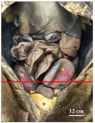
In the eight female specimens of Didelphis marsupialis, the greater omentum and the intestinal loops were retracted laterally to expose the reproductive systems, which occupied the caudal region of the abdominal cavity and the entire pelvic cavity (Fig. 1). These structures were located dorsally to the rectum and cranio-ventrally to the urinary bladder (Fig. 2). The position of these organs in the caudal region of the abdominal cavity differs from that of domestic animals, in which they are found in the mid-abdominal region (Konig and Liebich, 2011). Consequently, the ovaries of common opossums are relatively distant from the kidneys.
In the pelvic cavity, part of the reproductive organs was covered and protected by a considerable amount of adipose tissue (Fig. 1), unlike in domestic animals, where this type of tissue is found surrounding the ovaries (Konig and Liebich, 2011).
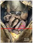
Photographic image of a female common opossum showing the reproductive organs (yellow asterisks) located in the caudal region of the abdominal cavity and in the pelvic cavity. The red line represents the boundary between these cavities. A considerable amount of adipose tissue covers these organs in the pelvic cavity (red asterisk). Ventral view.
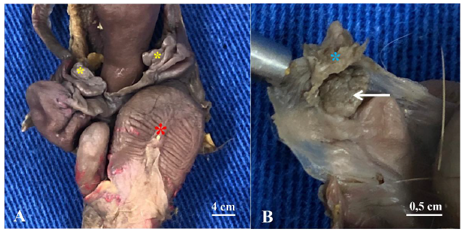
Macroscopically, it was possible to observe that the ovaries of common opossums are small and ovoid in shape, located inside the ovarian bursa, close to the uteri and laterally to the urinary bladder (Figures 3A and 3B). This finding is consistent with the descriptions by Reenfre and Shaw (2002) and Gonçalves et al. (2009) in other opossum species. In contrast, Yllera et al. (2023) reported that in the sugar glider (Petaurus breviceps), the ovaries are flattened and lack an ovarian bursa.
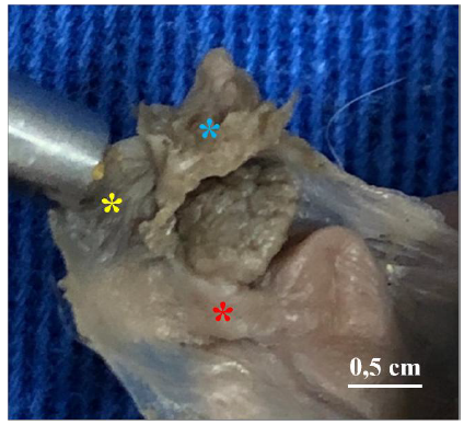
Similarly to what is described in South American marsupials popularly known as gray short-tailed opossums (Monodelphis domestica) (Wick and Kress, 2002a) and in other opossum species (Gonçalves et al., 2009), the uterine tubes (oviducts) in common opossums are attached to the ovarian bursae and located near and cranio-lateral to the uterine horns (Fig. 4).
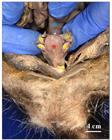
Photographic image of a female common opossum showing the reproductive system organs (yellow asterisks) located dorsally to the rectum (blue asterisk) and cranio-ventrally to the urinary bladder (red asterisk). Ventral view.
The right and left uterine horns are elongated and fusiform in shape (Figure 4); this anatomical characteristic of the common opossum coincides with the descriptions by Wick and Kress (2002b) in gray short-tailed opossums, in which the uterine horns are completely separated from one another.
In accordance with the descriptions by Regli and Kress (2002), Freyer et al. (2002), Wick and Kress (2002a, 2002b) in gray short-tailed opossums, and by Gonçalves et al. (2009) in other opossum species, it was found that the uterus of the common opossum is double, parallel, and completely separated (Fig. 5). In contrast, this differs from female domestic animals, which have only a single uterus (Konig and Liebich, 2011).
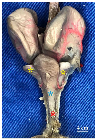
The uteruses of common opossums open into two small, independent internal cervices (Zeller and Freyer, 2001; Regli and Kress, 2002; Gonçalves et al., 2009), unlike domestic animals, which have only one cervix (Konig and Liebich, 2011). The cervices of marsupials open into the vaginal canal, which consists of two long, curved lateral vaginas, according to Fig. 6, located caudal to the uterus and lateral to the urinary bladder (Renfree and Shaw, 2002).
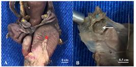
Photographic images of a female common opossum showing in A: small ovaries (yellow asterisks) located laterally to the urinary bladder (red asterisk) and in B: right ovary with an ovoid shape (arrow) and ovarian sac (blue asterisk). Ventral view.
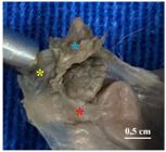
Photographic image of a female common opossum showing the right uterine tube (yellow asterisk) attached to the ovarian bursa (blue asterisk) and located near and cranio-lateral to the corresponding uterine horn (red asterisk). Ventral view.
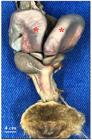
Photographic image of a female common opossum showing large, double, and parallel uteri (red asterisks). Ventral view.
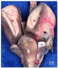
Photographic image of a female common opossum showing the lateral vaginas (blue asterisks), located caudally to the uteri (red asterisks) and laterally to the urinary bladder (yellow asterisk). Ventral view.
Pagliarani et al. (2023) also described the presence of two lateral vaginas in female marsupial koalas (Phascolarctos cinereus). According to Gonçalves et al. (2009), the size of the lateral vaginas varies depending on the maturity level of the female opossum, being more evident in reproductively mature or multiparous females and less developed in younger individuals.
According to Yllera et al. (2023), in the sugar glider (Petaurus breviceps), the function of the lateral vaginas is to facilitate the transport of sperm to the uteri, as the male deposits semen in the upper part of the common urogenital sinus, where the caudal ends of the lateral vaginas empty. Consequently, the existence of these vaginas influenced the development of the penis, and in some marsupials, the penile glans is a prominent and bifurcated structure adapted to them (Renfree, 1974; Harder et al., 1993; Behringer et al., 2006; Gonçalves et al., 2009).
The lateral vaginas extend to the pseudovagina, located in the median sagittal plane, which leads to the common urogenital sinus (Fig. 7), similarly to what has been reported in other opossum species (Gonçalves et al., 2009), including the white-eared opossum (Didelphis albiventris) (Schimming et al., 2018). On the other hand, these anatomical particularities differ when compared to domestic animals (Konig and Liebich, 2011).
According to Wick and Kress (2002b), in gray short-tailed opossums, the pseudovagina is not permanent and forms prior to each birth; for this reason, it is referred to as the birth canal.
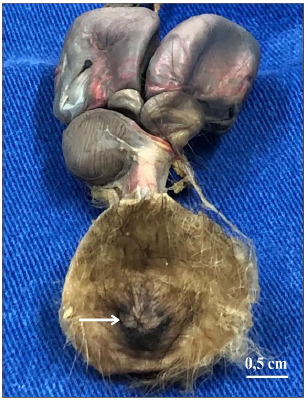
The common urogenital sinus is characterized by an elongated tube that opens internally into the genital ostium, urinary ostium, and intestinal ostium, which flow into a small cavity known as the cloaca. This structure eliminates the fetus, urine, and feces through a single external opening (Fig. 8), similar to what has been described in marsupials such as the quokka (Setonix brachyurus) (Sharmam, 1954), the tammar wallaby (Macropus eugenii) (Tyndale-Biscoe and Rodger, 1978), the brown antechinus (Antechinus stuartii) (Taggart and Temple-Smith, 1991), gray short-tailed opossums (Wick and Kress, 2002b), other opossum species (Gonçalves et al., 2009; Johnson-Delaney and Lenox, 2016), the sugar glider (Yllera et al., 2023), and koalas (Pagliarani et al., 2023). In this way, marsupials lack a vulva and anus. The urinary bladder connects to the urogenital sinus through a short urethra, consistent with the descriptions by Renfree and Shaw (2002).
Although Johnson-Delaney (2002), Pagliarani et al. (2023), and Yllera et al. (2023) described, respectively, in short-tailed opossums, koalas, and sugar gliders, the presence of a dorsoventrally flattened and bifurcated clitoris located at the caudal portion of the common urogenital sinus, near the opening of the cloaca, this anatomical structure was not identified in the eight specimens of common opossums analyzed.
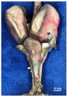
Photographic image of a female common opossum showing the lateral vaginas (yellow asterisks) extending to the pseudovagina (blue asterisk), which leads to the common urogenital sinus (red asterisk). Ventral view.
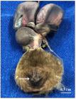
Photographic image of a female common opossum showing the presence of the cloaca (arrow), characterized by the convergence of the genital, urinary, and intestinal ostia. Ventral view.
CONCLUSION
The topography and external anatomy of the female reproductive organs of common opossums are similar to those of most other marsupials; however, they differ from those of domestic mammals. The data obtained in this study provides morphological insights for understanding the reproductive cycle and for developing future strategies for conservation, population control, management, and improvements in the quality of life and survival of these marsupials.
ACKNOWLEDGMENT
National Council for Scientific and Technological Development - Brazil (CNPq, process number 138993/2024-6).
REFERENCES
- BEHRINGER, R.R.; EAKIN, G.S.; RENFREE, M.B. Mamamalian diversity: gametes, embryos and reproduction. Reprod. Fertil. Dev., v.18, p.99-107, 2006.
- CÁCERES, N.C.; MONTEIRO-FILHO, E.L.A. Tamanho corporal em populações naturais de Didelphis (Mammalia: Marsupialia) do Sul do Brasil. Braz. J. Biol., v.59, p.461-469, 1999.
- FREYER, C.; ZELLER, U.; RENFREE, M.B. Ultrastucture of the placenta of the tammar wallaby, Macropus eugenii: comparison with the grey shorttailed opossum, Monodelphis domestica J. Anat., v.201, p.101-119, 2002.
- GONÇALVES, N.N.; MAÇANARES, C.A.F.; MIGLINO, M.A. et al. Aspectos morfológicos dos órgãos genitais femininos do gambá (Didelphis sp.). Braz. J. Vet. Res. Anim. Sci., v.46, p.332-338, 2009.
- HARDER, J.D.; STONEROOK, J.M.; PONDY, J. Gestation and placentation in two new world opossums: Didelphis virginiana and Monodelphis domestica J. Exp. Zool., v.266, p.463-479, 1993.
- JOHNSON-DELANEY, C.A. Reproductive medicine of companion marsupials. Vet. Clin. North Am. Exot. Anim. Pract., v.5, p.537-553, 2002.
- JOHNSON-DELANEY, C.A.; LENNOX, A.M. Reproductive disorders of marsupials. Vet. Clin. North Am. Exot. Anim. Pract, v.20, p.539-553, 2016.
- KONIG, H.E.; LIEBICH, H.G. Anatomia dos animais domésticos - texto e atlas colorido. 4.ed. Porto Alegre: Artmed, 2011. 788p.
- MACHADO, M.R.F.; REIS, A.C.G.; MARTINS, L. et al Arco aórtico do Saruê (Didelphis marsupialis - Linnaeus, 1758). Pubvet, v.4, p.1-6, 2010.
- NOMINA Anatomica Veterinaria. 6.ed. New York: WAVA, 2017.
- PAGLIARANI, S.; PALMIERI, C.; MCGOWAN, M. et al Anatomy of the female koala reproductive tract. Biology, v.12, p.1-34, 2023.
- REGLI, C.; KRESS, A. Changes in the epithelium of the vaginal complex during the estrous cycle of the marsupial Monodelphis domestica Cells Tissues Organs, v.172, p.276-296, 2002.
- RENFREE, M.B. Ovariectomy during gestation in the american opossum, Didelphis marsupials virginiana J. Reprod. Fertil., v.39, p.127-130, 1974.
- RENFREE, M.B.; SHAW, G. Reproduction in monotremes and marsupials. Encycl. Life Sci., v.2, p.1-5, 2002.
- RENFREE, M.B.; TYNDALE-BISCOE, C.H. Intrauterine development after diapause in the marsupial Macropus eugenii Dev. Biol., v.32, p.28-40, 1973.
- SCHIMMING, B.C.; CESÁRIO, M.D.; MATHEUS, S.M.M. Morphology of the vaginal complex in the white-eared opossum (Didelphis albiventris): gross anatomy and light microscopy. Anat. Histol. Embryol, v.47, p.566-572, 2018.
- SENOS, R.; RIBEIRO, M.S.; BENEDICTO, H.G. et al. Lumbosacral plexus in brazilian common opossum. Folia Morphol, v.75, p.300-305, 2016.
- SHARMAM, G.B. Studies on marsupial reproduction. 3. Normal and delayed pregnancy in Setonix brachyurus. Aust. J. Zool, v.3, p.56-70, 1954.
- TAGGART, D.A.; TEMPLE-SMITH, P.D. Transport and storage of spermatozoa in the female reproductive tract of the brown marsupial mouse, Antechinus stuartii (Dasyuridae). J. Reprod. Fertil., v.93, p.97-110, 1991.
- TYNDALE-BISCOE, C.H.; RODGER, J.C. Differential transport of spermatozoa into the two sides of the genital tract of a monovular marsupial, the tammar wallaby (Macropus eugenii). J. Reprod. Fertil., v.52, p.37-43, 1978.
- WICK, R.; KRESS, A. Ultrastructural changes in the cervical epithelium during the estrous cycle of the marsupial Monodelphis domestica (grey short-tailed opossum). Cells Tissues Organs, v.171, p.162-176, 2002b.
- WICK, R.; KRESS, A. Ultrastructural changes in the uterine luminal and glandular epithelium during the oestrous cycle of the marsupial Monodelphis domestica (grey short-tailed opossum). Cells Tissues Organs, v.170, p.111-131, 2002a.
- YLLERA, M.D.M.; ALONSO-PEÑARANDO, D.; LOMBARDERO, M. Gross anatomy of the female reproductive system of sugar gliders (Petaurus breviceps). Animals, v.13, p.2377, 2023.
- ZELLER, U.; FREYER, C. Early ontogeny and placentation of the grey short-tailed opossum, Monodelphis domestica (Didelphidae: Marsupialia): contribution to the reconstruction of the marsupial morphotype. J. Zool. Syst. Evol. Res, v.39, p.137-158, 2001.
Data in article.