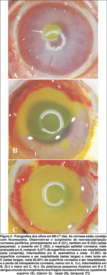
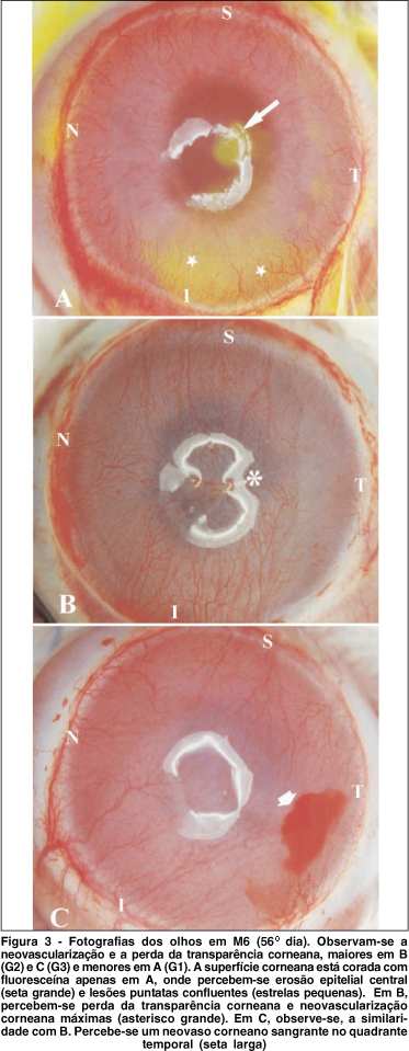
Purpose: To compare the clinical aspects of three experimental models of limbal stem cell (SC) deficiency in rabbits. Methods: In the present study, 54 rabbits (108 eyes) were divided into 3 experimental groups - (G1), (G2) and (G3). Each group consisted of 18 rabbits. The left eyes were submitted to 3 different experimental techniques - (T1), (T2) and (T3), respectively. A control group was formed with the 54 remaining eyes (RE). In all 3 techniques n-heptanol was used. In T1, the epithelium was removed by applying n-heptanol for 5 minutes. In T2, the n-heptanol application was followed by a 360º conjunctival peritomy at 4 mm beyond the limbus and scraping of the episcleral tissue. In T3, besides the procedure followed in T2, a lamellar dissection towards the limbus was performed, including 1.5 mm of the peripheral cornea and 2 mm of the scleral surface. Six clinical parameters of the corneas were studied: neovascularization, opacity, surface irregularity, epithelial wound healing, epithelial erosion, presence of granuloma and other injuries. Results: Corneal neovascularization started earlier in G1 and G2. It occurred heterogeneously, ranging from mild to intense, in 100% of the cases. It remained stable from the 28th day up to the end of the experiment (56th day). It was more intense in the upper and lower corneal quadrants and less intense in the nasal and temporal quadrants. Corneal opacity and irregularity of the corneal surface were less intense in G1 than in G2 and G3, which were similar. Reepithelialization started after a 3-day latency period in all the animals submitted to the three experimental techniques and was complete on day 7 in G1 and day 14 in G2 and G3. The rate of occurrence of corneal epithelial erosion and corneal stromal granuloma were similar in the 3 groups. Conclusions: T2 and T3 seemed to be suitable experimental models for total limbal stem cell deficiency and most of the parameters showed similar results. T1 was found to be an appropriate model for the partial-thickness removal of limbal SC. Conjunctivalization and neovascularization occurred in all experimental models.
Germ cells; Limbus corneal; Models, animal; Rabbits


