
 Investigation of the effects of nicotinamide mononucleotide on kidney damage in rats with cecal ligation and perforation sepsis model
ABSTRACT
Purpose: To evaluate the protective effects of nicotinamide mononucleotide (NMN), a NAD+ precursor, on kidney tissue in rats with sepsis induced by cecal ligation and puncture model.
Methods: In this study, 28 female Sprague Dawley rats were randomly divided into four equal groups: control, sham, sepsis, and sepsis+NMN (500 mg/kg, i.p.). Kidney tissues taken 24 hours after the sepsis model were histopathologically evaluated under light microscopy for tubular damage, glomerular congestion, and inflammatory infiltration parameters.
Results: Histopathological examination revealed severe tubular injury, marked glomerular congestion, and dense inflammatory cell infiltration in the kidney tissues of the sepsis group. In the group treated with NMN, a significant reduction in these pathological findings was observed, while the glomerular structure and tubular architecture were preserved. Semi-quantitative histopathological scoring confirmed significantly increased renal injury in the sepsis group and significant attenuation of tissue damage following NMN treatment.
Conclusion: The data obtained showed that NMN may play an important cytoprotective role in sepsis-induced acute kidney injury by preserving tissue architecture. More comprehensive molecular and biochemical studies are needed to fully elucidate the therapeutic potential and mechanisms of action of NMN.
Key words
Sepsis; Nicotinamide Mononucleotide; Kidney
Introduction
More than 19 million people worldwide are affected by sepsis each year, which is a type of systemic inflammatory disorder that is associated with very high mortality rates1. Sepsis can occur because of various types of insults (e.g., burns, severe trauma, hypotension/shock, peri-operational complications). The end result is that sepsis causes multiple organ system failure as a result of an overproduction of severe inflammatory responses after an inflammatory insult (e.g., heart, lung, brain, liver, kidney, pancreas), and there is usually also failure of other organs involved in the response2,3. The lung and kidney generally experience early injury during the sepsis cascade process3. Clinically, patients can exhibit a combination of symptoms such as confusion, fever (also known as pyrexia), low blood pressure (hypotension), and/or decreased urine volume (oliguria). If not treated rapidly, these symptoms can develop into catastrophic complications including but not limited to coagulopathy, acute respiratory distress syndrome, and acute kidney injury4,5.
The pathophysiology of sepsis depends on an altered permeability of endothelial cells. Macrophages and neutrophils are activated and recruited to sites of infection by the action of bacterial endotoxins. Once there, these immune cells produce an overwhelming amount of pro-inflammatory cytokines (chemokines) and nitrogen oxides (NO), which leads to vasodilation or vascular hypotension due to the loss of vascular tone6. Also, when blood vessels become leaky (increased blood vessel permeability), it creates small blood clots (microthrombi), which reduces blood flow to the tissues, allowing for accumulations of reactive oxygen species and ultimately leading to organ failure at the cellular level7,8.
Nicotinamide mononucleotide (NMN) has gained importance as a molecule that plays a crucial role as a nucleotide and serves as an important precursor to nicotinamide adenine dinucleotide (NAD+)9. Playing a vital role in cellular energy metabolism, NMN restores NAD+ levels by acting as a substrate for mitochondrial enzymes10. It has been reported that NMN can normalize the NAD+/NADH ratio, which has a protective effect against oxidative stress11. Nicotinamide treatment has been reported to improve mortality and provide protection against acute liver injury in endotoxemia and perforation-induced sepsis models12. Nicotinamide riboside (NR), found in milk-based products, is metabolized to NMN, a precursor of NAD, via the enzymes NRK1 and NRK213.
In the literature, it has been reported that NMN reduces lipopolysaccharide (LPS)-induced inflammation in macrophages14 and improves contractile function in mice15. NMN restored cardiac NAD levels in an ischemia-reperfusion model16 and improved metabolic balance in mice with type 2 diabetes17. It has also been found to increase NAD content and survival rate in rat hemorrhagic shock models18 and reduce LPS-induced lung19 and kidney damage20. In sepsis models induced by cecal ligation and puncture, NMN has been reported to reduce mortality by regulating the inflammatory response21.
The aim of this study was to investigate the potential protective effects of NMN administration on kidney tissue in experimental sepsis induced by cecal ligation and puncture (CLP) using histopathological methods.
Methods
This study was approved by the Burdur Mehmet Akif Ersoy University Local Animal Experiments Ethics Committee, with decision number 1,320 dated on May 6, 2024. In the study, 28 female Sprague Dawley rats (8 weeks old, 200–250 g) obtained from the Burdur Mehmet Akif Ersoy University Experimental Research Center were used. The animals were kept in standard cages at 22 ± 2°C, with 50–60% humidity and a 12-hour light/dark cycle. Ad libitum (free) access to water and standard feed were provided throughout the experiment. All experimental procedures were performed in accordance with the National Institutes of Health Guidelines for the Care and Use of Laboratory Animals.
The rats were randomly divided into four equal groups (n = 7):
-
Control group: healthy group with no procedures performed;
-
Sham group: group that underwent only laparotomy, but without CLP;
-
Sepsis (CLP) group: group in which sepsis was induced by cecal CLP;
-
CLP + NMN group: group in which NMN (500 mg/kg, intraperitoneal) was administered 30 minutes before and 12 hours after the CLP procedure22.
Sepsis model: cecal ligation and puncture procedure
The sepsis model was created using the CLP technique described in the literature23. Rats were anesthetized intraperitoneally (i.p.) with a combination of ketamine (80 mg/kg) and xylazine (10 mg/kg). After preparing the abdominal region under aseptic conditions, a 2-cm midline incision was made. The cecum was exposed and ligated with 4-silk sutures in the proximal 1/3 portion. The ligated cecum was punctured three times with an 18 G needle and gently squeezed to allow fecal contents to seep into the peritoneum. After the cecum was returned to its anatomical position, the laparotomy layers were closed with 4-sterile-silk sutures. The sham group underwent only laparotomy; the cecum was not ligated or punctured. All surgical procedures were performed by the same researcher to prevent experimental variability.
Twenty-four hours after the CLP procedure considering the peak period of cytokine storm and organ damage reported in the literature, all rats were painlessly euthanized by a high dose of thiopental sodium (0.5 g/kg, i.p.)24,25. Following euthanasia, decapitation was performed, and kidney tissues were rapidly excised.
Tissue processing
Kidney tissue samples were fixed in 10% neutral buffered formalin for 48 hours, gradually dehydrated to remove water content using graded concentrations of ethanol (from 50 to 70%, 96%, and finally absolute alcohol)26 and cleared with xylene to improve transparency for paraffin infiltration27. The tissues were then embedded into paraffin wax to form blocks that offered support for accurate sectioning28. Sections were cut from the paraffin blocks with a rotary microtome (Leica RM2125) at a thickness of 4 µm. Sections were placed on microscope slides, ensuring smooth and wrinkle-free adhesion.
Hematoxylin and eosin staining
The slides were placed in an oven at 60°C for 30 minutes and then went through deparaffinization, which was carried out by immersing the slides three times for 5 minutes each in xylene. This was followed by gradual rehydration of the sections through a series of alcohols consisting of 100% ethanol for 5 minutes; 96% ethanol for 3 minutes; 70% ethanol for 3 minutes; and finally distilled water for 2 minutes, which prepared the tissue for further staining. To stain acidic structures such as nucleic acid cytoplasmic components were immersed for 5 minutes in Mayer’s hematoxylin solution, which stains those structures blue or purple. After hematoxylin staining, the sections were rinsed off in running tap water for 1 minute to wash away the excess hue. The sections were differentiated with acid-alcohol (1% hydrochloric acid in 70% ethanol) for 1–2 seconds and rinsed off again in running water for 1 minute to remove the excess acid-alcohol mixture used for differentiation. Thereafter, sections were counter-stained with eosin Y solution for 1 minute, which colored the cytoplasm, and rinsed quickly with distilled water to remove additional staining solution while preventing much eosin from being lost29,30. The sections were dehydrated through a series of graded alcohols: 70% ethanol (1 min), 96% ethanol (1 min), 100% ethanol (2 exchanges, 2 minutes each). The sections were then cleared in xylene by immersing them three times for 5 minutes each to ensure adequate tissue transparency for mounting31. Finally, dehydrated and cleared sections were mounted with Entellan and permitted to dry at room temperature. The stained sections were evaluated under a light microscope (Nikon Eclipse i50, Tokyo, Japan) with various magnifications to check for tissue morphology31.
Semi-quantitative histopathological scoring
To provide an objective assessment of renal histopathological alterations, a semi-quantitative scoring system was used. Histopathological evaluation was performed independently by two blinded histologists using 10 randomly selected non-overlapping fields from each kidney section at 200× magnification. Tubular injury was graded on a scale of 0–4 according to the percentage of affected tubules (0 = normal histology, 1 = < 25% involvement, 2 = 25–50% involvement, 3 = 50–75% involvement, and 4 = > 75% involvement). Glomerular congestion and inflammatory cell infiltration were graded on a scale of 0–3 according to severity (0 = absent, 1 = mild, 2 = moderate, and 3 = severe). For each animal, the mean score obtained from all evaluated fields was calculated and used for statistical analysis.
Statistical analysis
Semi-quantitative histopathological scores are presented as mean ± standard deviation (SD). The normality of data distribution was assessed using the Shapiro–Wilk’s test. Differences in tubular injury, glomerular congestion, and inflammatory cell infiltration scores among groups were analyzed using one-way analysis of variance (ANOVA) followed by Tukey’s multiple comparison test. Statistical analyses were performed using GraphPad Prism 10 (GraphPad Software, San Diego, CA, United States of America). A p-value < 0.05 was considered statistically significant.
Results
Histopathological results
Control group glomeruli and proximal and distal tubules maintained their normal histological architecture. Glomeruli exhibited normal morphology with well-preserved capillary structures and a Bowman’s space that was within normal anatomical boundaries. Tubular epithelial cells were arranged regularly, with prominent, well-positioned nuclei. Structures of Henle’s loops and collecting ducts of medulla maintained normal histological features with normal cellular orientation.
Likewise, the sham group showed cortical structures maintained like the control group. No marked change was noted in either glomerular or tubular morphology. Medullary structures of Henle’s loops and collecting ducts remained intact without any signs of inflammation or cellular injury.
In the sepsis group, prominent structural alterations were noted in the glomeruli, such as glomerular congestion and infiltration of inflammatory cells. Most glomeruli showed fibrotic changes with a significant compromise of their structural integrity. Some atrophic glomeruli were identified, and coagulation was observed within the glomerular capillaries (shown in the inset picture). Bowman’s space was significantly dilated in most renal corpuscles. The proximal tubule diameter was reduced. Hypertrophy or degenerative changes with eosinophilic cytoplasm and pyknotic nuclei were seen in some proximal and distal tubular cells and juxtaglomerular cells were noted. Infiltration of inflammatory cells was detected throughout both cortical and medullary areas. Interstitial tissue showed fibrotic remodeling and infiltration of inflammatory cells. There were signs of mild vascular congestion and alteration of vascular wall structures. On the medulla, Henle’s loop and collecting ducts underwent degenerative changes, disorganization of cells, and marked infiltration of interstitial inflammatory cells.
In the NMN group, the general glomerular architecture was better preserved, with far less congestion and inflammatory cell infiltration compared to the sepsis group. Fewer atrophic glomeruli were observed, and the extent of Bowman’s space dilation was significantly decreased as compared to sepsis group. Overall, the integrity of tubular epithelial structures was mostly maintained, with minimal degenerative changes. In the medulla, Henle’s loops and collecting ducts maintained normal morphological features. However, inflammatory cell infiltration was found surrounding some Henle’s loops, while localized fibrotic changes were noted in the interstitial connective tissue. Cellular organization in the collecting ducts was mostly preserved, while focal luminal dilation was noted (Fig. 1).

Representative hematoxylin and eosin-stained kidney sections from control, sham, sepsis and sepsis + nicotinamide mononucleotide groups (100× and 400×).
Semi-quantitative histopathological results
Semi-quantitative histopathological scoring corroborated the qualitative microscopic findings. Tubular injury, glomerular congestion, and inflammatory cell infiltration scores were significantly higher in the sepsis group than in the control and sham groups (p < 0.001). Compared with the sepsis group, the sepsis + NMN group exhibited significantly lower tubular injury, glomerular congestion, and inflammatory cell infiltration scores (p < 0.05). No significant differences were observed between the control and sham groups (Fig. 2).
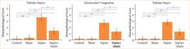
Semi-quantitative histopathological scoring of renal injury. Tubular injury score, glomerular congestion score, and inflammatory cell infiltration score in the control, sham, sepsis, and sepsis + nicotinamide mononucleotide groups. Data are presented as mean ± standard deviation (n = 7 animals per group). Statistical significance was determined using one-way analysis of variance followed by Tukey’s multiple comparison test.
Discussion
In this study, the protective effects of NMN on sepsis-induced acute kidney injury (AKI) were investigated at the histopathological level using a CLP model. The data obtained revealed that severe glomerular and tubular injury occurring in the sepsis group was significantly alleviated with NMN treatment, and tissue integrity was largely preserved.
Sepsis is a complex clinical condition leading to multiple organ failure because of the uncontrolled inflammatory response of the immune system to infection32. AKI is one of the most common complications of sepsis and directly affects mortality. In the pathogenesis of sepsis-related AKI, pro-inflammatory cytokine storm, oxidative stress, endothelial dysfunction, and mitochondrial damage play a key role33,34. These processes lead to a decrease in renal blood flow, tubulointerstitial fibrotic changes and consequently dramatic decreases in glomerular filtration rate.
The Bowman’s capsule dilation, tubular cell degeneration, and intense interstitial inflammatory infiltration observed in the sepsis group in our study parallel the findings of renal perfusion impairment and peritubular capillary damage reported by Seely et al.35.
NMN is a critical nucleotide that restores cellular metabolism and energy balance by increasing intracellular NAD+ levels. In the literature, it is known that a decrease in NAD+ levels is directly related to mitochondrial dysfunction, cellular senescence, and increased oxidative stress36. It is suggested that NMN application reduces cellular stress by supporting the NAD+ pool and has a cytoprotective effect in organs with high energy demand, such as the kidney16,37. The reduction in glomerular congestion and the preservation of tubular epithelial structure observed in the NMN group in our study support this metabolic supportive effect of NMN on renal parenchyma.
Our current findings are consistent with previous research14,38 showing that NMN suppresses macrophage-derived inflammation and slows the fibrotic process. Zhou et al.39 highlighted that NAD+ increase improves mitochondrial damage in systemic inflammation and heart failure. Similarly, in our study, the reduction in inflammatory cell infiltration observed following NMN treatment suggested that this molecule may modulate the detrimental effects of systemic inflammation on renal tissue.
The importance of oxidative damage in sepsis pathogenesis has also been highlighted in studies in which different treatment agents have been tested. For example, Çavuşoğlu et al.40 reported that oxytocin improved oxidative stress markers in sepsis-induced renal damage. Guo et al.41 reported the same for ginsenoside Rg1. The decrease in the number of pyknotic nuclei and the minimal level of eosinophilic cytoplasmic changes (degeneration) that we observed in the NMN group provided morphological evidence suggesting that NMN may protect tissue architecture. Su et al.42, while discussing the relationship between cellular stress and inflammation in the CLP model, stated that anti-inflammatory strategies are critical for kidney health.
In our study, the fact that NMN normalized the widening of the Bowman’s space, which was particularly evident in the sepsis group, and reduced the number of atrophic glomeruli suggested that renal microcirculation may have been preserved. The high bioavailability and low toxicity profile of NMN make this molecule a promising candidate for sepsis management. A recent study showed that NMN supplementation alleviated sepsis-related acute kidney injury by activating the NAD+/SIRT3 signaling pathway43. This finding was consistent with our observations in the NMN group, in which tubular epithelial integrity was preserved and cellular degeneration remained minimal. In the literature, NMN has been reported to reduce pro-inflammatory cytokine release and immune cell infiltration by inhibiting P38 and P65 (NF-κB) phosphorylation in sepsis models44.
The significant reduction in inflammatory cell infiltration in both cortical and medullary regions of rats treated with NMN in our study can be explained by the potential inhibitory effect of NMN on these molecular signaling pathways. It has been reported that NMN can inhibit DNA damage and cellular senescence in tubular cells45. This information is a critical mechanism supporting the near normalization of pyknotic nuclei and eosinophilic cytoplasmic changes that we detected in the sepsis group with NMN treatment. Semi-quantitative histopathological scoring provided objective support for the microscopic observations. Consistent with the qualitative findings, sepsis induced marked tubular injury, glomerular congestion, and inflammatory cell infiltration, whereas NMN administration significantly reduced all evaluated histopathological injury parameters. These findings further support the renoprotective effect of NMN in CLP-induced sepsis.
A limitation of the present study was the lack of biochemical renal function parameters, such as serum creatinine and blood urea nitrogen levels. Consequently, the histopathological findings could not be directly correlated with renal functional outcomes. Future studies incorporating histopathological, biochemical, and molecular assessments are warranted to provide a more comprehensive evaluation of the renoprotective effects of NMN in sepsis-associated acute kidney injury.
Conclusion
This study demonstrates that NMN protects kidney tissue architecture and significantly reduces morphological damage in an experimental sepsis model caused by CLP, as revealed by histopathological findings. The data obtained support the idea that NMN has cytoprotective and anti-inflammatory potential against sepsis-induced AKI and may be effective in maintaining glomerular and tubular integrity. NMN is a promising therapeutic agent candidate in the destructive process of sepsis leading to multiple organ failure. However, more comprehensive preclinical and clinical studies are needed to fully understand the clinical efficacy and renal protection mechanisms of NMN, which will examine different dose regimens, application durations, and advanced molecular signaling pathways.
Acknowledgements
Not applicable.
-
Research performed at the Experimental Research Center, Burdur Mehmet Akif Ersoy University, Burdur, and at the Department of Histology and Embryology, Faculty of Medicine, Ağrı İbrahim Çeçen University, Ağrı, Turkey.
-
Funding
Scientific Research Projects Coordination Unit of Ağrı İbrahim Çeçen UniversityGrant No.: TIP.24.012
-
Declaration of use of artificial intelligence tools
During the preparation of this work, the authors used ChatGPT-4o and DeepL for translation and grammar correction. After using these tools, the authors reviewed and edited the content.
Data availability statement
Upon request, researchers will be granted access to the dataset utilized in this study.
References
-
1 Cao T, Ni R, Ding W, Ji X, Fan GC, Zhang Z, Peng T. Nicotinamide mononucleotide as a therapeutic agent to alleviate multi-organ failure in sepsis. J Transl Med. 2023;21(1):883. https://doi.org/10.1186/s12967-023-04767-3
» https://doi.org/10.1186/s12967-023-04767-3 -
2 Rhodes A, Evans LE, Alhazzani W, Levy MM, Antonelli M, Ferrer R, Kumar A, Sevransky JE, Sprung CL, Nunnally ME, Rochwerg B, Rubenfeld GD, Angus DC, Annane D, Beale RJ, Bellinghan GJ, Bernard GR, Chiche JD, Coopersmith C, Backer DP, French CJ, Fujishima S, Gerlach H, Hidalgo JL, Hollenberg SM, Jones AE, Karnad DR, Kleinpell RM, Koh Y, Lisboa TC, Machado FR, Marini JJ, Marshall JC, Mazuski JE, McIntyre LA, McLean AS, Mehta S, Moreno RP, Myburgh J, Navalesi P, Nishida O, Osborn TM, Perner A, Plunkett CM, Ranieri M, Schorr CA, Seckel MA, Seymour CW, Shieh L, Shukri KA, Simpson SQ, Singer M, Thompson BT, Townsend SR, Van der Poll T, Vincent JL, Wiersinga WJ, Zimmerman JL, Dellinger RP. Surviving Sepsis Campaign: International Guidelines for Management of Sepsis and Septic Shock: 2016. Crit Care Med. 2017;45(3):486–552. https://doi.org/10.1097/CCM.0000000000002255
» https://doi.org/10.1097/CCM.0000000000002255 -
3 Lan KC, Chao SC, Wu HY, Chiang CL, Wang CC, Liu SH, Weng TI. Salidroside ameliorates sepsis-induced acute lung injury and mortality via downregulating NF-κB and HMGB1 pathways through the upregulation of SIRT1. Sci Rep. 2017;7(1):12026. https://doi.org/10.1038/s41598-017-12285-8
» https://doi.org/10.1038/s41598-017-12285-8 -
4 Angus DC, Linde-Zwirble WT, Lidicker J, Clermont G, Carcillo J, Pinsky MR. Epidemiology of severe sepsis in the United States: analysis of incidence, outcome, and associated costs of care. Crit Care Med. 2001;29(7):1303–10. https://doi.org/10.1097/00003246-200107000-00002
» https://doi.org/10.1097/00003246-200107000-00002 -
5 Alberti C, Brun-Buisson C, Burchardi H, Martin C, Goodman S, Artigas A, Sicignano A, Palazzo M, Moreno R, Boulmé R, Lepage E, Le Gall R. Epidemiology of sepsis and infection in ICU patients from an international multicentre cohort study. Intensive Care Med. 2002;28(2):108–21. https://doi.org/10.1007/s00134-001-1143-z
» https://doi.org/10.1007/s00134-001-1143-z -
6 Eaves-Pyles T, Murthy K, Liaudet L, Virág L, Ross G, Soriano FG, Szabó C, Salzman AL. Flagellin, a novel mediator of Salmonella-induced epithelial activation and systemic inflammation. J Immunol. 2001;166(2):1248–60. https://doi.org/10.4049/jimmunol.166.2.1248
» https://doi.org/10.4049/jimmunol.166.2.1248 - 7 Reinhart K, Bloos F, Brunkhorst F. Pathophysiology of sepsis and MOF. In: Fink MP, Abraham E, Vincent JL, Kochanek PM, editors. Textbook of critical care. 5th ed. Elsevier; 2011.
-
8 Souza AC, Yuen PS, Star RA. Microparticles: markers and mediators of sepsis-induced microvascular dysfunction, immunosuppression, and AKI. Kidney Int. 2015;87(6):1100–8. https://doi.org/10.1038/ki.2015.26
» https://doi.org/10.1038/ki.2015.26 -
9 Yoshino J, Baur JA, Imai SI. NAD+ intermediates: the biology and therapeutic potential of NMN and NR. Cell Metab. 2018;27(3):513–28. https://doi.org/10.1016/j.cmet.2017.11.002
» https://doi.org/10.1016/j.cmet.2017.11.002 -
10 Zapata-Pérez R, Tammaro A, Schomakers BV, Scantlebery AML, Denis S, Elfrink HL, Giroud-Gerbetant J, Cantó C, López-Leonardo C, McIntyre RL, van Weeghel M, Sánchez-Ferrer Á, Houtkooper RH. Reduced nicotinamide mononucleotide is a new and potent NAD+ precursor in mammalian cells and mice. FASEB J. 2021;35(4):e21456. https://doi.org/10.1096/fj.202001826R
» https://doi.org/10.1096/fj.202001826R -
11 Yuan H, Wan J, Li L, Ge P, Li H, Zhang L. Therapeutic benefits of the group B3 vitamin nicotinamide in mice with lethal endotoxemia and polymicrobial sepsis. Pharmacol Res. 2012;65(3):328–37. https://doi.org/10.1016/j.phrs.2011.11.014
» https://doi.org/10.1016/j.phrs.2011.11.014 -
12 Bieganowski P, Brenner C. Discoveries of nicotinamide riboside as a nutrient and conserved NRK genes establish a Preiss-Handler independent route to NAD+ in fungi and humans. Cell. 2004;117(4):495–502. https://doi.org/10.1016/s0092-8674(04)00416-7
» https://doi.org/10.1016/s0092-8674(04)00416-7 -
13 Liu J, Zong Z, Zhang W, Chen Y, Wang X, Shen J, Yang C, Liu X, Deng H. Nicotinamide mononucleotide alleviates LPS-induced inflammation and oxidative stress via decreasing COX-2 expression in macrophages. Front Mol Biosci. 2021;8:702107. https://doi.org/10.3389/fmolb.2021.702107
» https://doi.org/10.3389/fmolb.2021.702107 -
14 Mills KF, Yoshida S, Stein LR, Grozio A, Kubota S, Sasaki Y, Redpath P, Migaud ME, Apte RS, Uchida K, Yoshino J, Imai SI. Long-term administration of nicotinamide mononucleotide mitigates age-associated physiological decline in mice. Cell Metab. 2016;24(6):795–806. https://doi.org/10.1016/j.cmet.2016.09.013
» https://doi.org/10.1016/j.cmet.2016.09.013 -
15 Yamamoto T, Byun J, Zhai P, Ikeda Y, Oka S, Sadoshima J. Nicotinamide mononucleotide, an intermediate of NAD+ synthesis, protects the heart from ischemia and reperfusion. PLoS One. 2014;9(6):e98972. https://doi.org/10.1371/journal.pone.0098972
» https://doi.org/10.1371/journal.pone.0098972 -
16 Yoshino J, Mills KF, Yoon MJ, Imai S. Nicotinamide mononucleotide, a key NAD (+) intermediate, treats the pathophysiology of diet- and age-induced diabetes in mice. Cell Metab. 2011;14(4):528–36. https://doi.org/10.1016/j.cmet.2011.08.014
» https://doi.org/10.1016/j.cmet.2011.08.014 -
17 Sims CA, Guan Y, Mukherjee S, Singh K, Botolin P, Davila A Jr., Baur JA. Nicotinamide mononucleotide preserves mitochondrial function and increases survival in hemorrhagic shock. JCI Insight. 2018;3(17):e120182. https://doi.org/10.1172/jci.insight.120182
» https://doi.org/10.1172/jci.insight.120182 -
18 Wang X, Hu X, Yang Y, Takata T, Sakurai T. Nicotinamide mononucleotide protects against β-amyloid oligomer-induced cognitive impairment and neuronal death. Brain Res. 2016;1643:1–9. https://doi.org/10.1016/j.brainres.2016.04.060
» https://doi.org/10.1016/j.brainres.2016.04.060 -
19 Tian Y, Zhu CL, Li P, Li HR, Liu Q, Deng XM, Wang JF. Nicotinamide mononucleotide attenuates LPS-induced acute lung injury with anti-inflammatory, anti-oxidative and anti-apoptotic effects. J Surg Res. 2023;283:9–18. https://doi.org/10.1016/j.jss.2022.09.030
» https://doi.org/10.1016/j.jss.2022.09.030 -
20 He S, Gao Q, Wu X, Shi J, Zhang Y, Yang J, Li X, Du S, Zhang Y, Yu J. NAD+ ameliorates endotoxin-induced acute kidney injury in a sirtuin1-dependent manner via GSK-3β/Nrf2 signalling pathway. J Cell Mol Med. 2022;26(7):1979–93. https://doi.org/10.1111/jcmm.17222
» https://doi.org/10.1111/jcmm.17222 -
21 Cros C, Margier M, Cannelle H, Charmetant J, Hulo N, Laganier L, Grozio A, Canault M. Nicotinamide mononucleotide administration triggers macrophages reprogramming and alleviates inflammation during sepsis induced by experimental peritonitis. Front Mol Biosci. 2022;9:895028. https://doi.org/10.3389/fmolb.2022.895028
» https://doi.org/10.3389/fmolb.2022.895028 -
22 Sun C, Xiong H, Guo T. β-nicotinamide mononucleotide alleviates sepsis-associated acute kidney injury by activating NAD+/SIRT3 signaling. Cell Biochem Biophys. 2025;83(2):2089–99. https://doi.org/10.1007/s12013-024-01619-9
» https://doi.org/10.1007/s12013-024-01619-9 -
23 Hong G, Zheng D, Zhang L, Ni R, Wang G, Fan GC, Lu Z, Peng T. Administration of nicotinamide riboside prevents oxidative stress and organ injury in sepsis. Free Radic Biol Med. 2018;123:125–37. https://doi.org/10.1016/j.freeradbiomed.2018.05.073
» https://doi.org/10.1016/j.freeradbiomed.2018.05.073 -
24 Selli J, Vural Keles D, Keles ON, Celik M, Yetim Z. Nicotinamide riboside preserves ovarian injury in an experimental sepsis model in rats. Eurasian J Med. 2023;55(2):128–34. https://doi.org/10.5152/eurasianjmed.2023.22255
» https://doi.org/10.5152/eurasianjmed.2023.22255 -
25 Wang P, Ba ZF, Chaudry IH. Mechanism of hepatocellular dysfunction during early sepsis. Arch Surg. 1997;132(4):364–70. https://doi.org/10.1001/archsurg.1997.01430280038005
» https://doi.org/10.1001/archsurg.1997.01430280038005 - 26 Kiernan JA. Histological and histochemical methods: theory and practice. 5th ed. Banbury: Scion; 2015.
-
27 Wang Q, Gu R, Olm F, Bèchet NB, Lindstedt S. Xylene versus isopropanol for paraffin wax processing of lung tissue. Appl Sci (Basel). 2024;14(5):1726. https://doi.org/10.3390/app14051726
» https://doi.org/10.3390/app14051726 -
28 Sadeghipour A, Babaheidarian P. Making formalin-fixed, paraffin embedded blocks. In: Yong W, editor. Biobanking: methods in molecular biology (v. 1897). New York: Humana Press; 2019. p. 253–68. https://doi.org/10.1007/978-1-4939-8935-5_22
» https://doi.org/10.1007/978-1-4939-8935-5_22 - 29 Prophet EB, Mills B, Arrington JB, Sobin LH. Laboratory methods in histotechnology. Washington: American Registry of Pathology; 1992.
-
30 Titford M. A short history of histopathology technique. J Histotechnol. 2006;29(2):99–1102. https://doi.org/10.1179/his.2006.29.2.99
» https://doi.org/10.1179/his.2006.29.2.99 - 31 Luna LG. Manual of histologic staining methods of the Armed Forces Institute of Pathology. 3rd ed. New York: McGraw-Hill; 1968.
-
32 Singer M, Deutschman CS, Seymour CW, Shankar-Hari M, Annane D, Bauer M, Bellomo R, Bernard GR, Chiche JD, Coopersmith CM, Hotchkiss RS, Levy MM, Marshall JC, Martin GS, Opal SM, Rubenfeld GD, van der Poll T, Vincent JL, Angus DC. The Third International Consensus Definitions for Sepsis and Septic Shock (Sepsis-3). JAMA. 2016;315(8):801–10. https://doi.org/10.1001/jama.2016.0287
» https://doi.org/10.1001/jama.2016.0287 -
33 Bellomo R, Kellum JA, Ronco C, Wald R, Martensson J, Maiden M, Bagshaw SM, Glassford NJ, Lankadeva Y, Vaara ST, Schneider A. Acute kidney injury in sepsis. Intensive Care Med. 2017;43(6):816–28. https://doi.org/10.1007/s00134-017-4755-7
» https://doi.org/10.1007/s00134-017-4755-7 -
34 Hotchkiss RS, Sherwood ER. Immunology. Getting sepsis therapy right. Science. 2015;347(6227):1201–2. https://doi.org/10.1126/science.aaa8334
» https://doi.org/10.1126/science.aaa8334 -
35 Seely KA, Holthoff JH, Burns ST, Wang Z, Thakali KM, Gokden N, Rhee SW, Mayeux PR. Hemodynamic changes in the kidney in a pediatric rat model of sepsis-induced acute kidney injury. Am J Physiol Renal Physiol. 2011;301(1):F209–17. https://doi.org/10.1152/ajprenal.00687.2010
» https://doi.org/10.1152/ajprenal.00687.2010 -
36 Watanabe E, Thampy LK, Hotchkiss RS. Immunoadjuvant therapy in sepsis. Acute Med Surg. 2018;5(4):309–15. https://doi.org/10.1002/ams2.363
» https://doi.org/10.1002/ams2.363 -
37 Imai SI. The NAD World 2.0: the importance of the inter-tissue communication mediated by NAMPT/NAD+/SIRT1 in mammalian aging and longevity control. NPJ Syst Biol Appl. 2016;2:16018. https://doi.org/10.1038/npjsba.2016.18
» https://doi.org/10.1038/npjsba.2016.18 -
38 Cantó C, Menzies KJ, Auwerx J. NAD (+) metabolism and the control of energy homeostasis. Cell Metab. 2015;22(1):31–53. https://doi.org/10.1016/j.cmet.2015.05.023
» https://doi.org/10.1016/j.cmet.2015.05.023 -
39 Zhou B, Wang DD, Qiu Y, Airhart S, Liu Y, Stempien-Otero A, O’Brien KD, Tian R. Boosting NAD level suppresses inflammatory activation of PBMCs in heart failure. J Clin Invest. 2020;130(11):6054–63. https://doi.org/10.1172/JCI138538
» https://doi.org/10.1172/JCI138538 -
40 Çavuşoğlu T, Savran Y, Uyanıkgil Y, Gülle K, Acara AÇ, Yiğittürk G, Kılıç KD, Çetin EÖ, Meral A, Erbaş O. Oxytocin effect on sepsis-induced experimental rat renal failure model: a histological and biochemical study. Sdü Sağlık Bilimleri Enstitüsü Dergisi. 2018;9(1):28–33. https://doi.org/10.22312/sdusbed.305491
» https://doi.org/10.22312/sdusbed.305491 -
41 Guo J, Wang R, Min F. Ginsenoside Rg1 ameliorates sepsis-induced acute kidney injury by inhibiting ferroptosis in renal tubular epithelial cells. J Leukoc Biol. 2022;112(5):1065–77. https://doi.org/10.1002/JLB.1A0422-211R
» https://doi.org/10.1002/JLB.1A0422-211R -
42 Su J, Chen S, Xiao J, Feng Z, Hu S, Su Q, Chen Q, Chen D. Aloe-emodin ameliorates cecal ligation and puncture-induced sepsis. Int J Mol Sci. 2023;24(15):11972. https://doi.org/10.3390/ijms241511972
» https://doi.org/10.3390/ijms241511972 -
43 Sun C, Xiong H, Guo T. β-nicotinamide mononucleotide alleviates sepsis-associated acute kidney injury by activating NAD+/SIRT3 signaling. Cell Biochem Biophys. 2025;83(2):2089–99. https://doi.org/10.1007/s12013-024-01619-9
» https://doi.org/10.1007/s12013-024-01619-9 -
44 Li HR, Liu Q, Zhu CL, Sun XY, Sun CY, Yu CM, Li P, Deng XM, Wang JF. β-nicotinamide mononucleotide activates NAD+/SIRT1 pathway and attenuates inflammatory and oxidative responses in septic mice. Redox Biol. 2023;63:102745. https://doi.org/10.1016/j.redox.2023.102745
» https://doi.org/10.1016/j.redox.2023.102745 -
45 Jia Y, Kang X, Tan L, Ren Y, Qu L, Tang J, Liu G, Wang S, Xiong Z, Yang L. Nicotinamide mononucleotide attenuates renal interstitial fibrosis after AKI by suppressing tubular DNA damage and senescence. Front Physiol. 2021;12:649547. https://doi.org/10.3389/fphys.2021.649547
» https://doi.org/10.3389/fphys.2021.649547
Edited by
-
Section editor:
Adam Deak https://orcid.org/0000-0001-7516-3152

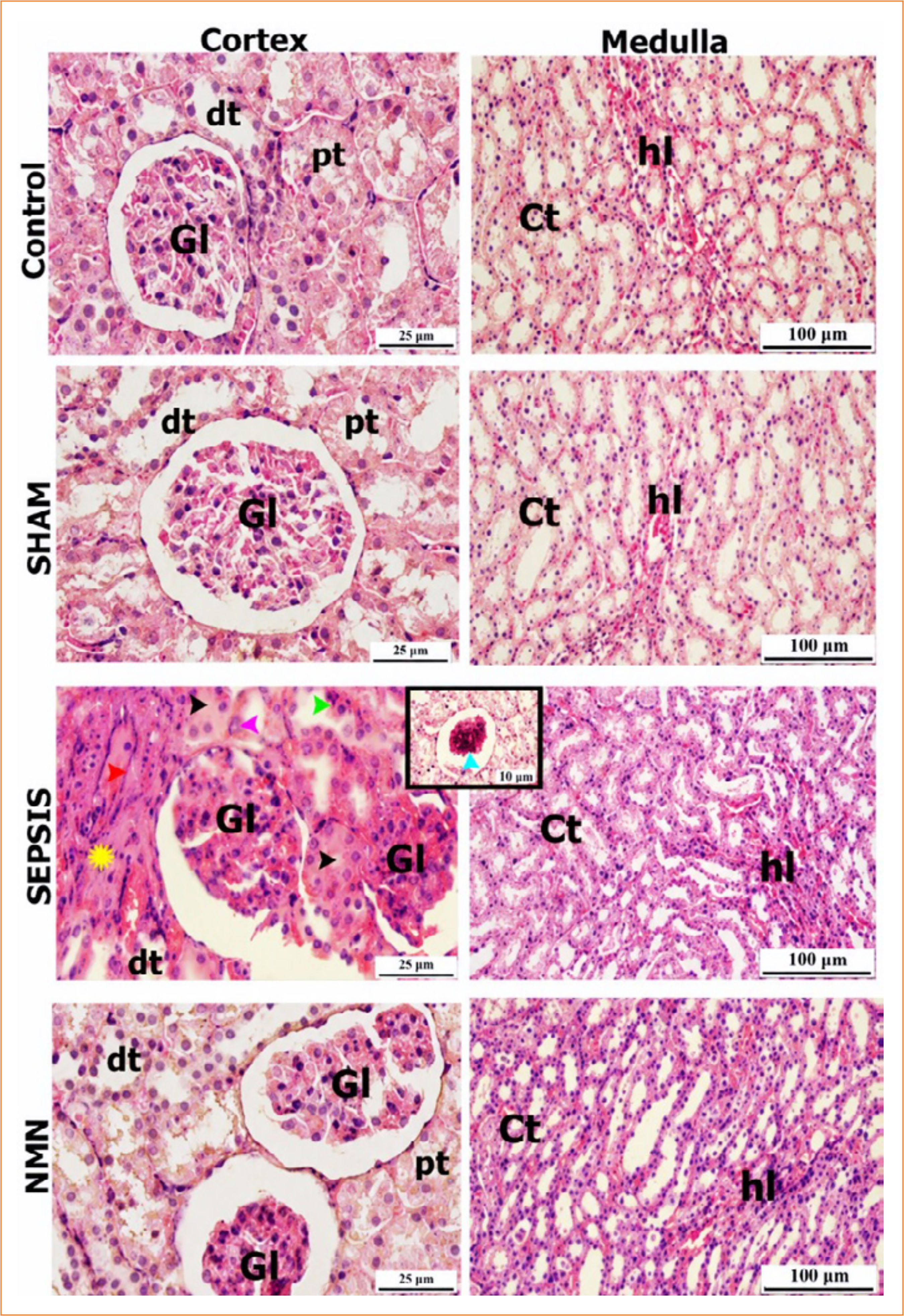 Gl: glomerulus; pt: proximal tubule; dt: distal tubule; hl: loops of Henle; Ct: collecting tubules; blue arrowhead: glomerular atrophy and thrombus formation; green arrowhead: cells with eosinophilic cytoplasm and pyknotic nuclei; pink arrowhead: hypertrophic cells; red arrowhead: impaired vascular wall; yellow star: fibrotic interstitial connective tissue. Source: Elaborated by the authors.
 ns: not significant; *p < 0.05 versus the control and sham groups; ***p < 0.001 versus the control and sham groups; #p < 0.05 versus the sepsis group. Source: Elaborated by the authors.