 IgM antiphospholipid antibodies are associated with a microvascular phenotype in antiphospholipid syndrome
Abstract
Background The clinical significance of IgM antiphospholipid antibodies (aPL) in antiphospholipid syndrome (APS) remains uncertain, while lupus anticoagulant is a well-established marker for thrombotic risk.
Methods The objective of the study is to evaluate the impact of IgM aPL on the clinical phenotype of primary APS (PAPS). In this retrospective multicenter study, patients meeting updated Sapporo classification criteria were categorized into three aPL profiles: isolated lupus anticoagulant (isolated-LA), isolated IgM anticardiolipin and/or IgM anti-β2-glycoprotein I antibodies (isolated-IgM-aPL), and LA and IgM antibodies (LA + IgM-aPL). Clinical features were compared between isolated-LA vs. isolated-IgM-aPL and isolated-LA vs. LA + IgM-aPL groups.
Results Among 202 patients, 17 (8.4%) had isolated-IgM-aPL, 145 (71.7%) isolated-LA, and 40 (19.8%) LA + IgM-aPL. Compared to isolated-LA, the isolated-IgM-aPL group had lower female prevalence (41.2% vs. 65.5%; p = 0.049), fewer venous thromboses (41.2% vs. 66%; p = 0.049), but more obstetric morbidity (58.8% vs. 26.2%; p = 0.005). There was a higher proportion of patients with livedo racemosa (47.1% vs. 20.7%; p = 0.015) and with white matter lesions (WML) (29.4% vs. 9.7%; p = 0.020) in the isolated-IgM-aPL group. In the multivariate analysis, WML remained independently associated (OR 3.7; p = 0.020). In the second comparison (isolated-LA vs. LA + IgM-aPL), a higher prevalence of livedoid vasculopathy (15.0% vs. 4.8%; p = 0.026) and WML (22.5% vs. 9.7%; p = 0.037) were observed in the LA + IgM-aPL group. Nonetheless, no independent associations were seen in the multivariate analysis.
Conclusion IgM aPL may be associated with a distinct APS phenotype characterized by microvascular involvement, including livedo and WML. These findings support the need for further research into the clinical implications of IgM isotype positivity in APS.
Keywords
Antiphospholipid syndrome; Antiphospholipid antibodies; Lupus anticoagulant
Introduction
Antiphospholipid syndrome (APS) is characterized by the presence of antiphospholipid antibodies (aPL) associated with arterial, venous, or microvascular thrombosis, pregnancy morbidity, or nonthrombotic manifestations [1]. APS can manifest as either a primary condition, occurring without an associated autoimmune disease, or as a secondary condition, which is linked to other autoimmune disorders, such as systemic lupus erythematosus (SLE) [2].
This wide range of clinical manifestations reflects the complexity of APS, which is further complicated by the variability in aPL profiles among affected individuals. The heterogeneous nature of these antibody profiles can influence the presentation and severity of APS, making diagnosis and management challenging [3, 4]. Among the aPL isotypes, lupus anticoagulant (LA) is the most well-established marker for thrombotic risk [5], whereas the clinical significance of IgM aPL remains a subject of ongoing debate [6]. The APS classification criteria, published in 2023, have introduced changes in how different aPL isotypes are weighted, reflecting the evolving understanding of their clinical relevance. Patients with isolated positivity to IgM isotypes do not fulfill the new criteria classification regardless of titers ≥ 40 units [1]. While IgG aPL are often emphasized due to its stronger association with thrombotic events, there is growing evidence suggesting that IgM aPL isotypes might also have important implications3. Recent studies have highlighted the distinct clinical subsets of thromboembolic events defined by IgM aPL, indicating that these antibodies might contribute significantly to APS [4]. Conversely, some researchers have proposed that IgM isotypes have weaker link to thrombosis and there is no added value for testing IgM-aPL in patients suspected of thrombotic APS [6]. This divergence in available evidence highlights a significant gap in the literature and underscores the need for further clarification and research.
Given the limited number of studies focusing specifically on the role of IgM aPL in APS, our study aims to analyze IgM aPL positive APS patients and compare them to LA positive APS patients to provide further insights on the impact of IgM aPL in the phenotypic expression of the disease. By that, we hope to contribute to the ongoing debate and enhance the understanding of APS subtypes and their management.
Patients and data collection
We retrospectively included patients with primary APS who fulfilled the updated Sapporo classification criteria [7] from three referral centers up to November 2023. Patients were categorized into three distinct aPL profiles: isolated IgM antiphospholipid antibodies (isolated-IgM-aPL), isolated lupus anticoagulant (isolated-LA), and combined LA and IgM antibodies (LA + IgM-aPL).
Isolated-IgM-aPL was defined as the presence of IgM anticardiolipin (aCL) and/or IgM anti-β2 glycoprotein I (aβ2GPI) antibodies in the absence of all other aPLs, including IgG aCL, IgG aβ2GPI, and lupus anticoagulant. Patients were considered IgM-positive when aCL and/or aβ2GPI levels were ≥ 40 units, and high IgM titers were defined as ≥ 80 units [1]. Given that low to moderate levels of IgG aPL may be clinically relevant and influence patient classification, a cutoff of < 40 units was applied to define IgG negativity.
Similarly, isolated-LA was defined as the presence of lupus anticoagulant in the absence of any aCL or aβ2GPI antibodies. Other aPL profiles were excluded, as were patients with secondary APS. Lupus anticoagulant testing was performed according to ISTH guidelines [8].
After categorization, two main comparisons were performed regarding clinical data: (1) isolated-LA vs. isolated-IgM-aPL and (2) isolated-LA vs. LA + IgM-aPL. Patients were compared regarding demographic data (mean age, sex, race); comorbidities (hypertension, diabetes, obesity, dyslipidemia, stroke, myocardial infarction (MI); clinical manifestations: macrovascular events (venous or arterial), pregnancy morbidity, microthrombotic and non-thrombotic manifestations (livedo racemosa, livedoid vasculopathy, echocardiography-proven cardiac valve disease, nephropathy and thrombocytopenia) and white matter lesions (WML).
Recurrent arterial or venous thrombosis were defined as more than 1 event not occurring at the same time. Recurrent pregnancy morbidity was defined as: (1) at least 3 consecutive abortions or (2) more than one fetal loss or more than one premature birth before 34th week due to pre-eclampsia or placental insufficiency or (3) any combination of updated Sapporo obstetrics criteria. Comorbidities definitions are described elsewhere [9, 10, 11] and clinical manifestations and aPL positivity of APS patients were defined according to updated Sapporo classification criteria [7].
Statistical analysis
Descriptive statistics were used to describe continuous variables (mean ± standard deviation - SD; median ± interquartile range - IQR) and categorical data were expressed as absolute number and percentage (%). The associations between clinical and laboratory variables in patients with isolated IgM isotype and those with other aPL antibodies were compared using the corrected χ2 test and Fisher's exact test for categorical data, and the Mann-Whitney U test for continuous variables. A significance level of 0.05 was applied. Odds-ratio (OR) were calculated with their 95% confidence interval (95% CI). A multivariate logistic regression analysis by Stepwise Backwards was performed to identify characteristics associated with IgM aPL profile; all variables with p < 0.1 in univariate analysis were included. All statistical analyses and Figures were conducted using SPSS software, version 26.0.
Results
Two-hundred and two patients were assessed; of these, 17 (8.4%) had isolated-IgM-aPL, 145 (71.7%) had isolated-LA and 40 (19.8%) had LA + IgM-aPL. Female sex accounted for 121 (59.9%) of the patients, 141 (69.8%) were white and 182 (90.1%) had thrombosis. The median age of them was 47 (37–57) years-old. Main characteristics of the patients are shown in Table 1.
In comparison to isolated-LA, patients with isolated-IgM-aPL had lower prevalence of female sex (41.2% vs. 65.5%; p = 0.049), lower prevalence of venous thrombotic events (41% vs. 66%; p = 0.049) and higher prevalence of obstetric events (58.8% vs. 26.2%; p = 0.005). When we analyzed recurrent obstetric events, the IgM-aPL profile remained significant (p = 0.02). Patients from the IgM isolated-aPL group had a higher proportion of livedo racemosa (47.1% vs. 20.7%; p = 0.015) and WML (29.4% vs. 9.7%; p = 0.020), (Table 2). In the multivariate analysis, WML was significantly correlated with the isolated-IgM-aPL profile (B-Coefficient 1.32; Odds Ratio 3.7; 95% CI 1.15–12.2; p = 0.020).
Patients with combined positivity for LA + IgM-aPL were predominantly males (47.5% vs. 65.5%; p = 0.038), with higher prevalence of livedoid vasculopathy (15.0% vs. 4.8%; p = 0.026) and higher prevalence of WML (22.5% vs. 9.7%; p = 0.037) when compared to isolated-LA (Table 2). No independent associations were seen in the multivariate analysis.
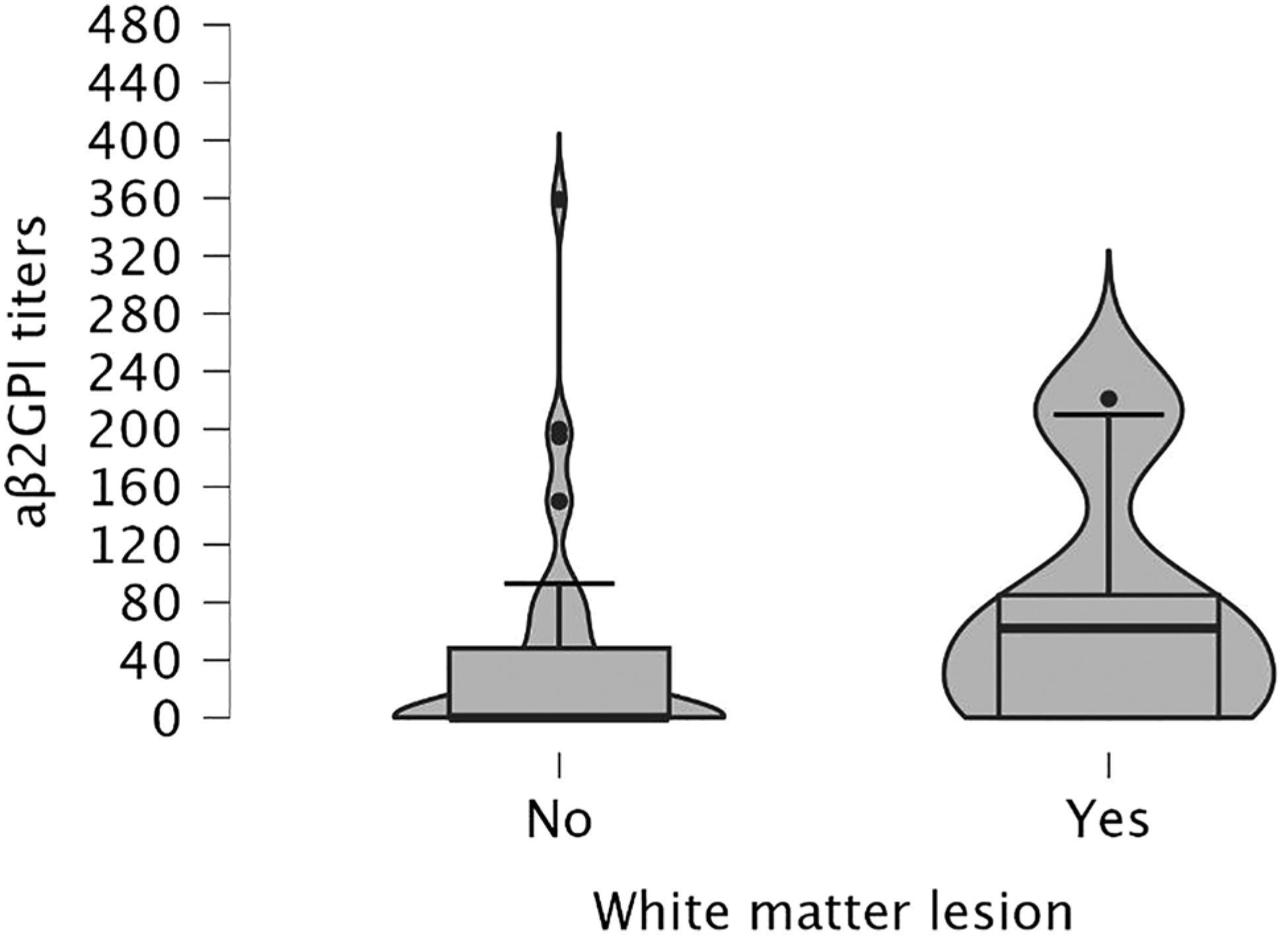
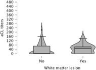
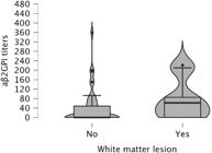
We also performed a subsequent analysis including all patients who tested positive for IgM, either isolated or associated with LA. When IgM isotype was analyzed as a continuous variable, increasing titers of both aCL IgM (p = 0.008) and anti-β2GPI IgM (p < 0.001) correlated with a higher prevalence of WML. The correlations between WML and IgM aCL and anti-β2GPI titers are shown in Figs. 1 and 2, respectively. These correlations were still observed after we categorized aCL IgM and anti-β2GPI IgM based on high titers (p = 0.02 and p < 0.001, respectively), ratifying the association between IgM and WML.
Discussion
Isolated IgM antiphospholipid syndrome (APS) patients constituted 6.5% of thrombotic APS cases in our cohort, a prevalence consistent with previous reported ranges of 3.5% to 12.3%6. Despite this relative low percentage, the morbidity associated with thrombotic events underscores the importance of these findings. Even though thrombosis is more associated with the IgG than the IgM isotype, a minority of studies found significant associations with IgM but not with IgG antibodies [12].
Our findings suggest a lower prevalence of venous thrombosis in patients with isolated IgM-aPL compared to isolated LA, but not for arterial thrombosis. A retrospective study found a small but significant association between high-titer IgM aβ2GPI and recurrence arterial thrombosis, while venous recurrence was associated with aβ2GPI and aCL of the IgG isotypes [13].
We observed a notable higher prevalence of obstetric events in the isolated-IgM-aPL group compared to those with isolated-LA (58.8% vs. 26.2%; p = 0.005). This association aligns with previous studies that have established a relation between IgM aPL antibodies and adverse obstetric outcomes. For instance, Chayoua et al. performed a multileft study involving 1,008 patients and found that isolated IgM was rare in thrombotic APS, but more frequent in obstetric APS, ranging from 3.5% to 5.4% and 5.7% to 12.3%, respectively. In the multivariate logistic regression analysis of aPL, IgM positivity was associated with pregnancy morbidity. Based on this data, the authors emphasize the potential benefit of screening for IgM aPL specifically in patients with obstetric complications [6]. This approach could improve early detection and management of APS in pregnant women, potentially reducing associated risks.
In our cohort we also observed an independent correlation between IgM aPL profile and WML. WML have recently been identified as more prevalent in APS patients with positive anti-neutrophil extracellular traps (NETs) IgG, adding another piece to the puzzle of APS pathogenesis [14]. The pathogenesis and etiology of WML is still unclear and one of the main contributors is known to be small vessel ischemic damage [15]. WML are considered to be a vascular contributor to various manifestations such as cognitive decline, dementia, depression, stroke and gait and balance problems. In a study performed by Urbanski et al., APS patients with isolated IgM anticardiolipin and/or aβ2GPI antibody had a higher rate of stroke, highlighting the macro thrombotic pathogenic role of this aPL. The positivity for isolated IgM anticardiolipin and/or aβ2GPI antibody remained an independent risk factor for stroke after adjustment for age, sex and cardiovascular risk factors [16]. Our study contributes to expanding the perception regarding not only macrovascular involvement, but also the potential relationship between IgM aPL and microthrombothic phenomena in the brain.
Moreover, a previous investigation reported a significant correlation between cognitive deficits and the presence of livedo racemosa and WML in APS patients [17]. Given that livedo racemosa and WML are both believed to be associated with microthrombotic events [1, 14], our findings showing a higher frequency of livedo racemosa (47.1% vs. 20.7%; p = 0.015) and with WML (29.4% vs. 9.7%; p = 0.020) in the isolated IgM-aPL group, support the hypothesis that IgM antibodies may be linked to a microthrombotic clinical phenotype. This aligns with the broader understanding that APS can manifest through various microvascular complications, including cognitive and neurological impairments.
When comparing patients with isolated-LA to those with LA + IgM-aPL, livedoid vasculopathy and white matter lesions (WML) appeared more frequent in the univariate analysis. However, these associations were not confirmed in the multivariate analysis. Notably, micro-vascular manifestations remained associated with the IgM aPL phenotype, independent of LA positivity.
Further research is needed to elucidate the mechanisms underlying these associations and to explore the potential benefits of early intervention strategies tailored to isolated IgM aPL patients. Understanding the full spectrum of clinical manifestations in this subgroup could lead to more effective management protocols and improved patient outcomes in APS.
Our research has limitations that must be considered when interpreting the results. First, this study has a retrospective design, which naturally restricts the ability to draw specific causal conclusions. Finally, since all patients derived from tertiary hospitals, the risk of selection bias and survivorship bias cannot be neglected. Conversely, this is the first study to suggest a correlation of microvascular manifestations linked to IgM-aPL. We also confirmed an important association with pregnancy morbidity.
Conclusion
This study highlights a potential significance of antiphospholipid antibody isotype IgM in the phenotypic expression of PAPS. Despite the recommendations of predominantly focusing on IgG isotypes and LA due to their stronger association with thrombotic events, our findings suggest that IgM antibodies also play a significant role and should not be neglected in patients’ assessment. The impact of thrombotic APS, even in isolated IgM patients, highlights the necessity for ongoing research and clinical awareness for this subset of patients.
A particularly relevant discovery in this study was the association between IgM antibodies and WML, advocating a possible relation with microvascular phenomena.
Future investigations should explore in more detail the mechanisms by which IgM antibodies contribute to APS pathogenesis and how they can be better integrated into clinical practice to optimize patient management.
-
Funding
No specific funding was received from any bodies in the public, commercial or not-for-profit sectors to carry out the work described in this article.
-
Declarations
Ethics approval and consent to participateThis study complies with the Declaration of Helsink, approved by the locally appointed Ethics Committee of University of São Paulo Faculty of Medicine Clinics Hospital under Certificate of Presentation for Ethical Consideration number: 70641023.9.0000.0068. The ethics committee waived the requirement for consent for publication.
-
Consent for publication
Not applicable.
-
Publisher's note
Springer Nature remains neutral with regard to jurisdictional claims in published maps and institutional affiliations.
Data availability
The data underlying this article cannot be shared publicly for the privacy of individuals that participated in the study]. The data will be shared on reasonable request to the corresponding author.
-
Abbreviations
- aβ2GPI Anti-β2-glycoprotein I antibodies
- aCL Anticardiolipin antibodies
- aPL Antiphospholipid antibodies
- APS Antiphospholipid syndrome
- CI Confidence interval
- IQR Interquartile range
- Isolated-IgM-aPL Isolated IgM anticardiolipin and/or IgM anti-β2-glycoprotein I antibodies ≥40 units
- Isolated-LA Isolated lupus anticoagulant
- ISTH International Society on Thrombosis and Haemostasis
- LA Lupus anticoagulant
- LA+IgM-aPL LA and IgM antibodies ≥40 units
- MI Myocardial infarction
- NETs Neutrophil extracellular traps
- OR Odds ratio
- SD Standard deviation
- SLE Systemic lupus erythematosus
- SPSS Statistical Package for the Social Sciences
- WML White matter lesions
Acknowledgments
Not applicable.
References
-
1 Barbhaiya M, Zuily S, Naden R, Hendry A, Manneville F, Amico MC, et al. 2023 ACR/EULAR antiphospholipid syndrome classification criteria. Ann Rheum Dis. 2023;82:1258–70. https://doi.org/10.1136/annrheumdis-2023-224123
» https://doi.org/10.1136/annrheumdis-2023-224123 -
2 Dabit JY, Valenzuela-Almada MO, Vallejo-Ramos S, Duarte-García A. Epidemiology of Antiphospholipid Syndrome in the General Population. Curr Rheumatol Rep. 2022;23:85. https://doi.org/10.1007/s11926-022-01099-2
» https://doi.org/10.1007/s11926-022-01099-2 -
3 Del Ross T, Ruffatti A, Cuffaro S, Tonello M, Calligaro A, Favaro M, et al. The clinical relevance of the IgM isotype of antiphospholipid antibodies in the vascular antiphospholipid syndrome. Thromb Res. 2015;136:883–6. https://doi.org/10.1016/j.thromres.2015.08.019
» https://doi.org/10.1016/j.thromres.2015.08.019 -
4 Locht H, Wiik A. IgG and IgM isotypes of anti-cardiolipin and anti-beta2-glycoprotein I antibodies reflect different forms of recent thrombo-embolic events. Clin Rheumatol. 2006;25:246–50. https://doi.org/10.1007/s10067-005-0044-6
» https://doi.org/10.1007/s10067-005-0044-6 -
5 Gebhart J, Posch F, Koder S, Perkmann T, Quehenberger P, Zoghlami C, et al. Increased mortality in patients with the lupus anticoagulant: the Vienna Lupus Anticoagulant and Thrombosis Study (LATS). Blood. 2015;125:3477–83. https://doi.org/10.1182/blood-2014-11-612746
» https://doi.org/10.1182/blood-2014-11-612746 -
6 Chayoua W, Kelchtermans H, Gris JC, Moore GW, Musiał J, Wahl D, et al. The (non-)sense of detecting anti-cardiolipin and anti-β2glycoprotein I IgM antibodies in the antiphospholipid syndrome. J Thromb Haemost. 2020;18:169–79. https://doi.org/10.1111/jth.14635
» https://doi.org/10.1111/jth.14635 -
7 Miyakis S, Lockshin MD, Atsumi T, Branch DW, Brey RL, Cervera R, et al. International consensus statement on an update of the classification criteria for definite antiphospholipid syndrome (APS). J Thromb Haemost. 2006;4:295–306. https://doi.org/10.1111/j.1538-7836.2006.01753.x
» https://doi.org/10.1111/j.1538-7836.2006.01753.x -
8 Devreese KMJ, Bertolaccini ML, Branch DW, de Laat B, Erkan D, Favaloro EJ, et al. An update on laboratory detection and interpretation of antiphospholipid antibodies for diagnosis of antiphospholipid syndrome: guidance from the ISTH-SSC Subcommittee on Lupus Anticoagulant/Antiphospholipid Antibodies. J Thromb Haemost. 2025;23:731–44. https://doi.org/10.1111/jth.14918
» https://doi.org/10.1111/jth.14918 -
9 Grundy SM, Stone NJ, Bailey AL, Beam C, Birtcher KK, Blumenthal RS, et al. 2018 AHA/ACC/AACVPR/AAPA/ABC/ACPM/ADA/AGS/APhA/ASPC/NLA/PCNA Guideline on the Management of Blood Cholesterol: A Report of the American College of Cardiology/American Heart Association Task Force on Clinical Practice Guidelines. J Am Coll Cardiol. 2019;139:e1082–e1143. https://doi.org/10.1016/j.jacc.2018.11.003
» https://doi.org/10.1016/j.jacc.2018.11.003 -
10 American Diabetes Association Professional Practice Committee; 2. Classification and diagnosis of diabetes: standards of medical care in diabetes—2022. Diabetes Care. 2022;45:S17–S38. https://doi.org/10.2337/dc22-S002
» https://doi.org/10.2337/dc22-S002 -
11 Whelton PK, Carey RM, Aranow WS, Casey DE Jr, Collins KJ, Dennison Himmelfarb C, et al. 2017 ACC/AHA/AAPA/ABC/ACPM/AGS/APhA/ASH/ASPC/NMA/PCNA Guideline for the prevention, detection, evaluation, and management of high blood pressure in adults: a report of the American College of Cardiology/American Heart Association Task Force on Clinical Practice Guidelines. Hypertension. 2018;71:e13–e115. https://doi.org/10.1161/HYP.000000000000-0065
» https://doi.org/10.1161/HYP.000000000000-0065 -
12 Kelchtermans H, Pelkmans L, de Laat B, Devreese KM. IgG/IgM antiphospholipid antibodies present in the classification criteria for the antiphospholipid syndrome: a critical review of their association with thrombosis. J Thromb Haemost. 2016;14:1530–48. https://doi.org/10.1111/jth.13379
» https://doi.org/10.1111/jth.13379 -
13 Niznik S, Rapoport MJ, Avnery O, Lubetsky A, Haj Yahia S, Ellis MH, Agmon-Levin N. Patterns of Recurrent Thrombosis in Primary Antiphospholipid Syndrome—Multicenter, Real-Life Long-Term Follow-Up. Front Immunol. 2022;13:843718. https://doi.org/10.3389/fimmu.2022.843718
» https://doi.org/10.3389/fimmu.2022.843718 -
14 Zuo Y, Navaz S, Tsodikov A, Kmetova K, Kluge L, Ambati A, et al. Anti–Neutrophil Extracellular Trap Antibodies in Antiphospholipid Antibody–Positive Patients: Results From the Antiphospholipid Syndrome Alliance for Clinical Trials and InternatiOnal Networking Clinical Database and Repository. Arthritis Rheumatol. 2023;75:1407–14. https://doi.org/10.1002/art.42489
» https://doi.org/10.1002/art.42489 -
15 Frey BM, Petersen M, Mayer C, Schulz M, Cheng B, Thomalla G. Characterization of White Matter Hyperintensities in Large-Scale MRI-Studies. Front Neurol. 2019;10:238. https://doi.org/10.3389/fneur.2019.00238
» https://doi.org/10.3389/fneur.2019.00238 -
16 Urbanski G, Yelnik CM, Maillard H, Launay D, Dubucquoi S, Hachulla E, et al. Antiphospholipid Syndrome With Isolated Isotype M Anticardiolipin and/or Anti-B2GPI Antibody Is Associated With Stroke. Stroke. 2018;49:2770–2. https://doi.org/10.1161/STROKEAHA.118.023021
» https://doi.org/10.1161/STROKEAHA.118.023021 -
17 Tektonidou MG, Varsou N, Kotoulas G, Antoniou A, Moutsopoulos HM. Cognitive Deficits in Patients With Antiphospholipid Syndrome: Association With Clinical, Laboratory, and Brain Magnetic Resonance Imaging Findings. Arch Intern Med. 2006;166:2278–84. https://doi.org/10.1001/archinte.166.20.2278
» https://doi.org/10.1001/archinte.166.20.2278
Edited by
-
Responsible Editor
Renan Rodrigues Neves Ribeiro do Nascimento