 MODULATING OXIDATIVE STRESS AND INFLAMMATION: SODIUM BUTYRATE’S THERAPEUTIC PROMISE IN EXPERIMENTAL COLITIS
ABSTRACT
Background: Oxidative stress and excessive cytokine signalling are central to the pathogenesis of ulcerative colitis (UC). Sodium butyrate (SB) has emerged as a potential therapeutic candidate due to its antioxidant and anti-inflammatory properties.
Methods: UC was induced in Swiss albino mice using dextran sulfate sodium (DSS). Experiment groups included normal control (NC), DSS, DSS + SB, and DSS + 5-ASA. Clinical severity was assessed by the disease activity index (DAI) and colon length. Oxidative stress (MDA, MPO), antioxidants (SOD, GSH), Pro-inflammatory cytokines (TNF-α, IL-6), and histopathological scoring were assessed.
Results: Increased DAI and shortening of the colon length with significantly elevated TNF-α and IL-6 levels and MDA and MPO, along with reduced GSH and SOD levels, which reflect oxidative damage and inflammation induced by DSS administration. Whereas treatment with SB therapy has been shown to lower DAI, preserve colon length, minimize MDA and MPO, restore GSH and SOD activity, and ameliorate cytokine levels, with effects comparable to those of 5ASA.
Conclusion: In DSS-induced colitis, the SB effectively attenuated oxidative stress and pro-inflammatory cytokine responses by restoring redox balance and preserving mucosal architecture. These findings suggest that administering SB as a supplementary treatment may be a viable option for managing UC.
Keywords:
Sodium Butyrate; ulcerative colitis; oxidative stress; inflammatory cytokines; dss-induced colitis
HIGHLIGHTS
• Oxidative stress and excessive inflammatory cytokine release are central to ulcerative colitis pathogenesis.
• Sodium butyrate exhibits known antioxidant and anti-inflammatory actions in gastrointestinal disorders.
• Sodium butyrate significantly ameliorated DSS-induced colitis by restoring antioxidant defenses and reducing inflammatory cytokines in mice.
• Its therapeutic efficacy was comparable to 5-ASA, highlighting butyrate’s promise as an adjunct therapy for ulcerative colitis.
RESUMO
Contexto: O estresse oxidativo e a sinalização excessiva de citocinas são centrais na patogênese da colite ulcerativa (CU). O butirato de sódio (BS) tem emergido como um potencial candidato terapêutico devido às suas propriedades antioxidantes e anti-inflamatórias.
Métodos: A CU foi induzida em camundongos albinos suíços utilizando sulfato de dextrano sódico (DSS). Os grupos experimentais incluíram controle normal (CN), DSS, DSS + BS e DSS + 5-ASA. A gravidade clínica foi avaliada pelo índice de atividade da doença (DAI) e pelo comprimento do cólon. Foram avaliados o estresse oxidativo (MDA, MPO), os antioxidantes (SOD, GSH), as citocinas pró-inflamatórias (TNF-α, IL-6) e o escore histopatológico.
Resultados: Observou-se aumento do DAI e encurtamento do cólon, com elevação significativa de TNF-α e IL-6, MDA e MPO, acompanhados de redução de GSH e SOD, refletindo dano oxidativo e inflamação induzidos pelo DSS. O tratamento com BS reduziu o DAI, preservou o comprimento do cólon, diminuiu MDA e MPO, restaurou a atividade de GSH e SOD e melhorou os níveis de citocinas, com efeitos comparáveis aos do 5-ASA.
Conclusão: Na colite induzida por DSS, o BS atenuou efetivamente o estresse oxidativo e as respostas de citocinas pró-inflamatórias ao restaurar o equilíbrio redox e preservar a arquitetura da mucosa. Esses achados sugerem que a administração de BS como terapia adjuvante pode ser uma opção viável para o manejo da CU.
Palavras-chave:
Butirato de sódio; colite ulcerativa; estresse oxidativo; citocinas inflamatórias; colite induzida por DSS
INTRODUCTION
Inflammatory processes within the gastrointestinal tract contribute to a variety of chronic disorders. Among these, inflammatory bowel disease (IBD), including ulcerative colitis (UC) and Crohn’s disease (CD), is one of the most prevalent and debilitating conditions globally1-4. IBD is characterized by alternating phases of clinical relapse and remission1,4. The etiology of IBD remains unknown, while its pathogenesis involves a complex interaction with some factors, including environmental factors, gut microbiome, genetics, and immune response, that disturb intestinal homeostasis and perpetuate the inflammatory cascade1,5,6. This chronic intestinal inflammation is profoundly linked with the overproduction of reactive oxygen species (ROS), increasing oxidative stress4,5,7. An imbalance between increased oxidants and decreased antioxidant activity is a hallmark of IBD Pathogenesis5. The disrupted intestinal epithelial barrier and increased permeability, commonly observed in IBD, allow the bacterial products to translocate, such as lipopolysaccharides (LPS), which trigger aberrant immune responses and exacerbate inflammation4,6-8. The pro-inflammatory cytokines such as Tumour Necrosis Factor-alpha (TNF-α), Interleukin-6 (IL-6), and Interleukin-1 beta (IL-1β) play pivotal roles in exacerbating tissue damage6,8-10. Oxidative stress is increasingly recognised as a significant characteristic of IBD pathogenesis, contributing to the initiation and perpetuation of mucosal injury11.
Monitoring the complex pathology of IBD using specific oxidative stress and inflammation biomarkers. Malondialdehyde (MDA) is a lipid peroxidation product whose elevated levels reflect cellular membrane damage caused by ROS4,7. Myeloperoxidase (MPO) is an enzyme primarily derived from neutrophils, which are massively infiltrated into the inflamed intestinal mucosa during IBD5,12, and promotes the generation of hypochlorous acid, exacerbating mucosal inflammation13. In contrast, antioxidants, including superoxide dismutase (SOD) and glutathione reductase (GSH), play a protective role. Together, SOD, GSH, and catalase make up the enzymatic antioxidant defense line that scavenges superoxide radicals and protects tissues from oxidative damage4,7,12. GSH is another critical endogenous antioxidant responsible for detoxifying lipid peroxides and playing a vital role in repairing oxidative injury1,7,12. Its depletion is often observed in active IBD1,3. TNF-α is a key pro-inflammatory cytokine that stimulates the production of ROS and recruits immune cells to the site of inflammation, leading to the disruption of tight junctions in the intestinal barrier2,14.
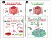
Although extensive research has been conducted on oxidative stress and inflammatory pathways in IBD, there remains a significant gap in demonstrating the correlation between oxidative stress and pro-inflammatory cytokines, TNF-α and IL-6, especially in the context of therapeutic intervention in an experimental model7,15. While prior studies have measured these markers individually, few have explored how their dynamic relationship is modulated by treatment. Sodium butyrate, a short-chain fatty acid derived from microbial fermentation of dietary fibre, is recognised for its known antioxidant and anti-inflammatory properties and ability to modulate the gut microbiota2,14. However, its ability to simultaneously regulate oxidative stress pathways and cytokine-mediated inflammation in IBD remains insufficiently explored. Therefore, the present study aims to evaluate the relationship between oxidative stress (MDA, MPO), antioxidants (SOD, GSH), concerning TNF-α levels in a dextran sulfate sodium (DSS)-induced colitis model, and to comprehensively assess the therapeutic impact of sodium butyrate on this complex interplay (Figure 1). Accordingly, the authors hypothesized that sodium butyrate would attenuate DSS-induced colitis primarily through its antioxidant and anti-inflammatory actions, and that improvements in redox balance and cytokine levels would translate into better mucosal preservation.
METHODS
Ethical approval and animals
The experimental protocol was reviewed and approved by the Institutional Animal Ethics Committee (IAEC) of KS Hegde Medical Academy, Nitte (Deemed to be University), Mangalore, India (Ref. KSHEMA/IAEC/2024/10). All possible measures were taken to minimize the number of animals used and to alleviate any pain or discomfort during the experimental procedures.
Swiss albino mice (6-8 weeks old) were obtained from the Institutional Animal House, KS Hegde Medical Academy. All mice were housed under standard conditions (12-hour light/dark cycle, 22±2°C, 50-60% humidity) with free access to standard chow and water.
Experimental and study design
After one week of acclimatization, mice were divided into four groups, each containing five mice: Normal Control (NC), followed by dextran sulfate sodium (DSS) induction, DSS-induced colitis treated with SB, and DSS-induced colitis treated with 5-aminosalicylic acid (5-ASA). Where the NC group received no treatment. In the IBD-DSS group, colitis was induced using DSS. The SB treatment group was given sodium butyrate for 7 days,andthe5-ASA group received 5-aminosalicylic acid for 7 days after DSS induction (Table 1).
Colitis was induced in all DSS groups (but not in the N. control group) by administering 2.5% (w/v) DSS (molecular weight 36,000-50,000 Da) in drinking water for 10 consecutive days, with fresh DSS prepared every 2 days. Sodium butyrate was administered at 1 mg/kg body weight and 5-ASA at 50-100 mg/kg body weight, both via oral gavage. Sacrificing the mice under anaesthesia and collecting the colonic and blood samples were processed and stored at −80°C until needed for use.
Evaluation of disease activity index (DAI) Score
The DAI score was used to assess the intensity of colon damage and clinical progression of colitisthrough daily evaluation of body weight change (%), stool consistency, and rectal bleeding, using a scoring system described by Kim et al.16 and illustrated in Table 2.
Assessment of biochemical markers
Malondialdehyde (MDA) level
MDA levels were determined in colon tissue homogenates using the thiobarbituric acid reactive substances (TBARS) assay, as described by Ohkawa et al. and Sravathi et al., with minor modifications17,18. Briefly, 500 µL of the sample was mixed with 1 mL of TCA-TBA-HCl reagent. The mixture was incubated and centrifuged to remove precipitated proteins. The absorbance of the resulting pink chromogen was measured at 535 nm using a UV-Vis spectrophotometer.
Myeloperoxidase (MPO) activity
MPO activity was measured using a colourimetric method based on the oxidation of 4-aminoantipyrine and phenol by hydrogen peroxide, catalyzed by MPO, to form a chromophore measurable at 510 nm. The sample was prepared as an enzyme extract. The reaction mixture contained 1.5 mL of 4-aminoantipyrine/phenol, 0.4 mL sodium phosphate buffer, 1.0 mL 1.7 mmol/L hydrogen peroxide, and 0.1 mL of the enzyme extract. The change in absorbance was monitored at 510 nm at 30-second intervals for 5 min. The procedure was adapted from established protocols with minor modifications19,20.
Reduced glutathione (GSH) level
GSH levels were estimated using the 5,5’-dithiobis-(2-nitrobenzoic acid) (DTNB) method, which is based on the formation of a stable yellow chromophore upon reaction of DTNB with sulfhydryl groups of reduced glutathione21. Briefly, onemillilitre of the supernatant was mixed with 1.5 mL of precipitating solution and allowed to stand for 10 min. The mixture was filtered, and 500 µL of the filtrate was combined with 2 mL of 0.3 M disodium hydrogen phosphate solution and 250 µL of freshly prepared DTNB reagent. Absorbance was measured at 412 nm against a reagent blank within 10 min. GSH concentration was calculated from a standard curve prepared using known GSH concentrations and expressed as µg/ml.
Superoxide dismutase (SOD) activity
SOD activity was determined according to the nitro blue tetrazolium (NBT) reduction method, which measures the inhibition of superoxide anion-mediated reduction of NBT to formazan in the presence of riboflavin and methionine as electron donors. Briefly, the reaction mixture consisted of 0.3 mL of riboflavin, 2.5 mL of methionine, 0.1 mL of NBT, 0.1 mL of the sample, and phosphate buffer to the required volume. After 10 min illumination in a fluorescent light chamber, absorbance was recorded at 560 nm. SOD activity was expressed as Units per Milligram (U/m) protein, where one unit of SOD was defined as the amount causing 50% inhibition of NBT reduction. The total protein concentration was determined using the biuret-based method for normalization. The technique was adapted from previous studies with minor modifications18,22.
Cytokine estimation (TNF-α and IL-6, ELISA)
The levels of tumour necrosis factor-alpha (TNF-α) and interleukin-6 (IL-6) were measured in serum using GENLISA Mouse ELISA kits (Krishgen Biosystems, India), following the manufacturer’s instructions. In brief, standards and samples were added to pre-coated microplate wells and incubated at 37°C. After washing, a biotin-conjugated detection antibody was added, followed by streptavidin-horseradish peroxidase and substrate solution. The stop solution halted the reaction, and absorbance was measured using a microplate reader at 450 nm. Cytokine concentrations were calculated and expressed in picograms per millilitre (pg/mL).
Histopathological analysis
Colon tissues were collected from all experimental groups and immediately fixed in 10% neutral buffered formalin for 24 to 48 hours. The fixed tissues were processed to make the paraffin blocks, whichweresectioned at 4-5 µm, and stained with hematoxylin and eosin (H&E).
Histopathological examination was conducted under a light microscope by a blind pathologist. Colon tissue alterations, including crypt damage, epithelial erosion, goblet cell depletion, and inflammatory cell infiltration, were evaluated. A murine-validated histological scoring system was applied, with scores ranging from 0 (unaffected) to 4 (very severe) as described previously23. Representative photomicrographs were captured using the Aperio CS2 digital pathology scanner (Leica Biosystems, Nussloch, Germany) for documentation and comparison across groups.
Statistical analysis
All data were expressed as mean ± standard deviation (SD). Statistical analyses were performed using GraphPad Prism version 8.0 software (GraphPad Software, CA, United States). Variations among multiple groups were analyzed using one-way analysis of variance (ANOVA) followed by Tukey’s post hoc test for multiple comparisons. A p-value < 0.05 was considered statistically significant.
RESULT
Disease activity index scoring
In the disease activity index, the DSS Group showed a marked increase compared to the NC group (P<0.0001). SB treatment significantly lowered DAI relative to the DSS group (P<0.01). In contrast, 5-ASA treatment caused only a modest, non-significant reduction (P>0.05). Overall, SB effectively alleviated DSS-induced disease activity, demonstrating efficacy comparable to the reference drug 5-ASA (Figure 2).

Effect of SB on colon length (cm)
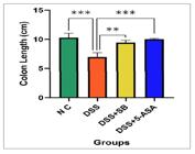
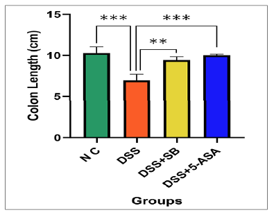
The DSS group resulted in a marked reduction in colon length compared with the NC group (P<0.001). Treatment with SB significantly preserved colon length relative to DSS (P<0.01), while 5-ASA produced a similar protective effect (P<0.001) (Figure 3).
Ameli--orative effect of SB on redox balance: attenuation of oxidative stress and promotion of antioxidants.
Malondialdehyde (MDA) level
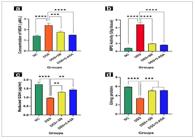
Malondialdehyde was markedly elevated in the DSS group (2.41±0.19 μM/L) compared with the NC (1.14±0.08 μM/L; P<0.0001). DSS + SB group (1.77±0.09 μM/L) significantly attenuated this increase. 5-ASA exerted the most pronounced suppression of MDA (1.51±0.00 μM/L, P<0.0001 vs DSS). These findings indicate that SB effectively reduces DSS-induced lipid peroxidation, with efficacy approaching that of 5-ASA (Figure 4A).
Myeloperoxidase (MPO) activity
MPO activity was significantly enhanced in the DSS group (6.84±0.16 U/g tissue, P<0.0001) relative to controls. The DSS+SB group (1.95±0.22 U/g tissue) and 5-ASA (1.63±0.06 U/g tissue) both markedly reduced MPO activity compared with DSS (all P<0.0001). These reductions demonstrate the anti-inflammatory potential of SB in limiting neutrophil-driven tissue injury (Figure 4B).
Reduced glutathione (GSH) level
Reduced glutathione levels were depleted by DSS (0.97±0.03 µg/ml) compared with control mice (1.17±0.21 µg/ml; P<0.0001). The DSS + SB group restored levels moderately (1.29±0.06 µg/ml; P=0.03), and 5-ASA demonstrated a more substantial restorative effect (1.43±0.09 µg/ml; P=0.003). These results highlight the capacity of sodium butyrate, especially when given after colitis induction, to support endogenous glutathione pools (Figure 4C).
Superoxide Dismutase (SOD) Activity
The DSS administration group significantly reduced SOD activity (3.42±0.41 U/mg protein) compared to the control group (5.96±0.98 U/mg protein; P<0.0001). DSS + SB group (5.05±0.33 U/mg protein, P<0.001) restored SOD activity, comparable to 5-ASA (5.15±0.23 U/mg protein). These data confirm that SB preserves enzymatic antioxidant capacity in DSS colitis (Figure 4D).
Mitigative effect of SB on pro-inflammatory cytokines
Tumor necrosis factor-α (TNF-α)
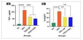
Serum TNF-α levels were sharply increased following DSS exposure (114.59±2.79 pg/mL) compared to controls (10.52±0.21 pg/mL; P<0.0001). DSS + SB group (62.82±4.60 pg/ml) significantly reduced TNF-α, with 5-ASA producing the most marked reduction (50.03±4.00 pg/ml; P<0.0001 vs DSS). These data underscore sodium butyrate’s ability to suppress a pivotal pro-inflammatory cytokine in colitis (Figure 5A).
Interleukin-6 (IL-6)
IL-6 levels were further significantly raised in DSS-treated mice (69.79±4.57 pg/ml) relative to controls (16.43±2.30 pg/ml; P<0.0001). DSS + SB group (42.80±2.82 pg/ml, P<0.01) significantly decreased IL-6, while 5-ASA (41.41±2.97 pg/ml, P<0.01) yielded the most substantial reduction. These findings confirm the anti-inflammatory efficacy of SB against DSS-induced elevation of cytokines (Figure 5B).
Histopathological findings
The histological examination of colonic sections revealed significant differences between groups (Figure 6). The NC group had intact mucosal architecture with well-preserved crypts, abundant goblet cells, and no signs of inflammatory infiltrates or ulceration. In contrast, the DSS group exhibited severe histopathological changes, including extensive crypt distortion, epithelial disruption, goblet cell depletion, prominent inflammatory cell infiltration, and mucosal ulceration, which corresponded to a significantly higher composite histological score (15.25±2.63, P<0.001 vs control; Figure 7).
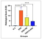
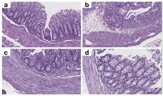
Treatment with SB (DSS+SB) partially ameliorated the DSS-induced damage, as evidenced by moderately preserved crypts, reduced infiltration, and improved goblet cell presence, with a lower composite score (8.25±2.22, P<0.01 vs DSS). The 5-ASA-treated group (DSS+5-ASA) demonstrated marked protection, characterized by a near-normal mucosal structure, minimal infiltration, and largely preserved goblet cells, resulting in the lowest composite score among the treated groups (4.50±0.58, P<0.001 vs DSS).
Overall, these findings confirm that DSS induces profound colonic injury, while both SB and 5-ASA exert protective effects, with 5-ASA showing the most substantial histological improvement.
Correlation analysis between oxidative stress, antioxidant status, and cytokines
Pearson’s correlation analysis was performed to explore further the relationship between oxidative stress and inflammatory responses (Table 3). Oxidative stress (MDA, MPO) positively correlated with pro-inflammatory cytokines TNF-α and IL-6 (r=0.85-0.90; P<0.001). In contrast, antioxidants (SOD, GSH) displayed strong negative correlations with cytokines (r=-0.75 to -0.92; P<0.001). These findings suggest that enhanced oxidative stress is closely linked to cytokine overproduction, whereas preserved antioxidant defences are inversely correlated with inflammatory mediators in DSS-induced colitis.
Table 3: the table demonstrates significant correlations between oxidative stress/antioxidant markers and pro-inflammatory cytokines in DSS-induced colitis mice, with elevated levels of MDA and MPO showing strong positive correlations with TNF-α and IL-6, while higher activities of SOD and concentrations of GSH exhibit strong negative correlations; all associations are statistically significant with P<0.001 except for SOD and IL-6, which is significant at P=0.001.
Correlation analysis between oxidative stress/antioxidant markers and pro-inflammatory cytokines in DSS-induced colitis.
DISCUSSION
The results of this current study demonstrate that SB has significant protective effects in DSS-induced colitis, as evidenced by changes in clinical severity, colon morphology, oxidative stress indicators, antioxidants, inflammatory cytokines, and histopathology. Further evidence of the interplay between redox imbalance and inflammation in driving colonic pathology came from correlation analysis. Together, these results suggest that SB has therapeutic potential as a complement to colitis treatment. Because SB treatment in this study was initiated only after DSS exposure, the effects observed represent post-induction therapeutic activity. This timing clarifies that the present findings reflect treatment of established inflammation rather than prevention of colitis development.
The increased DAI score and significant colon shortening, both known indicators, reflected the classical signs of colitis induced by DSS exposure, which is known to cause colon inflammation16,24. SB treatment significantly attenuated these clinical symptoms, with results equivalent to those of standard colitis treatment (5-ASA). These findings are consistent with a previous study, which indicated that butyrate treatment lowers clinical disease scores and prevents colon shortening in DSS-induced colitis model25,26. It is frequently used as a substitute for inflammation severity, and the preservation seen with SB confirms its ability to counter the structural effects of DSS injury6.
Our data clearly show that DSS disrupts intestinal redox equilibrium, as evidenced by elevated MDA and MPO levels, as well as lower GSH and SOD levels. These Changes align with earlier findings suggesting that central to colitis pathophysiology are oxidative stress, lipid peroxidation, and neutrophil infiltration27,28. SB treatment helped to rebalance this by enhancing antioxidant capacity while suppressing lipid peroxidation and neutrophil activity. Previous investigations have also shown that25,29. SB enhances antioxidant activity and reduces oxidative damage in experimental colitis, mechanistically through Nrf2 activation, which promotes antioxidant production and inhibits ROS-generating pathways, thereby attenuating oxidative injury1,3.
In UC, the pro-inflammatory cytokines TNF-α and IL-6 are key drivers of mucosal inflammation. As expected, DSS considerably raised inflammatory cytokines, while SB treatment significantly lowered their expression. These results are consistent with reports that butyrate encourages anti-inflammatory immunological responses and reduces cytokine synthesis by inhibiting NF-κB and HDAC activity29-31. The alleviation of clinical and histological symptoms observed in our investigation most likely resulted from the decrease in TNF-α and IL-6.
Histological examination revealed the degree of DSS-induced damage, including epithelial erosion, crypt damage, goblet cell depletion, and an inflammatory infiltrate, all consistent with DSS colitis16-23. SB Treatment significantly improved these characteristics, demonstrating maintained epithelial integrity and crypt structure as well as decreased inflammatory infiltration. Semiquantitative scoring supported these findings; SB showed 5-ASA-level protection. These results are consistent with previous studies, which indicate that butyrate supplementation supports mucosal integrity and reduces histopathological severity25,26.
Correlation analysis showed that oxidative stress markers (MDA and MPO) were positively correlated with pro-inflammatory cytokines (TNF-α and IL-6), while antioxidant parameters (GSH and SOD) were negatively correlated. This highlights the close interplay between oxidative stress and inflammation in the development of the pathogenesis of DSS colitis, thus supporting the notion of a vicious cycle between redox imbalance, injury, and cytokine upregulation4,15. The ability of SB to affect both redox and immune markers suggests a dual mechanism that interrupts this cycle and restores intestinal homeostasis.
Symbiotic processes are likely the origin of the multifaceted defence provided by SB. SB is an HDAC inhibitor that suppresses pro-inflammatory gene expression and controls chromatin structure29. Through receptor-mediated signalling (e.g., GPR109A, FFARs), it promotes anti-inflammatory responses and regulatory T-cell activity31. By activating Nrf2-dependent antioxidant pathways, SB enhances endogenous antioxidants1,3. Moreover, SB supports barrier integrity and goblet cell function, thereby limiting the translocation of luminal antigens26. Together, these actions explain the observed improvements across clinical, biochemical, and histological parameters.
SB showed notable efficacy comparable to 5-ASA, a standard therapy for UC32. This highlights its translational value both as a supplementary and alternative therapeutic modality. Future studies should focus on maximisingthe availability of butyrate in the colon by adjusting dosage schedules and delivery methods, given that it is rapidly metabolised in the proximal gut.
Limitations
Although our findings provide strong and compelling preclinical evidence, limitations persist. Though well-known, the DSS paradigm does not cover all facets of human IBD. A direct study of the molecular targets of SB, including specific HDAC isoforms or Nrf2-dependent genes, was not undertaken. Furthermore, the bioavailability of orally administered SB in humans is limited; therefore, tailored formulations or prodrug techniques may be necessary for clinical translation.
CONCLUSION
This study demonstrates that sodium butyrate attenuates DSS-induced colitis by reducing clinical severity, preserving colon morphology, restoring redox balance, suppressing pro-inflammatory cytokines, and improving histological outcomes. The strong correlations observed between oxidative markers and inflammatory mediators underscore the pivotal role of redox-inflammatory interactions in the pathogenesis of colitis. By exerting dual antioxidant and anti-inflammatory effects comparable to 5-ASA, sodium butyrate emerges as a promising microbiota-derived metabolite with translational potential for inflammatory bowel disease. Future investigations in clinical settings are warranted to validate these findings and explore long-term therapeutic potential.
REFERENCE
-
1 Chen J, Yin C, Zhang Y, Lai X, Liu C, Luo Y, et al. EGCG Alleviates DSS-Induced Colitis by Inhibiting Ferroptosis Through the Activation of the Nrf2-GPX4 Pathway and Enhancing Iron Metabolism. Nutrients 2025;17 :547. Doi:10.3390/nu17030547.
» https://doi.org/10.3390/nu17030547 -
2 Li L, Peng P, Ding N, Jia W, Huang C, Tang Y. Oxidative Stress, Inflammation, Gut Dysbiosis: What Can Polyphenols Do in Inflammatory Bowel Disease? Antioxidants (Basel). 2023;12:967. Doi:10.3390/antiox12040967.
» https://doi.org/10.3390/antiox12040967 -
3 Sahoo DK, Heilmann RM, Paital B, Patel A, Yadav VK, Wong D, et al. Oxidative stress, hormones, and effects of natural antioxidants on intestinal inflammation in inflammatory bowel disease. Front Endocrinol (Lausanne). 2023;14:1217165. Doi: 10.3389/fendo.2023.1217165.
» https://doi.org/10.3389/fendo.2023.1217165 -
4 Tratenšek A, Locatelli I, Grabnar I, Drobne D, Vovk T. Oxidative stress-related biomarkers as promising indicators of inflammatory bowel disease activity: A systematic review and meta-analysis. Redox Biol. 2024;77:103380. Doi: 10.1016/j.redox.2024.103380.
» https://doi.org/10.1016/j.redox.2024.103380 -
5 Balmus IM, Ciobica A, Trifan A, Stanciu C. The implications of oxidative stress and antioxidant therapies in Inflammatory Bowel Disease: Clinical aspects and animal models. Saudi J Gastroenterol. 2016;22:3-17. Doi: 10.4103/1319-3767.173753.
» https://doi.org/10.4103/1319-3767.173753 -
6 Kwon J, Lee C, Heo S, Kim B, Hyun CK. DSS-induced colitis is associated with adipose tissue dysfunction and disrupted hepatic lipid metabolism leading to hepatosteatosis and dyslipidemia in mice. Sci Rep. 2021;11:5283. Doi: 10.1038/s41598-021-84761-1.
» https://doi.org/10.1038/s41598-021-84761-1 -
7 Moura FA, de Andrade KQ, Dos Santos JCF, Araújo ORP, Goulart MOF. Antioxidant therapy for treatment of inflammatory bowel disease: Does it work? Redox Biol. 2015;6:617-39. Doi: 10.1016/j.redox.2015.10.006.
» https://doi.org/10.1016/j.redox.2015.10.006 -
8 Wang W, Han Y, Yin W, Wang Q, Wu Y, Du M. Intestinal and hepatic benefits of BBR-EVO on DSS-induced experimental colitis in mice. Front Microbiol. 2024;15:1428327. Doi: 10.3389/fmicb.2024.1428327.
» https://doi.org/10.3389/fmicb.2024.1428327 -
9 Franssen W. Targeting sedentary behaviour: an activity tracker approach to combat physical inactivity and improve cardiometabolic health. 2022. Doi: 10.26481/dis.20221121wf.
» https://doi.org/10.26481/dis.20221121wf -
10 Rohwer N, Jelleschitz J, Höhn A, Weber D, Kühl AA, Wang C, et al. Prevention of colitis-induced liver oxidative stress and inflammation in a transgenic mouse model with increased omega-3 polyunsaturated fatty acids. Redox Biol. 2023;64:102803. Doi: 10.1016/j.redox.2023.102803.
» https://doi.org/10.1016/j.redox.2023.102803 -
11 Tian T, Wang Z, Zhang J. Pathomechanisms of Oxidative Stress in Inflammatory Bowel Disease and Potential Antioxidant Therapies. Oxid Med Cell Longev. 2017;2017:4535194. Doi: 10.1155/2017/4535194.
» https://doi.org/10.1155/2017/4535194 -
12 Piechota-Polanczyk A, Fichna J. Review article: the role of oxidative stress in pathogenesis and treatment of inflammatory bowel diseases. Naunyn Schmiedebergs Arch Pharmacol. 2014;387:605-20. Doi: 10.1007/s00210-014-0985-1.
» https://doi.org/10.1007/s00210-014-0985-1 -
13 Chami B, Martin NJJ, Dennis JM, Witting PK. Myeloperoxidase in the inflamed colon: A novel target for treating inflammatory bowel disease. Arch Biochem Biophys. 2018;645:61-71. Doi: 10.1016/j.abb.2018.03.012.
» https://doi.org/10.1016/j.abb.2018.03.012 -
14 Liu H, Zhao W, Chen H, Wu H, Li X, Su A, et al. Highland Barley Improves DSS-Induced Ulcerative Colitis in C57BL/6J Mice. Food Sci Nutr. 2025;13:e70132. Doi: 10.1002/fsn3.70132.
» https://doi.org/10.1002/fsn3.70132 -
15 Krzystek-Korpacka M, Kempiński R, Bromke MA, Neubauer K. Oxidative Stress Markers in Inflammatory Bowel Diseases: Systematic Review. Diagnostics (Basel). 2020;10. Doi: 10.3390/diagnostics10080601.
» https://doi.org/10.3390/diagnostics10080601 -
16 Kim JJ, Shajib MS, Manocha MM, Khan WI. Investigating intestinal inflammation in DSS-induced model of IBD. J Vis Exp 2012. Doi: 10.3791/3678.
» https://doi.org/10.3791/3678 -
17 Ohkawa H, Ohishi N, Yagi K. Assay for lipid peroxides in animal tissues by thiobarbituric acid reaction. Anal Biochem. 1979;95:351-8. Doi: 10.1016/0003-2697(79)90738-3.
» https://doi.org/10.1016/0003-2697(79)90738-3 -
18 Sravathi V, Doppalapudi M, Yadala RK, Banothu A, Anumolu VK, Veera HDD, et al. Visnagin treatment attenuates DSS-induced colitis by regulating inflammation, oxidative, stress, and mucosal damage. Front Vet Sci. 2025;12:1558092. Doi: 10.3389/fvets.2025.1558092.
» https://doi.org/10.3389/fvets.2025.1558092 -
19 Simeonova R, Mihaylova R, Gevrenova R, Savov Y, Zheleva-Dimitrova D. Ulceroprotective Effects of Epilobium angustifolium Extract in DSS-Induced Colitis in Mice. Curr Issues Mol Biol. 2025;47. Doi: 10.3390/cimb47060444.
» https://doi.org/10.3390/cimb47060444 -
20 Bradley PP, Priebat DA, Christensen RD, Rothstein G. Measurement of cutaneous inflammation: estimation of neutrophil content with an enzyme marker. J Invest Dermatol. 1982;78:206-9. Doi: 10.1111/1523-1747.ep12506462.
» https://doi.org/10.1111/1523-1747.ep12506462 -
21 Moron MS, Depierre JW, Mannervik B. Levels of glutathione, glutathione reductase and glutathione S-transferase activities in rat lung and liver. Biochim Biophys Acta. 1979;582:67-78. Doi: 10.1016/0304-4165(79)90289-7.
» https://doi.org/10.1016/0304-4165(79)90289-7 - 22 Misra HP, Fridovich I. The role of superoxide anion in the autoxidation of epinephrine and a simple assay for superoxide dismutase. J Biol Chem 1972;247:3170-5.
-
23 Koelink PJ, Wildenberg ME, Stitt LW, Feagan BG, Koldijk M, van ‘t Wout AB, et al. Development of Reliable, Valid and Responsive Scoring Systems for Endoscopy and Histology in Animal Models for Inflammatory Bowel Disease. J Crohns Colitis. 2018;12:794-803. Doi: 10.1093/ecco-jcc/jjy035.
» https://doi.org/10.1093/ecco-jcc/jjy035 -
24 Khor B, Gardet A, Xavier RJ. Genetics and pathogenesis of inflammatory bowel disease. Nature. 2011;474:307-17. Doi: 10.1038/nature10209.
» https://doi.org/10.1038/nature10209 -
25 Li X, Wang C, Zhu J, Lin Q, Yu M, Wen J, et al. Sodium Butyrate Ameliorates Oxidative Stress-Induced Intestinal Epithelium Barrier Injury and Mitochondrial Damage through AMPK-Mitophagy Pathway. Oxid Med Cell Longev. 2022;2022:3745135. Doi: 10.1155/2022/3745135.
» https://doi.org/10.1155/2022/3745135 -
26 Jourova L, Satka S, Frybortova V, Zapletalova I, Anzenbacher P, Anzenbacherova E, et al. Butyrate Treatment of DSS-Induced Ulcerative Colitis Affects the Hepatic Drug Metabolism in Mice. Front Pharmacol. 2022;13:936013. Doi: 10.3389/fphar.2022.936013.
» https://doi.org/10.3389/fphar.2022.936013 -
27 Ansari MN, Rehman NU, Karim A, Soliman GA, Ganaie MA, Raish M, et al. Role of Oxidative Stress and Inflammatory Cytokines (TNF-α and IL-6) in Acetic Acid-Induced Ulcerative Colitis in Rats: Ameliorated by Otostegia fruticosa. Life (Basel). 2021;11. Doi: 10.3390/life11030195.
» https://doi.org/10.3390/life11030195 -
28 Korenaga D, Takesue F, Kido K, Yasuda M, Inutsuka S, Honda M, et al. Impaired antioxidant defense system of colonic tissue and cancer development in dextran sulfate sodium-induced colitis in mice. J Surg Res. 2002;102:144-9. Doi: 10.1006/jsre.2001.6314.
» https://doi.org/10.1006/jsre.2001.6314 -
29 Fang W, Xue H, Chen X, Chen K, Ling W. Supplementation with Sodium Butyrate Modulates the Composition of the Gut Microbiota and Ameliorates High-Fat Diet-Induced Obesity in Mice. J Nutr. 2019;149:747-54. Doi: 10.1093/jn/nxy324.
» https://doi.org/10.1093/jn/nxy324 - 30 Elias F, Mohammed A. Sodium Butyrate’s Antioxidant Effects Alleviate DSS-Induced Colitis in Rats. Azerbaijan Pharmaceutical and Pharmacotherapy Journal. 2024;23:1-5.
-
31 Yao W, Huo J, Liu K, Tao P. Exploring the health benefits of gut microbiota metabolites on combating ulcerative colitis via network pharmacology, bioinformatics and molecular docking. Sci Rep. 2025;15:25626. Doi: 10.1038/s41598-025-10851-z.
» https://doi.org/10.1038/s41598-025-10851-z -
32 Beiranvand M. A review of the biological and pharmacological activities of mesalazine or 5-aminosalicylic acid (5-ASA): an anti-ulcer and anti-oxidant drug. Inflammopharmacology. 2021;29:1279-90. Doi: 10.1007/s10787-021-00856-1.
» https://doi.org/10.1007/s10787-021-00856-1
Data-available-upon-request.

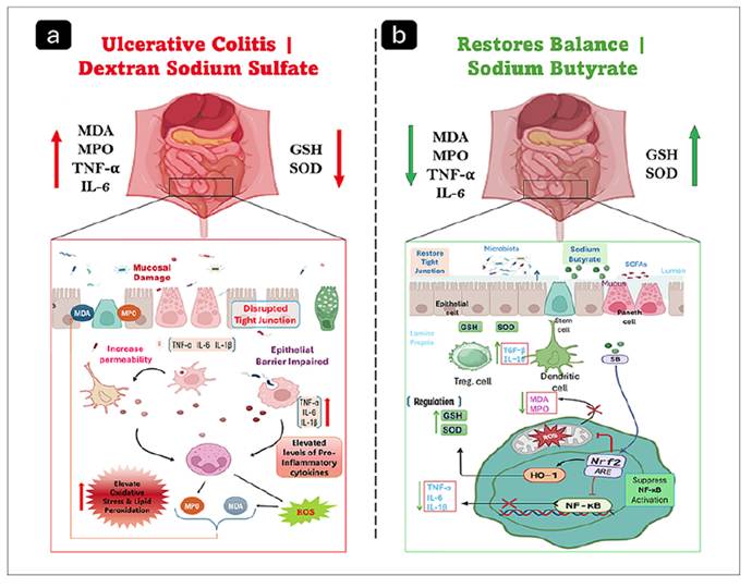 DSS administration induces colonic injury characterized by oxidative stress, increased pro-inflammatory cytokines, epithelial barrier disruption, and enhanced intestinal permeability (a). Sodium butyrate treatment ameliorates colonic damage by restoring antioxidant defenses, improving barrier integrity, and suppressing inflammatory signaling through activation of the Nrf2/HO-1 pathway and inhibition of NF-κB (b).
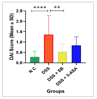 DAI scores are expressed as mean ± SD (n=5 per group). DSS treatment significantly increased DAI compared with the NC group (****P<0.0001). Sodium butyrate significantly reduced DAI compared with DSS (**P<0.01), while 5-ASA showed only a partial. Statistical significance was determined using one-way ANOVA followed by Tukey’s multiple comparison test.
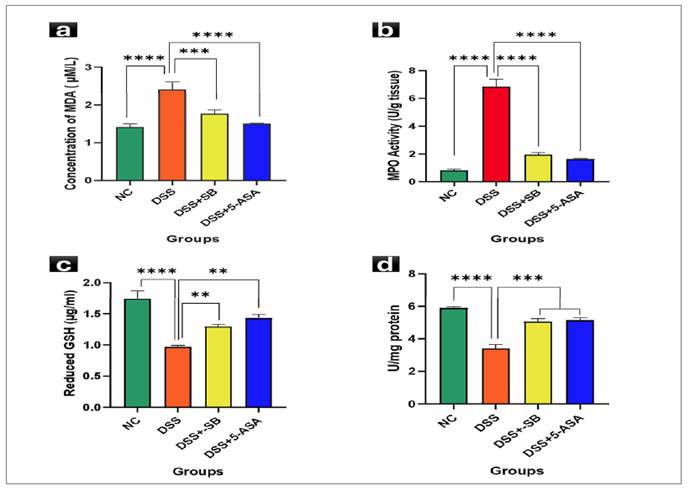 DSS markedly increased MDA, which was significantly attenuated by SB treatment and 5-ASA. Data are mean ± SD (n=5). **P<0.001, ***P<0.0001 vs DSS (A), MPO activity was significantly elevated in the DSS group, whereas in SB and 5-ASA markedly reduced. Data are mean ± SD (n=5). ****P<0.0001 vs DSS (B), DSS depleted GSH, which was significantly restored by SB treatment and 5-ASA. Data are mean ± SD (n=5). *P<0.05, **P<0.01 vs DSS (C), and DSS suppressed SOD activity, while SB and 5-ASA restored antioxidant defense. Data are mean ± SD (n=5). ***P<0.001 vs DSS (D).
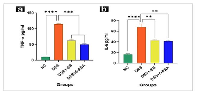 Serum TNF-α levels. DSS markedly elevated TNF-α, which was significantly reduced by SBtreatment and 5-ASA. Data are mean ± SD (n=5). ****P<0.0001 vs DSS (A). Serum IL-6 levels. DSS significantly increased IL-6; SB treatment and 5-ASA provided the strongest reductions. Data are mean ± SD (n=5). *P<0.05, **P<0.01 vs DSS (B).
 DSS administration resulted in a significant increase in histological score compared with NC, whereas both sodium butyrate (DSS+SB) and 5-ASA (DSS+5-ASA) significantly reduced the score. Data are presented as Mean ± SD (n=5 per group). ***P<0.001, **P<0.01 vs DSS
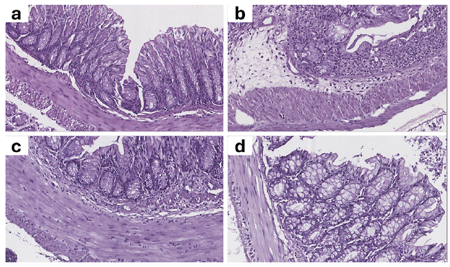 (A) NC: intact crypts, abundant goblet cells, no infiltration. (B) DSS: severe crypt distortion, epithelial damage, goblet cell loss, and heavy inflammatory infiltration. (C) DSS + SB: moderate preservation of mucosal architecture, reduced infiltration, partial goblet cell presence. (D) DSS + 5-ASA: near-normal mucosa, minimal infiltration, and preserved goblet cells.