The weight of genotype on the clinical presentation of COQ7-related hereditary motor axonal neuropathy: a case series and literature review
Abstract
Background Pathogenic variants of the COQ7 gene result in a spectrum of neurological diseases, mainly distal hereditary motor neuropathy (dHMN). We herein report cases of dHMN related to biallelic p.Met1? (c.3G > T [NM_016138]). We compare phenotypes among different COQ7variants reported in the literature.
Objective To describe and analyze our case series, review COQ7-related diseases, and compare our case series with the literature reports.
Methods We described 5 dHMN-p.Met1? patients and searched dHMN AND Brazil and COQ7 in the PubMed/MEDLINE, SciELO and Scopus databases. The categorical variables were expressed as absolute frequencies, and they were compared using the Fisher's exact test and odds ratios with 95%CIs; moreover, exploratory multivariate logistic models with penalization were applied to adjust the associations for genotype/geographic origin.
Results We analyzed four patients with dHMN plus (two with pyramidal syndrome [PS], one with cerebellar ataxia [CA] and PS, and one with cognitive impairment [CI]) and one with pure dHMN. Our search identified 47 cases of COQ7-related disorders, and The p.Met1? variant was more frequent in dHMN (p < 0.001). In exploratory multivariate models adjusting for genotype/geographic origin, the associations observed in the univariate analyses were partially sustained. The p.Met1? variant remained related to earlier age at onset, CI, and proximal lower limb weakness, whereas Brazilian origin continued to show association with cerebellar manifestations. The 95%CIs were wide due to the small sample, and the results should be interpreted as exploratory.
Conclusion In conclusion, our findings suggest that the p.Met1? variant is associated with selected phenotype, even after adjustment for genotype/geographic origin. Brazilian origin remained independently related to cerebellar involvement, indicating potential modifying factors beyond genotype.
Keywords
Charcot-Marie-Tooth Disease; Spastic Paraplegia; Muscular Atrophy, Spinal; Mitochondrial Diseases
INTRODUCTION
Mitochondrial diseases can occur due to genetic changes in nuclear or mitochondrial DNA, leading to defects in oxidative phosphorylation.1, 2 Coenzyme Q10 (CoQ10) is essential as an electron transporter and antioxidant.3 Consequently, CoQ10 is an important protein in cellular metabolism.3 Primary CoQ10 deficiency is a rare disease characterized by genetic and phenotypic heterogeneity.4 The condition is often treatable, usually responding to exogenous CoQ10 replacement,4 and new therapeutic targets are also under study.5–8 Several genes have already been correlated with primary CoQ10 deficiency in humans: PDSS1, PDSS2, COQ8A, COQ8B, COQ7, COQ9, COQ6, COQ4, COQ6, COQ2, and COQ5.9
The mitochondrial enzyme 5-demethoxyubiquinone hydroxylase, encoded by the COQ7 gene, is one of the enzymes involved in CoQ10 biosynthesis.10, 11 Mutations in COQ7 result in primary CoQ10 deficiency-8 (CoQ10D8).3 The first case of neurological disease related to COQ7 was published in 2015.6 It was a multisystemic form (MF) of the CoQ10 deficiency, with the mitochondrial encephalo-myo-nephro-cardiopathy phenotype. Subsequently, the COQ7 phenotype was expanded to include spastic paraplegia (SPG),5,10,12–14 demyelinating or axonal sensorimotor polyneuropathy,10,12,14,15 and motor neuron disease.16 In recent years, some case series6,12–15,17–27 have been published associating COQ7 variants with distal hereditary motor neuropathy (dHMN). Rebelo et al.15 reported cases in nine families with COQ7-related dHMN, and five Brazilian families harbored the p.Met1? variant, which shows signs of upper motor neuron dysfunction, cognitive impairment, and cerebellar ataxia (CA). Evidence for a founder effect of the p.Met1? variant in the region was also provided by Rebelo et al.15 We herein report the cases of 5 Brazilian patients from 3 unrelated families with the same p.Met1? (c.3G > T [NM_016138]) variant presenting different neurological findings associated with dHMN, reinforcing the link between this phenotype and the specific p.Met1? COQ7 variant. We also provide a narrative review of COQ7 disorders, comparing the phenotypes among different variants.
METHODS
In order to conduct the present observational and descriptive study, we retrospectively collect a series of cases from the Neuromuscular Outpatient Clinic at the tertiary Teaching Hospital of Faculdade de Medicina de São José do Rio Preto (FAMERP), in the state of São Paulo, Brazil. All cases were submitted to a genetic panel for neuromuscular disease involving 110 genes and using next-generation sequencing with the Illumina technology (Illumina, Inc.). Variant alignment and identification were performed using standard bioinformatics protocols, referencing the Genome Reference Consortium Human Build 38 (GRCh38). All procedures in the current study were performed under the ethical standards of the FAMERP Ethics in Research Committee and in accordance with the 1964 Helsinki Declaration and its later amendments or comparable ethical standards.
We compared the clinical features with the reported COQ7-patients and Brazilian dHMN cohorts in the literature. In July 2025, the PubMed/MEDLINE, SciELO, and Scopus databases were searched using the terms COQ7 and dHMN AND Brazil. The results were screened according to the type of study, and we only selected those involving humans. The categorical variables were expressed as absolute frequencies and compared using the Fisher's Exact test and odds ratios (OR), with 95%CIs. All tests were two-tailed. Statistical significance was set at p < 0.05 for all two-tailed tests. Descriptive tables and statistical computations were conducted using Google Sheets (Alphabet Inc.) and VassarStats software.
RESULTS
Case series
Patient 1. A 40-year-old man, born to consanguineous parents, reported difficulty walking since childhood and sporadic falls during sports activities. Over time, he developed imbalance, bilateral incoordination in the upper limbs, slow speech, and the need for aid to walk and to perform basic activities of daily living, with more frequent falls. The physical findings included fixed contracture of the ankle tendons, high-arched feet (Figure 1E), slurred speech, a wide-based gait, and preserved muscle strength, except for weakness in ankle dorsiflexion. The knee reflexes were increased, and those of the ankle were abolished. Appendicular ataxia, trunk-limb incoordination, and hypometric saccades were also noted. The mental status was preserved. A brain Magnetic Resonance Imaging (MRI) scan (Figure 1A,B) revealed mild cerebellar atrophy with a predominant involvement of the vermis and the upper half of the cerebellar hemispheres. Neurophysiological studies were consistent with motor axonal neuropathy. No abnormality was observed on general laboratory tests.

Brain magnetic resonance imaging (MRI) scan showing cerebellar atrophy, affecting predominantly the lobes: fluid-attenuated inversion recovery (FLAIR) sequence (A: axial; B: sagittal) in patient 1. Brain MRI scan with microangiopathy disease (Fazekas 3 on periventricular and deep white matter) in patient 4 (C,D). Pes cavus: patient 1 (E). Muscular atrophy of the foot and hand: patient 4 (F,G). Patient 5: lower limb atrophy affecting the tibialis anterior muscle and the medial portion of the thigh (H). Muscle biopsy of Patient 3 showed variation in muscle fiber diameter, with scattered small fibers and fibers presenting internal nuclei; multiple granular fibers (hematoxylin and eosin) (I) and ragged-red fibers were also noted." (Gomori's trichrome stain) (J). Patient 4's muscle biopsy: a predominance of type-1 fibers was observed in the reactions by adenosinetriphosphatases (ATPases), with some rare areas of type-2 fiber grouping without atrophy or hypertrophy (ATPase pH: 9.4) (K) Several cytochrome c oxidase (COX)-negative fibers were observed, demonstrating positivity on combined COX/succinate dehydrogenase (SDH) histochemistry.) in both cases (L,M).
Patient 2. A 25-year-old male patient presented with foot deformity, recurrent falls, and bilateral foot drop, which started at the age of 10 years. The patient reported a family history of consanguinity, with his parents being third cousins and his great-grandparents being first cousins. A neurological examination revealed high-arched feet, hammer toes, scoliosis, distally-predominant weakness in the upper limbs, fine postural tremor of the hands, and decreased proprioceptive sensation in the lower limbs. The reflexes were globally absent, except for those of the patella, which were normal. Electroneuromyography demonstrated symmetrically-decreased motor conduction amplitude, primarily suggestive of axonal compromise and signs of active denervation. A nerve biopsy exhibited no specific abnormalities, with only a mild increase in the endoneurial connective tissue observed. The levels of creatine kinase (CK) were mildly elevated, but no other serum laboratory tests were found to be abnormal.
Patient 3. A 72-year-old woman presented with slowly-progressive weakness in her lower limbs that had started at the age of 10 years. At 45 years, she required assisted ambulation, with the upper limbs also affected. Inability to walk and bulbar symptoms were present at 59 years old. There was moderate weakness in the hands, with intrinsic muscle atrophy; moderate distal and mild proximal weakness was found in the lower limbs. The electrophysiological examination revealed motor axonal neuropathy. The muscle biopsy demonstrated slight variation in the caliber of the muscle fibers, with predominance of type I, and mitochondrial abnormalities. The patient's condition deteriorated, and she passed away at the age of 72 years due to infectious complications.
Patient 4. A 67-year-old woman, the sister of patient 3. She reported a lifelong progressive weakness since childhood, initially on her lower limbs, but affecting her upper limbs throughout the decades since the onset of the symptom. At the age of 65 years, she was in a wheelchair after a traumatic brain injury, with contusion and intraparenchymal hematoma due to falling from her height. There was tetraparesis predominantly affecting the lower limbs, distal muscle atrophy in the four limbs, pes cavus, spastic hypertonia, and hyperreflexia in the upper limbs, accompanied by tendon retraction (Figure 1F,G). The electrophysiological study revealed a motor axonal neuropathy similar to that of patient 3. The muscle biopsy was suggestive of mitochondrial myopathy and chronic reinnervation, with variation in the diameter of the muscle fibers, slight disarrangement of the intermyofibrillar cytoarchitecture, with predominance of type I, and some areas of fiber type grouping (Figure 1J–M). A brain MRI scan revealed extensive microangiopathy (in the periventricular and deep white matter, classified as grade 3 on the Fazekas scale) (Figure 1C,D). There was no hypertension or diabetes, and the patient denied smoking.
Patient 5 is a 14-year-old female adolescent, granddaughter of patient 4. She presented a history of frequent falls and pain in the lower limb after exertion, starting at the age of 10 years. The mother confirmed mild intellectual disability and inattention early in childhood. The clinical examination revealed weakness in both proximal and distal muscles of the upper limbs and high-arched feet (Figure 1H). The serum laboratory tests were regular, except for slightly elevated CK (214 U/L). Electroneuromyography revealed motor axonal neuropathy with active denervation.
Patients 3, 4, and 5 had a family history of consanguinity (parents who were second cousins) and were from a small town in Brazil where consanguineous marriages were not rare.
Since patients 3 and 4 presented elevated serum CK levels and weakness, they underwent muscle biopsy at the time for differential diagnosis of mitochondrial disease, at a time when genetic testing was not yet available.
Except for patient 2, the remaining cases received CoQ10, at daily doses ranging from 200 to 400 mg for 1 or 2 years. No clear improvement or worsening was noted in any patient. The five patients underwent a genetic panel for neuromuscular disorders, and the p.Met1? variant of the COQ7 gene (chr16:19,067,667 G > T; c.3G > T - ENST00000321998) was identified in homozygosity in all reported cases.
Narrative review
A broad spectrum of phenotypic manifestations is associated with COQ7 deficiency, ranging from pure neurological forms to an MF. The most well-known form of MF related to COQ7 used to be a progressive encephalo-neuro-nephro-cardiopathy, frequently presenting with tubulopathy, lactic acidosis, hypertrophic cardiomyopathy, growth retardation, and delayed motor development.6,11,17,28,29 So far, this form has been reported to be associated with loss-of-function missense or frameshift variants. Both pure and complex forms of hereditary spastic paraplegia (HSP) have also been described in association with missense pathogenic COQ7 variants (3 cases with p.Leu111Pro, 3 cases with p.Arg54Gln, and 1 case with p.Pro108Thr).13,14,18,19,29 In the last few years, cases associated with the axonal phenotype Charcot-MarieTooth (CMT) disease were published:11,12,15 1 case with c.319C > T; Arg107Trp, one case with c.467T > G (p.Leu156Arg) and c.599_600delAGins TAATGCATC (p.Lys200IlefsTer56), and 1 case with c.446A > G;(p.Tyr149Cys) and c.3G > T (p.Met1?).
A Brazilian case of the COQ7 dHMN phenotype with the homozygous variant p.Met1? (c.1A > G) was presented as a poster at a national congress in 2021.23 In 2023, Smith et al.18 reported a consanguineous Syrian family with 3 affected siblings, all with the dHMN phenotype associated with the same biallelic variant. Jacquier et al.21 reported a Portuguese family with 3 dHMN cases associated with the biallelic variant p.Met1? (c.3G > T). In both studies, the variant's pathogenicity was demonstrated in in-vitro studies, which showed a severe decrease in COQ7 protein levels in the probands' fibroblasts and a decrease in CoQ10 production. In 2023, similar Brazilian cases were described related to p.Met1?: 2 unrelated women with infant-onset dHMN (presented as a conference poster),25 a case of youth-onset dHMN,10,11 1 case of the juvenile amyotrophic lateral sclerosis (ALS) form,16 and Rebelo et al.15 reported 6 cases in 5 Brazilian families with dHMN. Other dHMN variants were reported: c.161G > A;(p.Arg54Gln),10,15 c.253-2A > T/c.467T > A,22 c.160C > T/c.467T > G,22 c.197T > A (Ile66Asn)/c.446A > G (Tyr149Cys),24 c.197T > A (Ile66Asn)/c.319C > T (p.Arg107Trp),15 and c.197T > A (p.Ile66Asn) and c.446A > G (p.Tyr149Cy).15
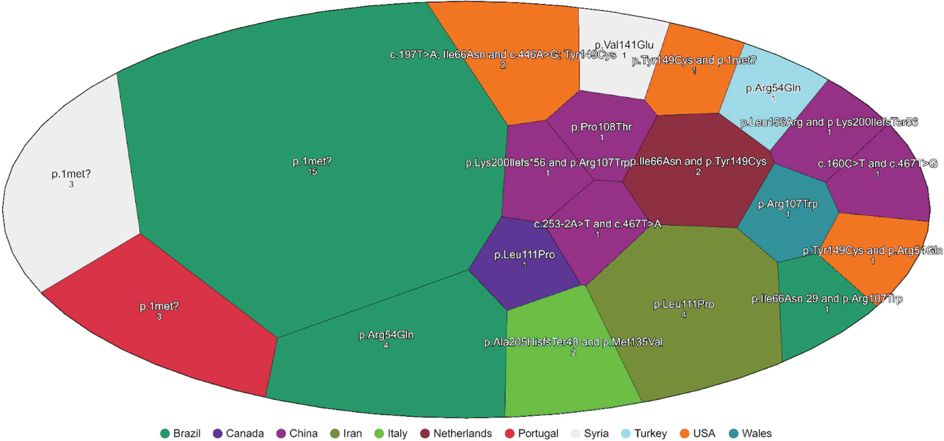
The different variants with phenotype and origin correlation are presented in Table 1 and illustrated in a Voronoi map in Figure 2.

Statistical analysis
In our search, we found 20 studies from 10 countries involving 47 patients with COQ7-related disorders. In total, 30 out of the 47 cases were related to dHMN (63.8%), 8, to SPG (17%), 6, to MF (12.7%), and 3, to CMT (6.3%). The p.Met1? variant was present in 21 COQ7 cases (44.6%) (Table 2). All patients presented dHMN: 9 (42.8% of the p.Met1? cases) with the pure dHMN phenotype, and 12 (57.2% of the p.Met1? cases) with dHMN plus. The dHMN plus cases were patients with the main clinical feature of motor axonal neuropathy but associated with other symptoms: 4 (19% of the p.Met1? cases) had dHMN associated with CA, 3 (14.2% of the p.Met1? cases) had dHMN and pyramidal signs (PS), another 3 (14%) had dHMN associated with CA and PS, 1 case (4.7% of the p.Met1? cases) was associated with cognitive impairment (the one reported in the present study), and 1 had juvenile ALS (Figure 3A). The origin of the p.Met1 cases were: 71.4% from Brazil, 14.2% from Portugal, and 14.2% from Syria. With the other variants, a high phenotypic heterogeneity was observed: 9 (34.6%) cases of dMHN (8 with pure and 1 with PS), 8 (30.7%) cases of SPG, 6 (23%) cases of MF, and 3 (11.5%) cases of CMT (Figure 3B). Regarding the Brazilian dHMN cohorts, 2 studies25,26 were included, amounting to 170 cases, with no COQ7 disease reported among them.

(A) Percentage of the phenotype in cases of Coenzyme Q10 deficiency-8 CoQ10D8 associated with the biallelic p.Met1 variant?. (B) Percentage of the phenotype in cases of CoQ10D8 associated with other variants reported in the literature.
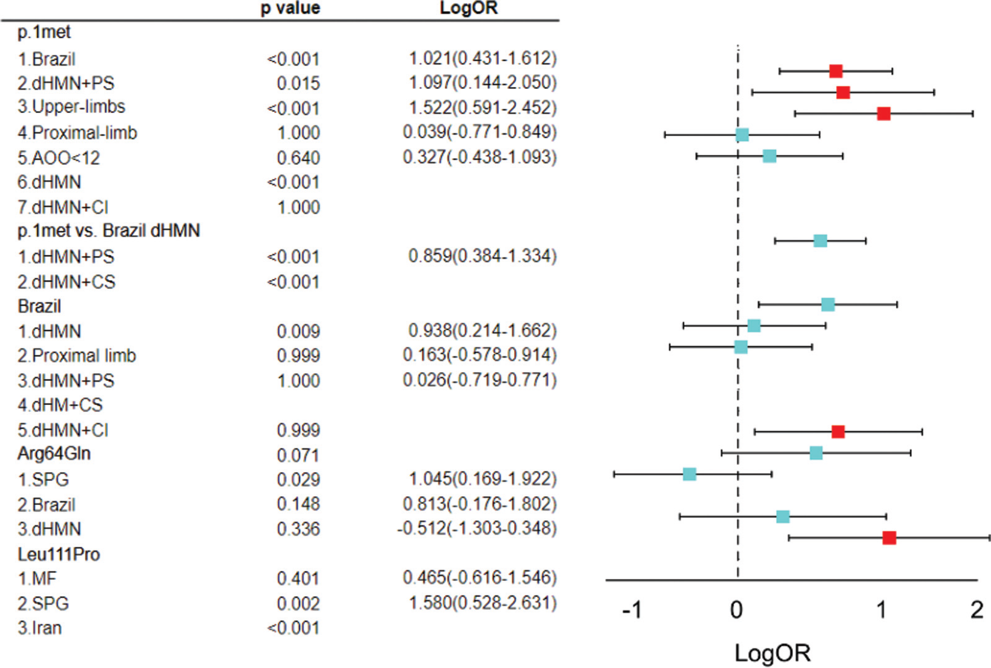
On the comparison of the phenotypes among the presence of variants, the p.Met1? was significantly more frequent in patients with dMHN, considering all cases of dHMN (pure and plus; 21 out 30; p-value < 0.001) (Figure 3). Brazilian origin was also significantly related to the p.Met1? variant compared to other regions (15 out 21; p-value < 0.001; OR = 10.5; 95%CI = 2.696–40.880). Brazilians have a statistically significant relationship with the dHMN phenotype (13 out of 28; p-value = 0.009; OR = 8.6; 95%CI = 1.63–45.86). All CA patients were from Brazil; this relationship was also statistically significant (p-value = 0.0063; Figure 4).

Values of p and LogOR (odds ratio) (Log10[Lower Odds Ratio] - Log10[Upper Odds Ratio]) of the categorical variables on the left side and forest plot on the right side.
Compared to the 170 cases of several types of dHMN from Brazil,25,26 PS (p-value < 0.001; OR = 7.2273; 95%CI = 2.4205–21.5797) and CA (pvalue < 0.001) associated with dHMN were statistically significant for p.Met1? COQ7 (Figure 3). The SPG-COQ7 variant is related to p.Leu111Pro, with statistical significance (p-value = 0.00181; OR = 34; 95%CI = 3.3741–427.9693). This variant had a strong association with Iran (p-value < 0.001). The p.Arg54Gln variant was statistically associated with SPG (p-value = 0.029; OR = 11.1; 95%CI = 1.475–83.5322) (Figure 4).
We also performed multivariate statistical analyses based on penalized logistic regression models, for each clinical outcome of interest and fitted separate logistic regression models using genotype and country of origin as predictors. The variants were binarized according to their presence or absence in each individual (p.Met1?, p.Arg54?, p.Leu111Pro), and geographic origin was coded as Brazil versus non-Brazil. Given the limited sample size and the presence of sparse cells in the contingency tables, a penalty term (L2-regularization) was applied to stabilize the coefficient estimation and reduce small-sample bias. The models were fitted using maximum likelihood estimation with ridge penalization and a fixed regularization parameter (C = 1.0), and effect sizes were derived as adjusted ORs. To obtain empirical 95%CIs, we generated bootstrap resampling distributions (500 iterations per model), computing percentile-based confidence limits for each predictor. Because of the sparse patterns in several outcomes, the results were considered exploratory and hypothesis-generating rather than confirmatory. The p.Met1? variant and Brazilian origin remained associated with selected phenotypic features after the multivariable adjustment, although the 95%CIs remained wide. These models were used strictly to test whether the observed univariate associations persisted after mutual adjustment among predictors. Complete data on the statistical analysis can be found in Tables 3 and 4.
Multivariate penalized logistic regression results (L2), adjusted for genotype and geographic origin
DISCUSSION
Although COQ7 deficiency can lead to different phenotypes, we herein reported 5 patients from 3 different kinships, all with dHMN. Until 2024, this presentation was not featured among the COQ7 spectrum, and it could be considered an uncommon manifestation of the disease.19,30 Among dHMNs, neither was COQ7 considered a possible etiology until recently, not even as a rare etiology for dHMNs.31 However, a new scenario with distal motor axonal neuropathy has been reported in several individuals with COQ7-related CoQ10 deficiency.12,13,15,18–23 The 5 additional cases herein reported strengthen the notion that this presentation is a crucial phenotype linked to the gene. This phenotype has now become the most reported phenotype related to COQ7 in the current literature, with or without concomitant involvement of the first motor neuron.
The p.Met1? (c.3G > T and c.1A > G) variant was found to be statistically linked to the dHMN phenotype (p < 0.001).All patients homozygous for this variant had dHMN: 9 (42.8% of the p.Met1? cases) with the pure dHMN phenotype and 12 (57.2% of the p.Met1? cases) with dHMN plus. This suggests that the clinical manifestations of COQ7-related CoQ10 deficiency may be correlated to specific variants, possibly varying according to the locus and type of the variant. The p.Met1? variant disrupts the translation initiation codon, leading to a loss of protein production. Thus, protein synthesis is prevented from beginning at the predicted point. A significantly-reduced translation of COQ7 isoform 1 was already demonstrated with this variant.21 Other isoforms of the protein can probably provide mitochondria with COQ7, which would partially compensate for the loss of isoform 1 with some grade of phenotype rescue. Another reported variant in the same codon (c.1A > G; p.Met1?) was also associated with pure dHMN, supporting this view. Thus, the hypothesis raised by Jacquier et al.21 in 2023 is reinforced here: restricted dHMN would be a milder presentation related to p.Met1?, due to its lower impact compared to variants located in the catalytic site.21
Patient 1 presented dHMN plus with cerebellar and pyramidal syndrome with cerebellar atrophy on MRI (Figure 1C,D). Patient 2 had pure dHMN. Patients 3 and 4, siblings, both had dHMN plus (PS), and patient 5 had dHMN plus with cognitive impairment. All patients presented onset of symptoms in the first decade of life. These findings are consistent with those of other reports of dHMN related to COQ7 in the literature. The phenotype with dHMN associated with CA was observed in 7 cases in the literature, all harboring the p.Met1? variant. However, CA was only reported in patients of Brazilian origin, which was also significantly linked to dHMN with CA (p < 0.001), Moreover, PS in Brazilian patients with dHMN was associated with COQ7. Therefore, among Brazilian patients, dHMN with CA and/or PS suggests a higher chance of COQ7 deficiency. These findings raise the hypothesis that the environment could also play a significant role in phenotype expression.
The neuroimaging features related to COQ7 deficiency were only reported in MF: hyperintensity in the periventricular white matter on T2-weighted and fluid-attenuated inversion recovery (FLAIR) sequences, multiple cystic changes involving bilateral corona radiata, the basal ganglia, and thalami, brainstem hypoplasia, mega cisterna magna, severe thinning of the corpus callosum, enlargement of the ventricular system, and progressive cortical atrophy.28,32 In the present study, Patient 4, with dHMN and PS, had a prominent periventricular and deep white matter T2 hyperintensity, besides her age and absence of other microangiopathy risk factors. This image finding would be worthy of search on other cases of COQ7 deficiency, to verify if could be another milder manifestation of the disease, or even of the specific p.Met1? variant.
Except for patient 3, our cases were put on CoQ10 reposition. Neither improvement nor deterioration was found in this short observation period. Furthermore, all treated cases started on CoQ10 late in life and at low dosages (100–500 mg a day, due to the high cost), which hinders the observation of benefits. Oral CoQ10 treatment has been partially successful in some cases of primary CoQ10 deficiency.25,33 Nevertheless, no apparent improvement in COQ7 patients has been indisputably observed.11,15 This treatment presents deficient absorption and bioavailability due to high molecular weight and low aqueous solubility, which limits its therapeutic potential.34 Consensus regarding the dose of the CoQ10 replacement is lacking, and the recommended dosage ranges from 5 to 50 mg/kg/day, with 1,200mg a day as the maximum safest dose.33 Higher doses of CoQ10, with early beginning of treatment, could lead to better responses once in-vitro studies suggest improvement with the replacement.15,21 Furthermore, patients with peripheral manifestations may exhibit a better therapeutic response than those with central or multisystemic involvement.35
In conclusion, the present study supports the notion that COQ7 is an important cause of dHMN. The cases herein reported and data in the literature suggest that specific COQ7 variants may be associated with specific phenotypes: p.Met1?, with dHMN, and p.Arg54Gln and p.Leu111Pro, with SPG. Additionally, the p.Met1? variant seems to be linked to CA as well, especially in cases of Brazilian origin. Among Brazilian patients, dHMN with CA and/or PS suggests a higher chance of COQ7 deficiency. In Brazilian patients, CA raises the hypothesis of environmental influence in disease expression. Finally, white matter hyperintensity on MRI can also manifest in mild COQ7 cases. Even after multivariable adjustment for genotype distribution and geographic origin, the p.Met1? variant remained associated with earlier age at onset, cognitive impairment, and proximal lower-limb weakness, reinforcing its relevance as a phenotypic driver within COQ7-related disorders. Likewise, Brazilian origin continued to demonstrate an independent association with cerebellar involvement, suggesting the influence of contextual or modifying factors that extend beyond genotype alone. Although these findings were derived from exploratory penalized models and the 95%CIs remained wide due to the small sample size, they provide additional support for genotype–phenotype clustering and highlight the need for larger, systematically-characterized cohorts to validate these observations.
Data Availability Statement
Data will be available upon request to the corresponding author.
References
-
1 Rocha EBdS, Rodrigues KdL, Montouro LAM, et al. A case of mitochondrial DNA depletion syndrome type 11 - expanding the genotype and phenotype. Neuromuscul Disord 2023;33 (08):692–696. Doi: 10.1016/j.nmd.2023.06.004
» https://doi.org/10.1016/j.nmd.2023.06.004 -
2 Gorman GS, Chinnery PF, DiMauro S, et al. Mitochondrial diseases. Nat Rev Dis Primers 2016;2:16080. Doi: 10.1038/nrdp.2016.80
» https://doi.org/10.1038/nrdp.2016.80 -
3 Mantle D, Millichap L, Castro-Marrero J, Hargreaves IP. Primary Coenzyme Q10 Deficiency: An Update. Antioxidants 2023;12(08): 1652. Doi: 10.3390/antiox12081652
» https://doi.org/10.3390/antiox12081652 -
4 Galosi S, Barca E, Carrozzo R, et al. Dystonia-Ataxia with early handwriting deterioration in COQ8A mutation carriers: A case series and literature review. Parkinsonism Relat Disord 2019; 68:8–16. Doi: 10.1016/j.parkreldis.2019.09.015
» https://doi.org/10.1016/j.parkreldis.2019.09.015 -
5 Wang Y, Smith C, Parboosingh JS, Khan A, Innes M, Hekimi S. Pathogenicity of two COQ7 mutations and responses to 2,4dihydroxybenzoate bypass treatment. J Cell Mol Med 2017;21 (10):2329–2343. Doi: 10.1111/jcmm.13154
» https://doi.org/10.1111/jcmm.13154 -
6 Freyer C, Stranneheim H, Naess K, et al. Rescue of primary ubiquinone deficiency due to a novel COQ7 defect using 2,4dihydroxybensoic acid. J Med Genet 2015;52(11):779–783. Doi: 10.1136/jmedgenet-2015-102986
» https://doi.org/10.1136/jmedgenet-2015-102986 -
7 Hidalgo-Gutiérrez A, González-García P, Díaz-Casado ME, et al. Metabolic targets of coenzyme Q10 in mitochondria. Antioxidants 2021;10(04):520. Doi: 10.3390/antiox10040520
» https://doi.org/10.3390/antiox10040520 -
8 Staiano C, García-Corzo L, Mantle D, et al. Biosynthesis, Deficiency, and Supplementation of Coenzyme Q. Antioxidants 2023;12 (07):1469. Doi: 10.3390/antiox12071469
» https://doi.org/10.3390/antiox12071469 -
9 Alcázar-Fabra M, Rodríguez-Sánchez F, Trevisson E, Brea-Calvo G. Primary Coenzyme Q deficiencies: A literature review and online platform of clinical features to uncover genotype-phenotype correlations. Free Radic Biol Med 2021;167:141–180. Doi: 10.1016/j.freeradbiomed.2021.02.046
» https://doi.org/10.1016/j.freeradbiomed.2021.02.046 -
10 Wongkittichote P, Lasio LD, Magistrati M, et al. P360: Phenotypic and molecular characterization of a cohort of patients with primary CoQ10 deficiency caused by pathogenic variants in COQ7. Genet Med Open 2023;1(01):100388. Doi: 10.1016/j. gimo.2023.100388
» https://doi.org/10.1016/j.gimo.2023.100388 -
11 Wongkittichote P, Lasio MLD, Magistrati M, et al. Phenotypic, molecular, and functional characterization of COQ7-related primary CoQ10 deficiency: Hypomorphic variants and two distinct disease entities. Mol Genet Metab 2023;139(04):107630. Doi: 10.1016/j.ymgme.2023.107630
» https://doi.org/10.1016/j.ymgme.2023.107630 -
12 Zhang XY, Dong HL, Wu ZY. Axonal Charcot-Marie-Tooth disease due to COQ7 mutation: expanding the genetic and clinical spectrum. Brain 2023;146(12):e117–e119. Doi: 10.1093/brain/awad212
» https://doi.org/10.1093/brain/awad212 -
13 Sadr Z, Zare-Abdollahi D, Rohani M, Alavi A. A founder mutation in COQ7, p.(Leu111Pro), causes pure hereditary spastic paraplegia (HSP) in the Iranian population. Neurol Sci 2023;44(07): 2599–2602. Doi: 10.1007/s10072-023-06707-x
» https://doi.org/10.1007/s10072-023-06707-x -
14 Qiu Y, Xiong Y, Wang L, Zhu M, Tan D, Hong D. Homozygous variant in COQ7 causes autosomal recessive hereditary spastic paraplegia. Ann Clin Transl Neurol 2024;11(04):1067–1074. Doi: 10.1002/acn3.52037
» https://doi.org/10.1002/acn3.52037 -
15 Rebelo AP, Tomaselli PJ, Medina J, et al. Biallelic variants in COQ7 cause distal hereditary motor neuropathy with upper motor neuron signs. Brain 2023;146(10):4191–4199. Doi: 10.1093/brain/awad158
» https://doi.org/10.1093/brain/awad158 -
16 Souza PVS, Serrano PL, Farias IB, et al. Clinical and Genetic Aspects of Juvenile Amyotrophic Lateral Sclerosis: A Promising Era Emerges. Genes (Basel) 2024;15(03):311. Doi: 10.3390/ genes15030311
» https://doi.org/10.3390/genes15030311 -
17 Kwong AK, Chiu AT, Tsang MH, et al. A fatal case of COQ7associated primary coenzyme Q10 deficiency. JIMD Rep 2019;47 (01):23–29. Doi: 10.1002/jmd2.12032
» https://doi.org/10.1002/jmd2.12032 -
18 Smith IC, Pileggi CA, Wang Y, et al; Care4Rare Canada Consortium. Novel Homozygous Variant in COQ7 in Siblings With Hereditary Motor Neuropathy. Neurol Genet 2023;9(01):e200048. Doi: 10.1212/NXG.0000000000200048
» https://doi.org/10.1212/NXG.0000000000200048 -
19 Hashemi SS, Zare-Abdollahi D, Bakhshandeh MK, et al. Clinical spectrum in multiple families with primary COQ10 deficiency. Am J Med Genet A 2021;185(02):440–452. Doi: 10.1002/ajmg.a.61983
» https://doi.org/10.1002/ajmg.a.61983 -
20 Souza PVSd, Farias IB, Serrano PdL, et al. COQ7-Related Juvenile-Onset Motor Neuronopathy: A New Pathogenetic Dysfunction Associated with Motor Neuron Disease. Sclerosis. 2023;1(01): 22–26. Doi: 10.3390/sclerosis1010004
» https://doi.org/10.3390/sclerosis1010004 -
21 Jacquier A, Theuriet J, Fontaine F, et al. Homozygous COQ7 mutation: a new cause of potentially treatable distal hereditary motor neuropathy. Brain 2023;146(08):3470–3483. Doi: 10. 1093/brain/awac453
» https://doi.org/10.1093/brain/awac453 -
22 Liu XX, Wang N, Chen YK, et al. Biallelic variants in the COQ7 gene cause distal hereditary motor neuropathy in two Chinese families. Brain 2023;146(05):e27–e30. Doi: 10.1093/brain/awad040
» https://doi.org/10.1093/brain/awad040 - 23 Santos DF, Légora DB. A case of COQ7-associated primary coenzyme Q10 deficiency related to spastic paraparesis and hereditary motor neuropathy. Arq Neuro-Psiquiatr 2021;9(S1):79
-
24 Theunissen TEJ, Nguyen M, Kamps R, et al. Whole Exome Sequenc-ing Is the Preferred Strategy to Identify the Genetic Defect in Patients With a Probable or Possible Mitochondrial Cause. Front Genet 2018;9:400. Doi: 10.3389/fgene.2018.00400
» https://doi.org/10.3389/fgene.2018.00400 - 25 Tomaselli PJ, Frezatti RS, Barbosa FF, et al. Distal hereditary motor neuropathy: clinical and molecular characterization of Brazilian population. Arq Neuro-Psiquiatr 2022;80(S2):295
- 26 Sampaio PHMA, Fonseca ATQSM, Moreno CAM, et al. Clinical and genotypical spectrum of hereditary motor neuropathies. Arq Neuro-Psiquiatr 2022;80(S2):345
-
27 Alvarenga TN, Sousa-Santos PEM, Santos PEMS, et al. Primary coenzyme Q10 (COQ10) deficiency: Clinical presentation of a new variant in COQ7 gene. São Paulo Med J 2023;141(Suppl 1):56. Doi: 10.5327/1516-3180.141s1.613
» https://doi.org/10.5327/1516-3180.141s1.613 -
28 Pettenuzzo I, Carli S, Sánchez-Cuesta A, et al. COQ7 defect causes prenatal onset of mitochondrial CoQ10 deficiency with cardiomyopathy and gastrointestinal obstruction. Eur J Hum Genet 2024; 32(08):938–946. Doi: 10.1038/s41431-024-01615-w
» https://doi.org/10.1038/s41431-024-01615-w -
29 Wang Y, Gumus E, Hekimi S. A novel COQ7 mutation causing primarily neuromuscular pathology and its treatment options. Mol Genet Metab Rep 2022;31:100877. Doi: 10.1016/j.ymgmr.2022.100877
» https://doi.org/10.1016/j.ymgmr.2022.100877 -
30 Salviati L, Trevisson E, Agosto C, Doimo M, Navas P. Primary Coenzyme Q10 Deficiency Overview. In: Adam MP, Feldman J, Mirzaa GM, et al., editors. GeneReviews®. Seattle: University of Washington; 1993–2026. Available from: https://www.ncbi.nlm.nih.gov/books/NBK410087/
» https://www.ncbi.nlm.nih.gov/books/NBK410087/ -
31 Frasquet M, Sevilla T. Hereditary motor neuropathies. Curr Opin Neurol 2022;35(05):562–570. Doi: 10.1097/WCO.0000000000001087
» https://doi.org/10.1097/WCO.0000000000001087 -
32 Münch J, Prasuhn J, Laugwitz L, et al. Neuroimaging in Primary Coenzyme-Q10-Deficiency Disorders. Antioxidants 2023;12(03):718. Doi: 10.3390/antiox12030718
» https://doi.org/10.3390/antiox12030718 -
33 Stefely JA, Pagliarini DJ. Biochemistry of Mitochondrial Coenzyme Q Biosynthesis. Trends Biochem Sci 2017;42(10):824–843. Doi: 10.1016/j.tibs.2017.06.008
» https://doi.org/10.1016/j.tibs.2017.06.008 -
34 García PG. Pathological and therapeutic mechanisms in CoQ deficiency: the role of the proteins involved in the Q-Junction. [Ph. D. thesis (Biomedicine)] Granada: Centro de Investigación Biomédica, Departamento de Fisiología, Facultad de Medicina, Universidad De Granada; 2023. Available from: https://digibug.ugr.es/bitstream/handle/10481/81257/87234.pdf?sequence=4&isAllowed=y
» https://digibug.ugr.es/bitstream/handle/10481/81257/87234.pdf?sequence=4&isAllowed=y -
35 Neergheen V, Chalasani A, Wainwright L, et al. Coenzyme Q10 in the Treatment of Mitochondrial Disease. J Inborn Errors Metab Screen 2017;5:1–8. Doi: 10.1177/2326409817707771
» https://doi.org/10.1177/2326409817707771
Edited by
-
Editor-in-Chief:
Hélio A. G. Teive. https://orcid.org/0000-0003-2305-1073
-
Associate Editor:
Marcondes Cavalcante França Jr. https://orcid.org/0000-0003-0898-2419