Immunolocalization of anti-Mullerian hormone in bovine testes and ovaries during fetal development
Abstract
The anti-Mullerian hormone (AMH) or Mullerian Inhibiting Substance (MIS), produced by the Sertoli cells induces the regression of Mullerian ducts in male embryos. Granulosa cells from ovarian follicle are homologous to Sertoli cells and produce AMH. The aim of this study was the immunolocalization of AMH in the testicles and ovaries of bovine fetuses at different ages. Gonads from bovine fetuses between 4 and 7 months (24-98 CRL) were collected in a local slaughterhouse, fixed in 10% formaldehyde, and processed for conventional histology and embedded in paraffin cut with a rotative microtome, deparaffinized, and subjected to immunohistochemistry using the anti- AMH 1:50 (SC 28912, Santa Cruz Biotechnology) according to the manufacturer's instructions. Immunostaining was observed in the cytoplasm of Sertoli cells, within the sexual cords (developing seminiferous tubules) at all ages and was not observed in gonocytes and interstitial tissue. In the ovary there was light staining in granulosa cells but not in the theca cells, however, intense staining was observed in the cytoplasm of the oocyte of primordial, primary, secondary and antral follicles, at all ages analyzed, suggesting that despite being produced by granulosa cells, AMH concentrates in the oocyte cytoplasm. The importance of AMH in sexual differentiation and action in Muller's ducts is well understood, however there are no studies on the role of this protein in gonadal development, this being the first report of AMH immunolocalization in fetal bovine testis and ovaries, requiring further studies on the action and importance of AMH in the gonadal development of bovine fetuses.
Keywords:
AMH; immunohistochemistry; testis; ovary; bovine fetus
Introduction
Anti-Müllerian Hormone (AMH) is a protein molecule, belonging to the Transforming Growth Factor beta superfamily (TGF-β), along with other substances such as inhibin, activin and GDF-9 that act during gonadal development and present effects stimulants or inhibitors in the division and differentiation of germ cells, in such a way that they help in the regulation of fertility (Josso and Di Clemente, 1999: Rey et al., 2003) and while most members of this superfamily have several functions, the action of AMH is restricted to the reproductive organs (Lee and Donahoe, 1993), however, its action and receptors have been observed more recently in other organs (Wang et al., 2005; Clarkson et al., 2011; Silva and Giacobini, 2021; Gowkielewicz et al., 2024). It is a homodimeric disulfide-linked glycoprotein with a molecular weight of 140 kD, strongly expressed in Sertoli cells, more specifically in the rough and Golgi complex endoplasmic reticulum (Tran and Josso, 1982; Hayashi et al., 1984), since the testicular differentiation until puberty and to a much lesser degree in granulosa cells from birth to menopause (Josso et al., 2001) and both Sertoli and granulosa cells express the AMH type II receptor (Di Clemente et al., 1994; Baarends et al., 1995), suggesting that AMH has an autocrine role, being inhibitory of the proliferation of Leydig cells (Behringer et al., 1994; Bergadá et al., 2006 ), and in ovaries of mice function as a regulator of the initial and cyclic recruitment of ovarian follicles (Durlinger et al., 2002).
In rats, the Müllerian ducts are sensitive to AMH on the 14th day of gestation, and regression occurs between the 16th and 20th day of gestation, being insensitive after this period (Picon, 1969). AMH expression by Sertoli cells remains at high levels during fetal and prepubertal life then decreases to levels like ovarian levels in contrast to granulosa cells which maintain a low and constant level of AMH expression throughout life reproductive system (Josso et al., 2001).
The expression of AMH by Sertoli cells is regulated by androgens, which are the most important (Rey et al., 1994; Boukari et al., 2009); by gonadotropins that stimulate AMH expression by Sertoli cells, (Rey et al., 1993; Aksglaede et al., 2010) and by germ cell maturation which has an effect on AMH expression as it was observed that Sertoli cells from tubules where germ cells have started meiosis stop producing AMH earlier than those in which germ cells have not yet entered meiosis (Al-Attar et al., 1997).
In adult male mice it was shown that high levels of AMH affect the development of the reproductive system causing cryptorchidism, underdeveloped epididymis and absence of seminal vesicles and high levels of AMH after birth can interfere with the regulation of steroidogenesis of Leydig cells affecting development and maintenance of spermatogenesis (Behringer et al., 1990; Lyet et al., 1995).
In cattle, the freemartin sexual abnormality causes infertility in females born from twin-to-male births because the fetuses divide the placental membranes and fluids, with blood and hormone exchange, changing the characteristics of the female fetus, with the AMH being responsible for the gonadal changes observed in this condition (Vigier et al., 1984).
Granulosa cells, homologous to Sertoli cells, also produce AMH, but expression begins in the perinatal period, being restricted to granulosa cells on the sixth day after birth (Vigier et al., 1984). Granulosa cells from primary and early antral follicles show AMH expression and in antral follicles only granulosa cells close to the oocyte and some surrounding the antrum produce, however, atretic follicles and theca cells do not express (Münsterberg and Lovell-Badge, 1991). Costa et al. (2025) observed AMH immunostaining in granulosa cells and Sertoli cells of ovarian and dorsal testers of buffalo fetuses at 5-8 months.
In humans, AMH blocks meiosis in the fetal ovary leading to loss of germ cells and inhibits aromatase and LH receptors (Josso et al., 1998) observed in granulosa cells from preantral follicles of fetal ovaries only from week 36 (Rajpert-De Meyts et al., 1999). In sheep, it was detected in pre- and post-birth granulosa cells (Bézard et al., 1987) and Durlinger et al. (1999) mention that females without AMH present early follicular depletion, with its expression in human females GC induced by BMP -2, -6, -7 and -15 (Ogura-Nose et al., 2012).
Adult human ovaries show AMH restricted to granulosa cells from primary, secondary, preantral and antral follicles and GC from primordial follicles, oocyte and ovarian stroma show no marking for AMH (Weenen et al., 2004) however, the presence of AMH has been observed in oocytes, ovarian stroma, and theca cells along the granulosa layer of growing follicles (Stubbs et al., 2005).
In ovaries of rodent fetuses, AMH is not detected (Ueno et al., 1989; Durlinger et al., 2001) and in ovaries of human fetuses AMH expression occurs in granulosa cells around the 13th week and continues throughout the week of follicular formation where GC envelops the oocyte to form primordial follicles, suggesting that AMH may play a role during fetal ovarian development and these differences in AMH expression pattern between species and this contrast with that observed in humans make it difficult to understand the role of AMH in ovarian physiology (Modi et al., 2006).
AMH inhibits the growth of primordial follicles and by immunohistochemistry it was observed that AMH is located in ovarian stromal cells that surround the nests of germ cells and neo-formed primordial follicles and that in ovaries of adult animals AMH acts by inhibiting the transition of primordial to primary and is produced by granulosa cells of secondary and early antrum to antral follicles formation, and the role of AMH in neonatal and adult ovaries is to inhibit the progression of the primordial follicle (Nilsson et al., 2011) and in prepubertal and adult pigs, in immunohistochemical studies, the expression of AMH was observed in the granulosa cells of primordial follicles, primary follicles, preantral and preovulatory and in the oocyte (Almeida et al., 2012).
AMH is one of the most important factors in controlling the rate of activation of primordial follicles and, like BMP's, also members of the TGFβ superfamily, AMH plays a key role in suppressing the recruitment of primordial follicles by reducing SCF expression (Stem Cell Factor), also known as Kit Ligand, a cytokine produced by the granulosa cells of primordial follicles, and Fibroblast Growth Factor β (FGFβ) produced by the oocyte that appears to stimulate the transition from primordial to primary in several species (Dunlop and Anderson, 2015) and inhibits VEGF production by granulosa cells and GDF-9 in human ovaries by modulating follicular vascularization on which follicular maturation depends (Detti et al., 2015). In mice, it inhibits the effect of FSH on follicular growth in vitro, negatively affecting the expression of a large number of genes involved in folliculogenesis and decreasing estradiol levels, reducing follicular atresia, aromatase expression and intracellular cAMP levels (Hayes et al., 2016).
AMH inhibits GC proliferation in in vitro culture and oocyte-produced factors such as BMP-15 increase the expression of AMH, AMH-R2 and transition factor WT1 in GC from 5 to 8mm follicles in cattle and this increase may initiate AMH signaling that can suppress the expression of CYP19A1 (responsible for the production of aromatase that converts androgens into estrogen) and the proliferation of GC, and together these actions keep the follicles in a primordial stage (Poole et al., 2016) and regulates estrogen levels through its ability to modulate ovarian sensitivity to FSH (Jurczak et al., 2016).
Mammals bi-directionally differentiate their reproductive organs during early embryonic development and the Müllerian ducts give rise to the upper third of the vagina, the cervix, the body of the uterus, uterine tubes and the ovarian tunica albuginea (Renaud et al., 2005) and the Wolffian ducts differentiate into seminal vesicles, the vas deferens and the epididymis (Goodfellow and Lovell-Badge, 1993) in female and male embryos, respectively. The genesis of one set of ducts, along with the atrophy of the other, is controlled by hormones AMH and testosterone. In female embryos, the lack of testosterone is responsible for the atrophy of the Wolffian ducts. In male embryos, the presence of AMH results in the regression of structures derived from the Müllerian ducts (Goodfellow and Lovell-Badge, 1993). AMH also affects other types of cells in fetal life, for example, it can have disadvantageous effects on the maturation of type II pneumocytes, which explains the more frequent occurrence of respiratory distress syndrome in prematurely born male infants (Catlin et al., 1990).
AMH is synthesized both in the testes and the ovaries but with a different timing. During development of the male fetuses, AMH is expressed in the Sertoli cells of gonads as early as 8 weeks of pregnancy (48 days post conception) (Rajpert-De Meyts et al., 1999; Mamsen et al., 2017; Lucas-Herald and Mitchell, 2022), and then it reaches high serum values (~50ng/mL) (MacLaughlin and Donahoe, 2010; Kim et al., 2014). In post-natal males, the AMH level is inversely proportional to the testosterone level (Teixeira et al., 1999).
In female fetuses, AMH gene expression starts in GCs of preantral ovarian follicles at 36 weeks of gestation (Rajpert-De Meyts et al., 1999) and AMH proteins are not detected in the serum until the 37th week of gestation (Guibourdenche et al., 2003). In contrast, a more recent study indicated that AMH gene expression on GCs started from 23 weeks of gestation and serum AMH levels are detectable in female newborns born after 25 weeks and 6 days of pregnancy (Kuiri-Hänninen et al., 2011).
AMH being secreted by preantral and small antral follicles (up to 8 mm in diameter) as a paracrine factor inhibits the transition of primordial follicles into the primary follicle (Dewailly et al., 2014; Kumariya et al., 2021).
The effect of AMH is opposite to that of other members of the TGFb family, such as GDF and BMPs, which promote primordial follicles transition into primary follicle. Phosphorylation of the transcription factor FOXO3 (forkhead box O3) promotes the conversion of primordial follicles into primary follicle, while AMH inhibits FOXO3 phosphorylation and induces autophagy in the oocyte (Castro et al., 2016; Sonigo et al., 2019) and the chronic expression of AMH in early stages of female fetal development may exercise a harmful effect on primordial follicles leading to apoptosis via autophagy (Kumariya et al., 2021)
There seems to be a feedback loop between the gonads and the pituitary gland, in which AMH increases follicle-stimulating hormone (FSH) and luteinizing hormone (LH) synthesis in the pituitary gland, while FSH reduces the production of AMH in the ovaries (Bédécarrats et al., 2003; Grynberg et al., 2012; Devillers et al., 2019) and increases in the testes (Josso and Picard, 2022; Lucas-Herald and Mitchell, 2022). In males FSH positively controls AMH secretion by activating the AMH promoter through phosphorylation of different transcription factors, like SF1 (steroidogenic factor 1) and SOX9 (Josso and Picard, 2022; Lasala et al., 2011; Josso and Rey, 2020)
AMH appears to play an important role in the pathogenesis of PCOS which is characterized by chronic anovulation, hyperandrogenism and distinctive ovarian morphology (Pellatt et al., 2011; Norman et al., 2007). In women with PCOS, the plasma concentration of AMH is 2-3 times higher (Wang et al., 2017) or even 12 times more than the norm (Tal et al., 2014). Hence, the concentration of AMH inside the ovarian follicle is higher in patients with PCOS (Pellatt et al., 2007).
It was confirmed that AMHR2 is expressed in hepato-carcinomas (HCC), colorectal (CRC), non-small-cell lung (NSCLC) and renal cancer cells (RCC) and was also detected in melanoma and head and neck cancer cells (Barret et al., 2021).
For a long time, it was recognized that AMH did not cross the blood-brain barrier, and its action at the brain level was linked to the paracrine secretion of neurons, however, recent studies showed a point of contact at the level of the median eminence, outside the blood-brain barrier, where the GnRH afferents of neurons can come into contact with the circulating AMH (Weenen et al., 2004). It has been demonstrated in mice that the number of motoneurons and cerebella Purkinje cells were sex-dependent and that AMH could be involved in this phenomenon.
In 13-15-day-old mouse fetuses and 5-8-week-old adult mice showed that AMH determines physiological densification of neurons during fetal life, in adult mice, AMH ensures proper autocrine and paracrine functions, especially in the case of neuron damage (Chang et al., 2000; Wang et al., 2005) and in motor neuron disorders AMH has a protective effect and could be therapeutically advantageous being the gene expression of AMH is highest in in motor neurons with levels comparable to that in Sertoli cells and ovarian GCs and the expression strength of AMHR2 is 30 times higher in motor neurons than in other parts of the brain, the spinal cord or muscles (Wang et al., 2005).
In mice, Wang et al. (2005) suggests that MIS is a regulator of mature motor neurons. Motor neurons produce MISRII, which is the unique receptor for MIS, as well as the three types I receptors that associate with MISRII. Furthermore, MIS supported the survival and differentiation of embryonic motor neuronsin vitro, indicating that activation of MIS receptors leads to downstream functional consequences in motor neurons and in immature motor neurons, also observed in vitro an action of AMH on neuronal differentiation and survival (Clarkson et al., 2011).
AMH appears to play a role in brain development (Wang et al., 2009) and sexual dimorphism has been demonstrated in mouse models in the neuronal composition of the bed of the nucleus of the stria terminalis (BNST) (Wittmann and McLennan, 2013). Male mice have a higher number of BNST neurons than female mice, with males having a higher number of neurons in mice expressing AMH compared to the mutated model for this gene. Mice deficient in AMH or its receptor would also show discreet signs of feminization of their spinal motor neurons and their exploration behaviors.
In mice, GnRH neurons themselves have been shown to produce AMH from embryonic life and GnRH neurons also express AMH in human fetus (Silva and Giacobini, 2021) and AMHR2 is expressed in both GnRH neurons in mice and human fetuses (Cimino et al., 2016). Neurons also synthesize AMH suggesting that it acts centrally to control the activity of neurons that produce GnRH and with the finding that AMH only regulates FSH and that this regulation is dimorphic between immature male and female rats it is suggested that AMH plays a key role in the hormonal complex that precedes puberty in females, which is a key factor in male sexual differentiation and regulates pituitary gonadotropic activity, giving new perspectives on understanding reproduction control, and although AMH receptivity has been identified in the brain and pituitary, the ability of the AMH to interfere with the hypothalamic-pituitary control of reproduction still needs clarification (Garrel et al., 2016).
AMH is considered a factor regulating the synaptic transmission in the hippocampus and with its paracrine/autocrine function it may influence processes, such as learning and memory, and functions as a protective and growth factor, and may support the researchs and treatment of Alzheimer’s disease (Wang et al., 2020).
The presence of AMHRII receptors in the human heart, and in the heart and aorta of mouse models, supports the hypothesis that AMH may also play a role in cardiovascular disease. AMHRII receptor interacts with the BMP protein pathway, whose role in angiogenesis and vascular maintenance and, due to its ability to inhibit the cell cycle and induce apoptosis, it has also garnered interest in oncology, with antibodies targeting AMHR2 are being investigated for their potential in diagnosing and treating various cancers (Gowkielewicz et al., 2024).
Some studies on gonadal development in bovine fetuses were carried out by several authors in female (Monteiro et al., 2003; Diniz et al., 2005; Antonino et al., 2017; ) and male fetuses (Souza et al., 2017) evaluating the morphological development, follicular population, testicular development and testosterone concentration, however, there are no studies on the presence of AMH during gonadal development in male and female bovine fetuses, which is the objective of this work.
Methods
Fetal gonads were collected from animals that had already been slaughtered at the SOCIPE slaughterhouse (Belém, Pará, Brazil), thus eliminating the need for approval by the university's research ethics committee.
Testicles and ovaries of bovine fetuses were collected in a slaughterhouse and processed at the Laboratory of Histological Techniques at the Federal University of Pará. Fetal age was determined by the craniocaudal length (CRL), and the gonads were fixed in 10% formalin for 24h, dehydrated in dedincreasing concentrations of alcohol, diaphanized in xylene, infiltrated and embedded in paraffin and 5µm cuts were made in a rotating microtome. A slide from the gonads of each animal was stained with H.E to analyze the efficiency of histological processing.
AMH immunolocalization
For AMH immunolocalization, the slides with histological cut of the testes and ovaries were deparaffinized in xylene, rehydrated in ethanol, washed in buffered saline (PBS) and incubated in 3% hydrogen peroxide in methanol for 30 minutes, immersed in citrate buffer of sodium heated at 70°C for 25 minutes and blocked with 10% normal goat serum (16210072, Invitrogen, Burlington, ONT, Canada) for 1 hour and incubated "overnigth" with primary anti-AMH antibody (MIS - H- 300) (SC 28912) at a dilution of 1:200 for 12 hours.
After that, they were washed in 0.05% tween PBS (TWEEN 20, 25564, EMS, Hatfield, PA.) and post-incubated in peroxidase-conjugated anti-rabbit IgG secondary antibody (SC 2030) diluted 1:200 for 2 hours. The reaction was developed in a solution of DAB (3,3' diaminobenzidine) (750118, Invitrogen, Burlington, ONT, Canada) for 5 minutes and then washed in distilled water, stained with HE, dehydrated and mounted with Entelan (Merck) and coverslip. For the negative control, PBS was used instead of the primary antibody. This entire procedure was in accordance with the manufacturers' instruction.
Microscopic analysis
The AMH immunolocalization analyzes were performed in an Eclipse Ci-E photomicroscope (Nikon Corporation, Tokyo, Japan) coupled to a NIKON DS-Ri1 digital camera (Nikon Corporation, Tokyo, Japan) and NIS-Elements Basic Research software - NIKON Version 4.0.
Results
The results show immunolocalization of AMH in testis and ovaries of bovine fetus at different ages described below.
Testis
Thirteen pairs of testes were collected from bovine fetuses aged 4 to 7 months (24-79cm CRL) according to Table 1.
Distribution of male bovine fetuses collected according to age determined by CRL and immunolocalization of AMH.
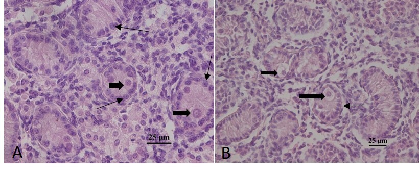
Staining with H.E showed sexual cords (developing seminiferous tubules) well preserved with the presence of a tubular lining epithelium formed by Sertoli cells and the presence of few gonocytes (Figure 1).

Histological aspects of bovine fetal testis at 4 (A) and 6 (B) months showing Sertoli Cells (SC ) and Gonocytes (G ).
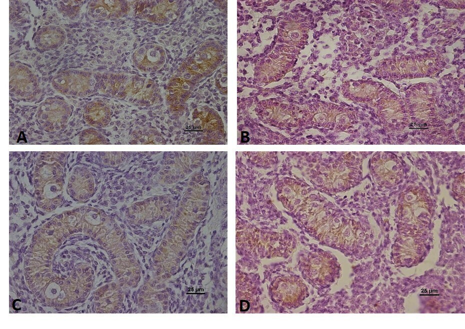
AMH immunostaining was observed in the cytoplasm of Sertoli cells, within the sexual cords at all ages analyzed, and the presence of AMH was not observed inside the gonocytes and interstitial tissue at any age analyzed (Figure 2).

Immunolocalization of Anti-Mullerian Hormone (AMH) in the testes of bovine fetuses at 4 months (A), 5 months (B), 6 months (C), and 7 months (D) of gestational ages.
Ovaries
Ovaries from bovine fetuses between 4 and 7 months of age were collected as shown in the Table 2 below.
AMH immunolocalization
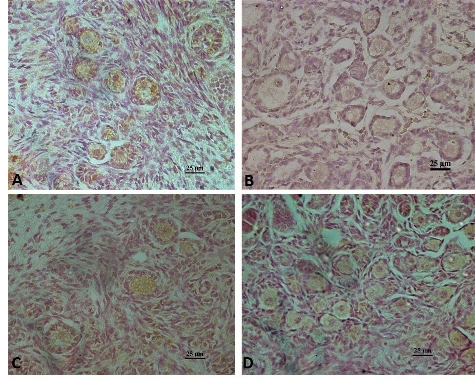
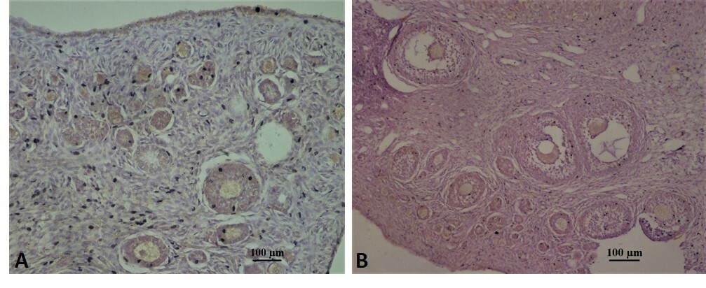
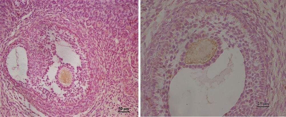
AMH immunostaining was observed between 4 and 7 months in primordial and primary follicles, the location being specifically stronger in the cytoplasm of oocytes (Figure 3). Secondary and antral follicles were observed at 7 months of age, with strong AMH labeling in the oocyte cytoplasm and light labeling in the granulosa cells of these follicles (Figures 4 and 5).
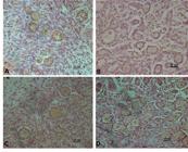
Immunolocalization of Anti-Mullerian Hormone (AMH) in the ovaries of bovine fetuses at 4 months (A), 5 months (B), 6 months (C), and 7 months (D) of gestational ages.
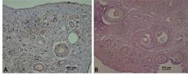
AMH immunolocalization in ovaries of 2 bovine fetuses (A and B animals) at 7 months were is observed secundary and antral follicles with immunolocalization in the oocyte citoplasm.

Antral follicles of bovine fetus at 7 months showing the immunolocalization of AMH in the oocyte citoplasm, and a light immunoreaction in the granulosa cells, primordial follicles and stroma.
Discussion
Immunolocalization of AMH in fetal bovine testes
Considering that the role of AMH in gonadal development is not well understood, this is the first report of the immunolocalization of AMH in fetal bovine testis showing the production of AMH during gonadal development in the male fetus.
The marking of AMH in the testes at different fetal ages demonstrates that testicular development, as well as sexual differentiation, is influenced by the action of this hormone, and this continuous expression observed throughout development demonstrates the activity of Sertoli cells leading us to infer that some type of interaction between these two cell types occurs, such as in the regulation of receptors and/or cell proliferation. The presence of AMH did not occur in the interstitial space where the Leydig cells responsible to produce testosterone are found.
Indeed, SRY induces the expression of the AMH gene via the SOX 9, SF1, WT1, DAX1, GATA4 genes which will directly regulate the expression of AMH in the fetal testis. the temporal expression of key regulatory genes governing sex-differentiation showed that in testis aged 48—64 days, AMH was highly expressed whereas in ovary no expression was detected. In contrast, there was higher expression of AMHR in ovary than testis (Mamsen et al., 2017) and the induction of AMH secretion will thereafter be dependent on FSH regulation.
It is the binding of AMH type II receptor in the mesenchymal cells of the Müllerian ducts that induces the morphological changes leading to degeneration of the Müllerian duct system. It may be related to an apoptotic mechanism of epithelial cells associated with changes in the basal membrane in response to paracrine signals (Mullen and Behringer, 2014). AMH is one of the earliest testis-specific proteins expressed by the embryonic male gonad from the 8th week of amenorrhea and provokes irreversible Müllerian duct regression, which is completed by the end of week 9 (Rey et al., 1996).
AMH rises quickly after birth, with a peak at three to six months, then the level stabilizes and lasts until puberty, establishment of a negative feedback control on the secretion of AMH by intratesticular testosterone, which level increases sharply at puberty (Boukari et al., 2009). At puberty, the polarity of Sertoli cells changes with the formation of the blood-testis barrier and the development of tight junctions between Sertoli cells: the secretion of AMH then becomes luminal and no longer basal. A plasma concentration is then obtained which decreases in favor of that of the seminal fluid (Holst et al., 2020).
Estradiol upregulates AMH expression by increasing the activity of the human AMH promoter in the prepubertal Sertoli cell line SMAT1 by signaling through Estrogen Receptor ER, which binds to a specific ERE (Estrogen Response Element) sequence (Valeri et al., 2020). Persistant Müllerian duct syndrome (PMDS), an autosomal recessive genetic syndrome occurring in 46, XY individuals, is the consequence of mutations in the AMH gene (PMDS type I) located on chromosome 19 (19p13) or in the gene for its type II receptor located at 12q13 (PMDS type II) and these patients present testes but most often in an intra-abdominal ectopic position because the testicular descent is AMH-dependent (Xu et al., 2019).
The presence of AMH observed in this work corroborates the work of Ball et al. (2008) who observed, using the immunofluorescence technique, the presence of AMH in the testes of equine fetuses at 5.5; 10 and 11 months of gestational age, indicating that AMH expression by Sertoli cells remains at high levels during fetal development.
Our results were like what was observed in other mammalian species, showing the presence of AMH in Sertoli cells during gonadal development (Behringer, 1995; Lee et al., 2003; Costa et al., 2025). Sertoli cells produce one or more factors that act on Leydig cells during fetal life, which are AMH and Desert hedgehog (Dhh) (Migrenne et al., 2012). AMH remains elevated until puberty, decreasing through the maturation of Sertoli cells (Josso et al., 1993) and decreasing in the seminiferous tubules with spermatocytes or later stages of spermatogenesis (Rajpert-De Meyts et al., 1999).
Although FSH is known to have a stimulating effect on Sertoli cell mitosis (Sharpe, 1994), it also contributes to the process of differentiation (Walker and Cheng, 2005) and synthesis of growth factors by Sertoli cells, which promote the maturation of Leydig cells (Sharpe, 1994; Walker and Cheng, 2005), making them able to respond to increasing LH stimuli and primarily synthesize testosterone.
Inactivation of AMH expression has been associated with the appearance of primary spermatocytes (Rajpert-De Meyts et al., 1999) and when androgen receptors reach adequate levels in Sertoli cells, AMH is inhibited, testosterone reaches the seminiferous tubules and spermatogonia enter meiosis (Al-Attar et al., 1997) and the expression of AMH can be used as a marker to determine the state of differentiation of Sertoli cells, as its expression drops sharply during the perinatal and prepubertal period (Al-Attar et al., 1997).
Immunolocalization of AMH in fetal bovine ovaries
The immunolocalization of AMH in fetal ovaries showed a different pattern from that mentioned in the literature, with strong immunostaining in the cytoplasm of oocytes from primordial, primary, and secondary follicles and in granulosa cells in all fetal ages analyzed, contrary to what Vigier et al. (1984) mention that expression starts in the perinatal period. These results also differ from those observed in adult human ovaries, which show AMH restricted to granulosa cells of primary, secondary, preantral and antral follicles, not being present in GC of primordial follicles, oocyte, and ovarian stroma (Weenen et al., al., 2004). growing follicles. These differences may be due to physiological differences between species, but they may be due to variations in the methodology of the immunohistochemical technique.
Secondary and antral follicles were observed at 7 months showing a strong AMH labeling in the oocyte cytoplasm suggesting that, despite being produced by granulosa cells, this hormone is concentrated in the oocyte cytoplasm. The granulosa cells of these follicles also showed marking, although no marking was observed in the theca cells, which agrees with Münsterberg and Lovell-Badge (1991) who observed the expression of AMH in the granulosa cells of antral follicles and mention that theca cells do not express the AMH.
According to Nilsson et al. (2011), AMH inhibits the growth of primordial follicles and is in ovarian stromal cells that surround the nests of germ cells and neo-formed primordial follicles and in ovaries of adult animals, it inhibits the primordial transition to primary and is produced by granulosa cells of secondary follicles and in early antrum to antral formation. In this work we observed the presence of AMH in all ovarian follicles, from primordial to antral, both in the granulosa cells and in the oocyte cytoplasm, which agrees with what was observed in pigs by Almeida et al. (2012).
According to Dunlop and Anderson (2015), AMH is one of the most important factors in controlling the rate of activation of primordial follicles and plays a role in suppressing the recruitment of primordial follicles by reducing the expression of Kit Ligand and Fibroblast Growth Factor β (FGFβ) produced by the oocyte that seem to stimulate the transition from primordial to primary in several species however in this work we observed the presence of AMH in different phases of follicular growth suggesting other functions of this hormone in oocyte and follicular growth and also in ovarian development and, According to Modi et al. (2006), differences in AMH expression patterns between species make the understanding of AMH role in ovarian physiology difficult and considering that there are no studies in the literature on the presence of AMH in fetal bovine ovaries more studies are necessary for better understanding of the role of this protein in ovarian development.
In the ovary, AMH is expressed by preantral and small antral follicles (Weenen et al., 2004). AMH expression is detected in granulosa cells of activated primordial follicles but is absent in follicular stages following follicle-stimulating hormone (FSH)-dependent selection. Thus, AMH acts as a follicular gatekeeper ensuring that each small antral follicle (up to 8 mm) will produce little E2 prior to selection of the follicle that will undergo ovulation (Dewailly et al., 2014). AMH synthesized during the early ovarian follicular stages provides negative retrocontrol over folliculogenesis. It acts on two key mechanisms of folliculogenesis: first, on follicular recruitment: AMH has been shown to be a brake on the activation of primordial follicles, which prevents the recruitment of too large a follicle cohort in each menstrual cycle (La Marca and Volpe, 2006); secondly, on the stimulating effect of FSH on the follicles: AMH has a negative feedback on FSH and will decrease its level. The more follicles grow, the less AMH they synthesize. We therefore observe a decrease in this negative feedback, which allows mature follicles to be sensitive to FSH (Chang et al., 2013).
In pregnant buffaloes, the plasma AMH concentrations showed no correlation with AMH concentrations in their respective fetuses male or female, where the AMH concentration in males fetuses was 169-813 times greater than that in maternal circulation suggesting that AMH does not cross the placental barrier and AMH concentrations were significantly higher in male, with an increase according to fetal age, than in female fetuses. Also observed Immunolocalization of AMH buffaloes fetuses ovaries from the fifth month onwards and displayed intense immunostaining within the granulosa cells and in the cytoplasm of primordial follicle oocytes at 5, 6, 7, and 8 months (35–98cm CRL) and in the cytoplasm of pre-Sertoli cells within the sex cords (developing seminiferous tubules) at all ages (Costa et al., 2025).
Although the expression levels of AMH and AR in the ovary were lower compared to the testis, they still play roles in ovarian development and function and (Zhou et al., 2021; Liu et al., 2022). As an early marker of testicular differentiation, AMH is expressed in the embryonic testis but not the ovary in mammals, and its expression in the ovary starts after birth (37). In non-mammalian vertebrates such as birds and fish, AMH is expressed in the developing gonads of both sexes but predominantly in the testis. AMH expression in the early fetal testis is regulated by several key transcription factors, including SRY, SOX9, SF-1, WT1, and GATA4, suggesting an important role for AMH in male differentiation. (Cutting et al., 2014; Chen et al., 2017).
Conclusion
In conclusion, Anti-Müllerian Hormone (AMH) is strongly expressed in Sertoli cells of testis and in granulosa cells of primordial, primary, secondary, and antral follicles of ovaries of bovine fetuses between 4 and 7 months, where the presence of AMH was also observed in the cytoplasm of the oocyte from these follicles, however further studies are needed to elucidate the molecular mechanisms and role of AMH in the development and gonadal physiology of bovine fetuses.
Data availability statement
Research data is only available upon request.
-
Financial support:
Conselho Nacional de Desenvolvimento Científico e Tecnológico (CNPQ) for financial suporte awarded to Maria Auxiliadora Pantoja Ferreira (308895/2022-3).
-
How to cite:
Brito VC, Cruz GS, Lima MM, Souza EB, Ramos AS, Ferreira MAP, Miranda MS, Almeida NNC, Cordeiro MS, Santos SSD. Immunolocalization of anti-Mullerian hormone in bovine testes and ovaries during fetal development. Anim Reprod. 2026;23(3):e20250088. https://doi.org/10.1590/1984-3143-AR2025-0088
REFERENCES
-
Aksglaede L, Sørensen K, Boas M, Mouritsen A, Hagen CP, Jensen RB, Petersen JH, Linneberg A, Andersson AM, Main KM, Skakkebæk NE, Juul A. Changes in anti-Mullerian hormone (AMH) throughout the life span: a population-based study of 1027 healthy males from birth (cord blood) to the age of 69 years. J Clin Endocrinol Metab. 2010;95(12):5357-64. https://doi.org/10.1210/jc.2010-1207 PMid:20843948.
» https://doi.org/10.1210/jc.2010-1207 -
Al-Attar L, Noël LK, Dutertre M, Belville C, Forest MG, Burgoyne PS, Josso N, Rey R. Hormonal and cellular regulation of Sertoli cell anti-Mullerian hormone production in the postnatal mouse. J Clin Invest. 1997;100(6):1335-43. https://doi.org/10.1172/JCI119653 PMid:9294098.
» https://doi.org/10.1172/JCI119653 - Almeida F, Teerds KJ, Auler P, Soede NM. Anti-Mullerian hormone: expression and possible roles in the porcine ovary. In: Proceedings of the 17th International Congress on Animal Reproduction (ICAR); 2012 Jul 29-2 Aug; Vancouver, Canada. Hoboken: John Wiley & Sons; 2012. p. 496-7.
-
Antonino DC, Silva GV, Mohallem RFF, Oliveira M, Rocha CD, Antonino ML, Soares MM, Alves KA, Jacomini JO. Morfometria de ovários de fetos bovinos: isolamento e quantificação de folículos pré-antrais. Revista UNINGÁ. 2017;52(1):18-22. https://doi.org/10.46311/2318-0579.52.eUJ1389
» https://doi.org/10.46311/2318-0579.52.eUJ1389 -
Baarends WM, Hoogerbrugge JW, Post M, Visser JA, De Rooij DG, Parvinen M, Themmen APN, Grootegoed JA. Anti-Mullerian hormone and anti-Mullerian Hormone type II receptor messenger ribonucleic acid expression during postnatal testis development and in adult testis of the rat. Endocrinology. 1995;136(12):5614-22. https://doi.org/10.1210/endo.136.12.7588316 PMid:7588316.
» https://doi.org/10.1210/endo.136.12.7588316 -
Ball BA, Conley AJ, Grundy SA, Sabeur K, Liu IKM. Expression ofanti-Müllerian hormone (AMH) in the equine testis. Theriogenology. 2008;69(5):624-31. https://doi.org/10.1016/j.theriogenology.2007.11.009
» https://doi.org/10.1016/j.theriogenology.2007.11.009 -
Barret JM, Nicolas A, Jarry A, Dubreuil O, Meseure D, Passat T, Perrial E, Deleine C, Champenois G, Gaillard S, Duchalais E, Ray-Coquard I, Lahmar M, Dumontet C, Bennouna J, Bossard C, Roman-Roman S, Prost JF. The expression of anti-Müllerian hormone type II receptor (AMHRII) in non-gynecological solid tumors offers potential for broad therapeutic intervention in cancer. Biology. 2021;10(4):305. https://doi.org/10.3390/biology10040305 PMid:33917111.
» https://doi.org/10.3390/biology10040305 -
Bédécarrats GY, O’Neill FH, Norwitz ER, Kaiser UB, Teixeira J. Regulation of gonadotropin gene expression by Müllerian inhibiting substance. Proc Natl Acad Sci USA. 2003;100(16):9348-53. https://doi.org/10.1073/pnas.1633592100 PMid:12878721.
» https://doi.org/10.1073/pnas.1633592100 -
Behringer RR, Cate RL, Froelick GJ, Palmiter RD, Brinster RL. Abnormal sexual development in transgenic mice chronically expressing Müllerian inhibiting substance. Nature. 1990;345(6271):167-70. https://doi.org/10.1038/345167a0 PMid:2336108.
» https://doi.org/10.1038/345167a0 -
Behringer RR, Finegold MJ, Cate RL. Müllerian-inhibiting substance function during mammalian sexual development. Cell. 1994;79(3):415-25. https://doi.org/10.1016/0092-8674(94)90251-8 PMid:7954809.
» https://doi.org/10.1016/0092-8674(94)90251-8 -
Behringer RR. The Müllerian inhibitor and mammalian sexual development. Philos Trans R Soc Lond B Biol Sci. 1995;350(1333):285-9. https://doi.org/10.1098/rstb.1995.0163
» https://doi.org/10.1098/rstb.1995.0163 -
Bergadá I, Milani C, Bedecarrás P, Andreone L, Ropelato MG, Gottlieb S, Bergadá C, Campo S, Rey RA. Time course of the serum gonadotropin surge, inhibins, and anti-Mullerian hormone in normal newborn males during the first month of life. J Clin Endocrinol Metab. 2006;91(10):4092-8. https://doi.org/10.1210/jc.2006-1079 PMid:16849404.
» https://doi.org/10.1210/jc.2006-1079 -
Bergadá I, Milani C, Bedecarrás P, Andreone L, Ropelato MG, Gottlieb S, Bergadá C, Campo S, Rey RA. Time course of the serum gonadotropin surge, inhibins, and anti-Mullerian hormone in normal newborn males during the first month of life. J Clin Endocrinol Metab. 2006;91(10):4092-8. https://doi.org/10.1210/jc.2006-1079 PMid:16849404.
» https://doi.org/10.1210/jc.2006-1079 -
Bézard J, Vigier B, Tran D, Mauléon P, Josso N. Immunohistochemical study of anti-Mullerian hormone in sheepovarian follicles during fetal and post-natal development. J Reprod Fertil. 1987;80(2):509-16. https://doi.org/10.1530/jrf.0.0800509 PMid:3309279.
» https://doi.org/10.1530/jrf.0.0800509 -
Boukari K, Meduri G, Brailly-Tabard S, Guibourdenche J, Ciampi ML, Massin N, Martinerie L, Picard JY, Rey R, Lombès M, Young J. Lack of androgen receptor expression in sertoli cells accounts for the absence of anti-Mullerian hormone repression during early human testis development. J Clin Endocrinol Metab. 2009;94(5):1818-25. https://doi.org/10.1210/jc.2008-1909 PMid:19276236.
» https://doi.org/10.1210/jc.2008-1909 -
Castro FC, Cruz MHC, Leal CLV. Role of Growth differentiation factor 9 and bone morphogenetic protein 15 in ovarian function and their importance in mammalian female fertility - A review. Asian-Australas J Anim Sci. 2016;29(8):1065-74. https://doi.org/10.5713/ajas.15.0797 PMid:26954112.
» https://doi.org/10.5713/ajas.15.0797 -
Catlin EA, Powell SM, Manganaro TF, Hudson PL, Ragin RC, Epstein J, Donahoe PK. Sex-specific fetal lung development and Mullerian inhibiting substance. Am Rev Respir Dis. 1990;141(2):466-70. https://doi.org/10.1164/ajrccm/141.2.466 PMid:2301861.
» https://doi.org/10.1164/ajrccm/141.2.466 -
Chang H-M, Klausen C, Leung PCK. Antimüllerian hormone inhibits follicle-stimulating hormone-induced adenylyl cyclase activation, aromatase expression, and estradiol production in human granulosa-lutein cells. Fertil Steril. 2013;100(2):585-92.e1. https://doi.org/10.1016/j.fertnstert.2013.04.019 PMid:23663993.
» https://doi.org/10.1016/j.fertnstert.2013.04.019 -
Chang Q, Pereda A, Pinter MJ, Balice-Gordon RJ. Nerve injury induces gap junctional coupling among axotomized adult motor neurons. J Neurosci. 2000;20(2):674-84. https://doi.org/10.1523/JNEUROSCI.20-02-00674.2000 PMid:10632597.
» https://doi.org/10.1523/JNEUROSCI.20-02-00674.2000 -
Chen W, Liu L, Ge W. Expression analysis of growth differentiation factor 9 (Gdf9/gdf9), anti-mullerian hormone (Amh/amh) and aromatase (Cyp19a1a/cyp19a1a) during gonadal differentiation of the zebrafish,Danio rerio. Biol Reprod. 2017;96(2):401-13. https://doi.org/10.1095/biolreprod.116.144964 PMid:28203731.
» https://doi.org/10.1095/biolreprod.116.144964 -
Cimino I, Casoni F, Liu X, Messina A, Parkash J, Jamin SP, Catteau-Jonard S, Collier F, Baroncini M, Dewailly D, Pigny P, Prescott M, Campbell R, Herbison AE, Prevot V, Giacobini P. Novel role for anti-Müllerian hormone in the regulation of GnRH neuron excitability and hormone secretion. Nat Commun. 2016;7(1):10055. https://doi.org/10.1038/ncomms10055 PMid:26753790.
» https://doi.org/10.1038/ncomms10055 -
Clarkson AN, Talbot CL, Wang P-Y, MacLaughlin DT, Donahoe PK, McLennan IS. Müllerian inhibiting substance is anterogradely transported and does not attenuate avulsion-induced death of hypoglossal motor neurons. Exp Neurol. 2011;231(2):304-8. https://doi.org/10.1016/j.expneurol.2010.12.019 PMid:21195071.
» https://doi.org/10.1016/j.expneurol.2010.12.019 -
Costa NN, Ferreira MAP, Ramos PCA, Santos AX, Cordeiro MS, Santana PPB, Brito VC, Silva TVG, Homobono BP, Souza EB, King WA, Miranda MS, Santos SSD. Anti-mullerian hormone (AMH) in pregnant buffaloes: maternal and fetal plasma concentrations and immunolocalization in the fetal gonads. Anim Reprod. 2025;22(1):e20230152. https://doi.org/10.1590/1984-3143-ar2023-0152 PMid:40013122.
» https://doi.org/10.1590/1984-3143-ar2023-0152 -
Cutting AD, Ayers K, Davidson N, Oshlack A, Doran T, Sinclair AH, Tizard M, Smith CA. Identification, expression, and regulation of anti-Müllerian hormone type-II receptor in the embryonic chicken gonad. Biol Reprod. 2014;90(5):106. https://doi.org/10.1095/biolreprod.113.116491 PMid:24621923.
» https://doi.org/10.1095/biolreprod.113.116491 -
Detti L, Williams LJ, Fletcher NM, Saed GM. Anti-Mullerian hormone (AMH) may inhibit oocyte maturation and follicular vascularization in human ovarian cortex. Fertil Steril. 2015;104(3 Suppl):91. https://doi.org/10.1016/j.fertnstert.2015.07.422
» https://doi.org/10.1016/j.fertnstert.2015.07.422 -
Devillers MM, Petit F, Cluzet V, François CM, Giton F, Garrel G, Cohen-Tannoudji J, Guigon CJ. FSH inhibits AMH to support ovarian estradiol synthesis in infantile mice. J Endocrinol. 2019;240(2):215-28. https://doi.org/10.1530/JOE-18-0313 PMid:30403655.
» https://doi.org/10.1530/JOE-18-0313 -
Dewailly D, Andersen CY, Balen A, Broekmans F, Dilaver N, Fanchin R, Griesinger G, Kelsey TW, La Marca A, Lambalk C, Mason H, Nelson SM, Visser JA, Wallace WH, Anderson RA. The physiology and clinical utility of anti-Müllerian hormone in women. Hum Reprod Update. 2014;20(3):370-85. https://doi.org/10.1093/humupd/dmt062 PMid:24430863.
» https://doi.org/10.1093/humupd/dmt062 - Di Clemente N, Goxe B, Remy JJ, Cate R, Josso N, Vigier B, Salesse R. Inhibitory effect of AMH upon the expression of aromatase and LH receptors by cultured granulose cells of rat and porcine immature ovaries. Endocrine. 1994;2:553-8.
-
Diniz EG, Esper CR, Jacomini JO, Vieira RC. Desenvolvimento morfológico dos ovários em embriões e fetos bovinos da raça Nelore. Arq Bras Med Vet Zootec. 2005;57(1):70-6. https://doi.org/10.1590/S0102-09352005000100010
» https://doi.org/10.1590/S0102-09352005000100010 -
Dunlop CE, Anderson RA. Uses of anti-Mullerian hormone (AMH) measurement before and after cancer treatment in women. Maturitas. 2015;80(3):245-50. https://doi.org/10.1016/j.maturitas.2014.12.005 PMid:25596814.
» https://doi.org/10.1016/j.maturitas.2014.12.005 -
Durlinger AL, Gruijters MJ, Kramer P, Karels B, Kumar TR, Matzuk MM, Rose UM, de Jong FH, Uilenbroek JT, Grootegoed JA, Themmen AP. Anti-Mullerian hormone attenuates the effects of FSH on follicle development in the mouse ovary. Endocrinology. 2001;142(11):4891-9. https://doi.org/10.1210/endo.142.11.8486 PMid:11606457.
» https://doi.org/10.1210/endo.142.11.8486 -
Durlinger AL, Kramer P, Karels B, de Jong FH, Uilenbroek JT, Grootegoed JA, Themmen AP. Control of primordial follicle recruitment by anti-Müllerian hormone in the mouse ovary. Endocrinology. 1999;140(12):5789-96. https://doi.org/10.1210/endo.140.12.7204 PMid:10579345.
» https://doi.org/10.1210/endo.140.12.7204 -
Durlinger ALL, Visser JA, Themmen APN. Regulation of ovarian function: the role of anti-Müllerian hormone. Reproduction. 2002;124(5):601-9. https://doi.org/10.1530/rep.0.1240601 PMid:12416998.
» https://doi.org/10.1530/rep.0.1240601 -
Garrel G, Racine C, L’Hôte D, Denoyelle C, Guigon CJ, di Clemente N, Cohen-Tannoudji J. Anti-Müllerian hormone: a new actor of sexual dimorphism in pituitary gonadotrope activity before puberty. Sci Rep. 2016;6(1):23790. https://doi.org/10.1038/srep23790 PMid:27030385.
» https://doi.org/10.1038/srep23790 -
Goodfellow PN, Lovell-Badge R. Sry and sex determination in mammals. Annu Rev Genet. 1993;27(1):71-92. https://doi.org/10.1146/annurev.ge.27.120193.000443 PMid:8122913.
» https://doi.org/10.1146/annurev.ge.27.120193.000443 -
Gowkielewicz M, Lipka A, Zdanowski W, Was’niewski T, Majewska M, Carlberg C. Anti-Müllerian hormone: biology and role in endocrinology and cancers. Front Endocrinol (Lausanne). 2024;15:1468364. https://doi.org/10.3389/fendo.2024.1468364 PMid:39351532.
» https://doi.org/10.3389/fendo.2024.1468364 -
Grynberg M, Pierre A, Rey R, Leclerc A, Arouche N, Hesters L, Catteau-Jonard S, Frydman R, Picard J-Y, Fanchin R, Veitia R, di Clemente N, Taieb J. Differential regulation of ovarian anti-müllerian hormone (AMH) by estradiol through a- and bestrogen receptors. J Clin Endocrinol Metab. 2012;97(9):E1649-57. https://doi.org/10.1210/jc.2011-3133 PMid:22689696.
» https://doi.org/10.1210/jc.2011-3133 -
Guibourdenche J, Lucidarme N, Chevenne D, Rigal O, Nicolas M, Luton D, Léger J, Porquet D, Noël M. Anti-Müllerian hormone levels in serum from human foetuses and children: pattern and clinical interest. Mol Cell Endocrinol. 2003;211(1-2):55-63. https://doi.org/10.1016/j.mce.2003.09.011 PMid:14656477.
» https://doi.org/10.1016/j.mce.2003.09.011 -
Hayashi M, Shima H, Hayashi K, Trelstad RL, Donahoe PK. Immunocytochemical localization of Müllerian inhibiting substance in the rough endoplasmic reticulum and Golgi apparatus in Sertoli cells of the neonatal calf testis using a monoclonal antibody. J Histochem Cytochem. 1984;32(6):649-54. https://doi.org/10.1177/32.6.6373916 PMid:6373916.
» https://doi.org/10.1177/32.6.6373916 -
Hayes E, Kushnir V, Ma X, Biswas A, Prizant H, Gleicher N, Sen A. Intra-cellular mechanism of Anti-Müllerian hormone (AMH) in regulation of follicular development. Mol Cell Endocrinol. 2016;433:56-65. https://doi.org/10.1016/j.mce.2016.05.019 PMid:27235859.
» https://doi.org/10.1016/j.mce.2016.05.019 -
Holst BS, Panzani D, Camillo F, Svensson A, Rota A. Anti-Müllerian hormone (AMH) concentrations are maximal at puberty in male donkeys and secretion is redirected from the blood stream to seminal plasma. Anim Reprod Sci. 2020;218:106484. https://doi.org/10.1016/j.anireprosci.2020.106484 PMid:32507264.
» https://doi.org/10.1016/j.anireprosci.2020.106484 -
Josso N, Di Clemente N, Gouedard L. Anti-Mullerian hormone and its receptors. Mol Cell Endocrinol. 2001;179(1-2):25-32. https://doi.org/10.1016/S0303-7207(01)00467-1 PMid:11420127.
» https://doi.org/10.1016/S0303-7207(01)00467-1 -
Josso N, Di Clemente N. TGF-β family members and gonadal development. Trends Endocrinol Metab. 1999;10(6):216-22. https://doi.org/10.1016/S1043-2760(99)00155-1 PMid:10407395.
» https://doi.org/10.1016/S1043-2760(99)00155-1 -
Josso N, Lamarre I, Picard JY, Berta P, Davies N, Morichon N, Peschanski M, Jeny R. Anti-Mullerian hormone in early human development. Early Hum Dev. 1993;33(2):91-9. https://doi.org/10.1016/0378-3782(93)90204-8 PMid:8055780.
» https://doi.org/10.1016/0378-3782(93)90204-8 -
Josso N, Picard JY. Genetics of anti-Müllerian hormone and its signaling pathway. Best Pract Res Clin Endocrinol Metab. 2022;36(1):101634. https://doi.org/10.1016/j.beem.2022.101634 PMid:35249805.
» https://doi.org/10.1016/j.beem.2022.101634 -
Josso N, Racine C, di Clemente N, Rey R, Xavier F. The role of anti-Mullerian hormone in gonadal development. Mol Cell Endocrinol. 1998;145(1-2):3-7. https://doi.org/10.1016/S0303-7207(98)00186-5 PMid:9922092.
» https://doi.org/10.1016/S0303-7207(98)00186-5 -
Josso N, Rey RA. What does AMH tell us in pediatric disorders of sex development? Front Endocrinol. 2020;11:619. https://doi.org/10.3389/fendo.2020.00619 PMid:33013698.
» https://doi.org/10.3389/fendo.2020.00619 -
Jurczak A, Szkup M, Grzywacz A, Safranow K, Grochans E. The relationship between AMH and AMHR2 polymorphisms and the follicular phase in late reproductive stage women. Int J Environ Res Public Health. 2016;13(2):185. https://doi.org/10.3390/ijerph13020185 PMid:26848671.
» https://doi.org/10.3390/ijerph13020185 -
Kim JH, MacLaughlin DT, Donahoe PK. Müllerian inhibiting substance/antiMüllerian hormone: A novel treatment for gynecologic tumors. Obstet Gynecol Sci. 2014;57(5):343-57. https://doi.org/10.5468/ogs.2014.57.5.343 PMid:25264524.
» https://doi.org/10.5468/ogs.2014.57.5.343 -
Kuiri-Hänninen T, Kallio S, Seuri R, Tyrväinen E, Liakka A, Tapanainen J, Sankilampi U, Dunkel L. Postnatal developmental changes in the pituitary-ovarian axis in preterm and term infant girls. J Clin Endocrinol Metab. 2011;96(11):3432-9. https://doi.org/10.1210/jc.2011-1502 PMid:21900380.
» https://doi.org/10.1210/jc.2011-1502 -
Kumariya S, Ubba V, Jha RK, Gayen JR. Autophagy in ovary and polycystic ovary syndrome: role, dispute and future perspective. Autophagy. 2021;17(10):2706-33. https://doi.org/10.1080/15548627.2021.1938914 PMid:34161185.
» https://doi.org/10.1080/15548627.2021.1938914 -
La Marca A, Volpe A. Anti-Müllerian hormone (AMH) in female reproduction: is measurement of circulating AMH a useful tool? Clin Endocrinol. 2006;64(6):603-10. https://doi.org/10.1111/j.1365-2265.2006.02533.x PMid:16712660.
» https://doi.org/10.1111/j.1365-2265.2006.02533.x -
Lasala C, Schteingart HF, Arouche N, Bedecarrás P, Grinspon RP, Picard JY, Josso N, di Clemente N, Rey RA. SOX9 and SF1 are involved in cyclic AMP-mediated upregulation of anti-Müllerian gene expression in the testicular prepubertal Sertoli cell line SMAT1. Am J Physiol Endocrinol Metab. 2011;301(3):E539-47. https://doi.org/10.1152/ajpendo.00187.2011 PMid:21693691.
» https://doi.org/10.1152/ajpendo.00187.2011 - Lee MM, Donahoe PK. Mullerian inhibiting substance: a gonadal hormone with multiple functions. Endocr Rev. 1993;14(2):152-64. PMid:8325249.
-
Lee MM, Misra M, Donahoe PK, Maclaughlin DT. MIS/AMH in the assessment of cryptorchidism and intersex conditions. Mol Cell Endocrinol. 2003;211(1-2):91-8. https://doi.org/10.1016/j.mce.2003.09.014 PMid:14656481.
» https://doi.org/10.1016/j.mce.2003.09.014 -
Liu X, Dai S, Wu J, Wei X, Zhou X, Chen M, Tan D, Pu D, Li M, Wang D. Roles of anti-Mullerian hormone and its duplicates in sex determination and germ cell proliferation of Nile tilapia. Genetics. 2022;220(3):iyab237. https://doi.org/10.1093/genetics/iyab237 PMid:35100374.
» https://doi.org/10.1093/genetics/iyab237 -
Lucas-Herald AK, Mitchell RT. Testicular Sertoli cell hormones in differences in sex development. Front Endocrinol. 2022;13:919670. https://doi.org/10.3389/fendo.2022.919670 PMid:35909548.
» https://doi.org/10.3389/fendo.2022.919670 -
Lyet L, Louis F, Forest MG, Josso N, Behringer RR, Vigier B. Ontogeny of reproductive abnormalities induced by deregulation of anti-Mullerian hormone expression in transgenic mice. Biol Reprod. 1995;52(2):444-54. https://doi.org/10.1095/biolreprod52.2.444 PMid:7711213.
» https://doi.org/10.1095/biolreprod52.2.444 -
MacLaughlin DT, Donahoe PK. Müllerian inhibiting substance/anti-Müllerian hormone: A potential therapeutic agent for human ovarian and other cancers. Future Oncol. 2010;6(3):391-405. https://doi.org/10.2217/fon.09.172 PMid:20222796.
» https://doi.org/10.2217/fon.09.172 -
Mamsen LS, Ernst EH, Borup R, Larsen A, Olesen RH, Ernst E, Richard AA, Kristensen SG, Andersen CY. Temporal expression pattern of genes during the period of sex differentiation in human embryonic gonads. Sci Rep. 2017;7(1):15961. https://doi.org/10.1038/s41598-017-15931-3 PMid:29162857.
» https://doi.org/10.1038/s41598-017-15931-3 -
Migrenne S, Moreau E, Pakarinen P, Dierich A, Merlet J, Habert R, Racine C. Mouse testis development and function are differently regulated by follicle-stimulating hormone receptors signaling during fetal and prepubertal life. PLoS One. 2012;7(12):e53257. https://doi.org/10.1371/journal.pone.0053257
» https://doi.org/10.1371/journal.pone.0053257 -
Modi D, Bhartiya D, Puri C. Developmental expression and cellular distribution of Mullerian inhibiting substance in the primate ovary. Reproduction. 2006;132(3):443-53. https://doi.org/10.1530/rep.1.01178 PMid:16940285.
» https://doi.org/10.1530/rep.1.01178 -
Monteiro CMR, Carvalhal R, Perri SHV. bovine fetuses (Bos primigenius indicus) at various gestation stages. Braz J Vet Res Anim Sci. 2003;40:6. https://doi.org/10.1590/S1413-95962003000600004
» https://doi.org/10.1590/S1413-95962003000600004 -
Mullen RD, Behringer RR. Molecular genetics of Müllerian duct formation, regression and differentiation. Sex Dev. 2014;8(5):281-96. https://doi.org/10.1159/000364935 PMid:25033758.
» https://doi.org/10.1159/000364935 -
Münsterberg A, Lovell-Badge R. Expression of the mouse anti-Müllerian hormone gene suggests a role in both male and female sexual differentiation. Development. 1991;113(2):613-24. https://doi.org/10.1242/dev.113.2.613 PMid:1782869.
» https://doi.org/10.1242/dev.113.2.613 -
Nilsson EE, Schindler R, Savenkova MI, Skinner MK. Skinner inhibitory actions of Anti-Mu llerian Hormone (AMH) on ovarian primordial follicle assembly. PLoS One. 2011;6(5):e20087. https://doi.org/10.1371/journal.pone.0020087 PMid:21637711.
» https://doi.org/10.1371/journal.pone.0020087 -
Norman RJ, Dewailly D, Legro RS, Hickey TE. Polycystic ovary syndrome. Lancet. 2007;370(9588):685-97. https://doi.org/10.1016/S0140-6736(07)61345-2 PMid:17720020.
» https://doi.org/10.1016/S0140-6736(07)61345-2 -
Ogura-Nose S, Yoshino O, Osuga Y, Shi J, Hiroi H, Yano T, Taketani Y. Anti-Mullerian hormone (AMH) is induced by bone morphogenetic protein (BMP) cytokines in human granulosa cells. Eur J Obstet Gynecol Reprod Biol. 2012;164(1):44-7. https://doi.org/10.1016/j.ejogrb.2012.05.017 PMid:22682966.
» https://doi.org/10.1016/j.ejogrb.2012.05.017 -
Pellatt L, Hanna L, Brincat M, Galea R, Brain H, Whitehead S, Mason H. Granulosa cell production of anti-Müllerian hormone is increased in polycystic ovaries. J Clin Endocrinol Metab. 2007;92(1):240-5. https://doi.org/10.1210/jc.2006-1582 PMid:17062765.
» https://doi.org/10.1210/jc.2006-1582 -
Pellatt L, Rice S, Dilaver N, Heshri A, Galea R, Brincat M, Brown K, Simpson ER, Mason HD. Anti-Müllerian hormone reduces follicle sensitivity to follicle-stimulating hormone in human granulosa cells. Fertil Steril. 2011;96(5):1246-1251.e1. https://doi.org/10.1016/j.fertnstert.2011.08.015 PMid:21917251.
» https://doi.org/10.1016/j.fertnstert.2011.08.015 - Picon R. Action du testicule foetal sur le développement in vitro des canaux de Müller chez le rat. Arch. Arch Anat Microsc Morphol Exp. 1969;58(1):1-19. PMid:5822330.
-
Poole DH, Ocón-Grove OM, Johnson AL. Anti-Müllerian hormone (AMH) receptor type II expression and AMH activity in bovine granulosa cells. Theriogenology. 2016;86(5):1353-60. https://doi.org/10.1016/j.theriogenology.2016.04.078 PMid:27268296.
» https://doi.org/10.1016/j.theriogenology.2016.04.078 -
Rajpert-De Meyts E, Jorgensen N, Graem N, Muller J, Cate RL, Skakkebaek NE. Expression of Anti-Mullerian hormone during normal and pathological gonadal development: association with differentiation of sertoli and granulosa cells. J Clin Endocrinol Metab. 1999;84(10):3836-44. https://doi.org/10.1210/jc.84.10.3836 PMid:10523039.
» https://doi.org/10.1210/jc.84.10.3836 -
Renaud EJ, MacLaughlin DT, Oliva E, Rueda BR, Donahoe PK. Endometrial cancer is a receptor-mediated target for Mullerian Inhibiting Substance. Proc Natl Acad Sci USA. 2005;102(1):111-6. https://doi.org/10.1073/pnas.0407772101 PMid:15618407.
» https://doi.org/10.1073/pnas.0407772101 - Rey R, al-Attar L, Louis F, Jaubert F, Barbet P, Nihoul-Fékété C, Chaussain JL, Josso N. Testicular dysgenesis does not affect expression of anti-müllerian hormone by Sertoli cells in premeiotic seminiferous tubules. Am J Pathol. 1996;148(5):1689-98. PMid:8623936.
- Rey R, Lordereau-Richard I, Carel JC, Barbet P, Cate RL, Roger M, Chaussain JL, Josso N. Anti-Mullerian hormone and testosterone serum levels are inversely related during normal and precocious pubertal development. J Clin Endocrinol Metab. 1993;77(5):1220-6. PMid:8077315.
-
Rey R, Lukas-Croisier C, Lasala C, Bedecarrás P. AMH/MIS: what we know already about the gene, the protein, and its regulation. Molecular and Cellular. Mol Cell Endocrinol. 2003;211(1-2):21-31. https://doi.org/10.1016/j.mce.2003.09.007 PMid:14656472.
» https://doi.org/10.1016/j.mce.2003.09.007 - Rey R, Mebarki F, Forest MG, Mowszowicz I, Cate RL, Morel Y, Chaussain JL, Josso N. Anti-Mullerian hormone in children with androgen insensitivity. J Clin Endocrinol Metab. 1994;79(4):960-4. PMid:7962305.
- Sharpe RM. Regulation of spermatogenesis. In Knobil E, Neil JD, editors. The Physiology of Reproduction. New York: Raven Press; 1994. p. 1363-1434.
-
Silva MSB, Giacobini P. New insights into anti-Müllerian hormone role in the hypothalamic-pituitary-gonadal axis and neuroendocrine development. Cell Mol Life Sci. 2021;78(1):1-16. https://doi.org/10.1007/s00018-020-03576-x PMid:32564094.
» https://doi.org/10.1007/s00018-020-03576-x -
Sonigo C, Beau I, Grynberg M, Binart N. AMH prevents primordial ovarian follicle loss and fertility alteration in cyclophosphamide-treated mice. FASEB J. 2019;33(1):1278-87. https://doi.org/10.1096/fj.201801089R PMid:30113879.
» https://doi.org/10.1096/fj.201801089R - Souza JD, Miguel MCV, Maioli MA, Trali Neto AN, Arana DG, Laurini AR, Nogueira GDP. Bovine fetal testicular development in the Nellore breed. Semina: Ciênc Agrár. 2017;38(4 Supl 1):2551-60. http://doi.org/10.5433/1679-0359.
-
Stubbs SA, Hardy K, Da Silva-Buttkus P, Stark J, Webber LJ, Flanagan AM, Themmen APN, Visser JA, Groome NP, Franks S. Anti-Müllerian hormone protein expression is reduced during the initial stages of follicle development in human polycystic ovaries. J Clin Endocrinol Metab. 2005;90(10):5536-43. https://doi.org/10.1210/jc.2005-0907 PMid:16030171.
» https://doi.org/10.1210/jc.2005-0907 -
Tal R, Seifer DB, Khanimov M, Malter HE, Grazi RV, Leader B. Characterization of women with elevated antimüllerian hormone levels (AMH): correlation of AMH with polycystic ovarian syndrome phenotypes and assisted reproductive technology outcomes. Am J Obstet Gynecol. 2014;211(1):59.e1. https://doi.org/10.1016/j.ajog.2014.02.026 PMid:24593938.
» https://doi.org/10.1016/j.ajog.2014.02.026 -
Teixeira J, Fynn-Thompson E, Payne AH, Donahoe PK. Mullerian-inhibiting substance regulates androgen synthesis at the transcriptional level. Endocrinology. 1999;140(10):4732-8. https://doi.org/10.1210/endo.140.10.7075 PMid:10499532.
» https://doi.org/10.1210/endo.140.10.7075 -
Tran D, Josso N. Localization of anti-Müllerian hormone in the rough endoplasmic reticulum of the developing bovine Sertoli cell using immunocytochemistry with a monoclonal antibody. Endocrinology. 1982;111(5):1562-7. https://doi.org/10.1210/endo-111-5-1562 PMid:6751796.
» https://doi.org/10.1210/endo-111-5-1562 -
Ueno S, Kuroda T, Maclaughlin DT, Ragin RC, Manganaro TF, Donahoe PK. Mullerian inhibiting substance in the adult rat ovary during various stages of the estrous cycle. Endocrinology. 1989;125(2):1060-6. https://doi.org/10.1210/endo-125-2-1060 PMid:2752965.
» https://doi.org/10.1210/endo-125-2-1060 -
Valeri C, Lovaisa MM, Racine C, Edelsztein NY, Riggio M, Giulianelli S, Venara M, Bedecarrás P, Ballerini MG, di Clemente N, Lamb CA, Schteingart HF, Rey RA. Molecular mechanisms underlying AMH elevation in hyperoestrogenic states in males. Sci Rep. 2020;10(1):15062. https://doi.org/10.1038/s41598-020-71675-7 PMid:32934281.
» https://doi.org/10.1038/s41598-020-71675-7 -
Vigier B, Picard J-Y, Tran D, Legeai L, Josso N. Production of anti-Mullerian hormone: another homology between sertoli and granulosa cells. Endocrinology. 1984;114(4):1315-20. https://doi.org/10.1210/endo-114-4-1315 PMid:6546716.
» https://doi.org/10.1210/endo-114-4-1315 -
Walker WH, Cheng J. FSH and testosterone signaling in Sertoli cells. Reproduction. 2005;130(1):15-28. https://doi.org/10.1530/rep.1.00358
» https://doi.org/10.1530/rep.1.00358 -
Wang F, Niu WB, Kong HJ, Guo YH, Sun YP. The role of AMH and its receptor SNP in the pathogenesis of PCOS. Mol Cell Endocrinol. 2017;439:363-8. https://doi.org/10.1016/j.mce.2016.09.023 PMid:27664518.
» https://doi.org/10.1016/j.mce.2016.09.023 -
Wang K, Xu F, Campbell SP, Hart KD, Durham T, Maylie J, Xu J. Rapid actions of anti-Müllerian hormone in regulating synaptic transmission and long-term synaptic plasticity in the hippocampus. FASEB J. 2020;34(1):706-19. https://doi.org/10.1096/fj.201902217R PMid:31914642.
» https://doi.org/10.1096/fj.201902217R -
Wang P-Y, Koishi K, McGeachie AB, Kimber M, MacLaughlin DT, Donahoe PK, Mc Lennan IS. Mullerian Inhibiting Substance acts as a motor neuron survival factor in vitro. Proc Natl Acad Sci USA. 2005;102(45):16421-5. https://doi.org/10.1073/pnas.0508304102 PMid:16260730.
» https://doi.org/10.1073/pnas.0508304102 -
Wang P-Y, Protheroe A, Clarkson AN, Imhoff F, Koishi K, McLennan IS. Müllerian inhibiting substance contributes to sex-linked biases in the brain and behavior. Proc Natl Acad Sci USA. 2009;106(17):7203-8. https://doi.org/10.1073/pnas.0902253106 PMid:19359476.
» https://doi.org/10.1073/pnas.0902253106 -
Weenen C, Laven JSE, von Bergh ARM, Cranfield M, Groome NP, Visser JA, Kramer P, Fauser BCJM, Themmen APN. Anti-Müllerian hormone expression pattern in the human ovary: potential implications for initial and cyclic follicle recruitment. Mol Hum Reprod. 2004;10(2):77-83. https://doi.org/10.1093/molehr/gah015 PMid:14742691.
» https://doi.org/10.1093/molehr/gah015 -
Wittmann W, McLennan IS. The bed nucleus of the stria terminalis has developmental and adult forms in mice, with the male bias in the developmental form being dependent on testicular AMH. Horm Behav. 2013;64(4):605-10. https://doi.org/10.1016/j.yhbeh.2013.08.017 PMid:24012942.
» https://doi.org/10.1016/j.yhbeh.2013.08.017 -
Xu H-Y, Zhang H-X, Xiao Z, Qiao J, Li R. Regulation of anti-Müllerian hormone (AMH) in males and the associations of serum AMH with the disorders of male fertility. Asian J Androl. 2019;21(2):109-14. https://doi.org/10.4103/aja.aja_83_18 PMid:30381580.
» https://doi.org/10.4103/aja.aja_83_18 -
Zhou L, Li M, Wang D. Role of sex steroids in fish sex determination and differentiation as revealed by gene editing. Gen Comp Endocrinol. 2021;313:113893. https://doi.org/10.1016/j.ygcen.2021.113893 PMid:34454946.
» https://doi.org/10.1016/j.ygcen.2021.113893
Edited by
-
Editor-in-Chief:
Carlos Eduardo Ambrósio.