Application of omics technologies to identify reproductive phenotypes in the context of assisted reproductive technologies for cattle
Abstract
Fertility is a key determinant of reproductive efficiency, economic performance, and sustainability in cattle production systems. Traditional fertility phenotypes that are still widely used today, such as conception rate, calving interval, non-return rate, and semen quality parameters, primarily describe reproductive outcomes rather than predict fertility. Consequently, important molecular and physiological differences between animals with contrasting fertility may remain undetected. Omics technologies provide new opportunities to identify reproductive phenotypes that more accurately reflect the biological mechanisms underlying fertility in cattle. Genomics enables the identification of single nucleotide polymorphisms, quantitative trait loci, and haplotypes associated with variation in fertility. Transcriptomics has revealed gene expression patterns related to spermatogenesis, sperm function, uterine receptivity, and embryo-maternal communication. Proteomics has identified protein profiles associated with oocyte developmental competence, fertilization capacity, and embryonic development, while metabolomics reflects the biochemical state most closely linked to the expressed phenotype, identifying biomarkers associated with oxidative balance and energy, lipid, and protein metabolism. This manuscript reviews selected studies that have applied omics technologies to identify measurable reproductive phenotypes that better capture the biological basis of fertility in cattle within the context of assisted reproductive technologies. While genomics remains the most widely applied omics approach due to the stability and accessibility of DNA, downstream omics may provide a more accurate representation of the dynamic biological processes defining fertility. Future research should focus on translating these findings into practical assays for rapid and accessible biomarker measurement, supporting informed decision-making at the farm level.
Keywords:
breeding; biomarkers; cattle production; reproductive potential
Introduction
Cattle are a core species in the global livestock industry and play an essential role in food security and economic development. Beef and dairy cattle contribute substantially to human nutrition by providing high-quality protein and dairy products to billions of people worldwide (Wang et al., 2025). In addition, cattle farming plays an important role in maintaining biodiversity, regulating soil processes, and supporting rural landscapes (Dumont et al., 2019). At the same time, the sector faces increasing social and regulatory pressure due to its significant contribution to greenhouse gas emissions, accounting for approximately 65% of total livestock-related emissions (Harindintwali et al., 2021). Notably, cattle production systems with lower productivity tend to exhibit higher emission intensities, primarily due to inefficiencies in feed conversion and herd performance. Therefore, strategies that increase productive output per animal represent one of the most effective approaches to reduce emission intensity (FAO, 2013). In this context, enhancing desirable genetic traits related to productivity and resilience, together with improving reproductive efficiency, have become central determinants for achieving sustainability in dairy and beef farm systems.
The development of assisted reproductive technologies (ART) in the 20th century enabled a deeper understanding of reproductive biology and facilitated the identification of key biological mechanisms underlying reproductive success or failure, making ART the most effective tools for improving genetics in cattle (Betteridge, 2003; Mapletoft and Hasler, 2005). The most widely used ART in cattle is artificial insemination (AI), which allows the dissemination of superior male genetics, as a single ejaculate can be divided into multiple doses and preserved for storage and global distribution. Another major technology is embryo transfer (ET), which enables the multiplication of genetic merit from both males and females. Embryos transferred into recipient or surrogate cows can be produced in vivo, within the female reproductive tract following Multiple Ovulation Embryo Transfer (MOET), or in vitro, under laboratory conditions through in vitro production (IVP) (Mapletoft and Hasler, 2005). While embryos derived after MOET were predominant when this technology expanded commercially in the 1990s, IVP embryos have surpassed them over the last decade, with more than 2 million IVP embryos transferred worldwide annually and current estimates indicating ~7-fold higher usage compared to in vivo embryos (Viana, 2025).
Because the primary objective of ART in cattle is genetic improvement rather than the treatment of infertility, animals included in these programs are expected to have sufficient baseline fertility to ensure optimal outcomes. Traditionally, fertility evaluation in cattle relies on phenotypic indicators reflecting observable reproductive performance. In bulls, fertility is typically assessed through breeding soundness examination, which includes parameters such as scrotal circumference, semen volume, sperm concentration, motility, and morphology. In cows, commonly used indicators include calving interval, non-return rate, days open, pregnancy rate, and conception rate. However, these conventional metrics do not directly capture the driving biological mechanisms of fertility, and important molecular differences between animals with variable fertility may remain undetected.
This limitation highlights the need for approaches that more accurately link reproductive phenotypes to their biological basis. A promising avenue lies in the application of omics technologies, which have become increasingly accessible in recent decades. Omics approaches enable comprehensive measurement of the molecular components defining a phenotype and can therefore be used to characterize the biological status of key tissues and cell types in both males and females.
The aim of this manuscript is to review studies that have applied omics technologies to help identify measurable phenotypes that better reflect the biological mechanisms underlying fertility in cattle, in the context of ART. Before delving into the omics technologies applied to males or females, a brief description of the main characteristics of each available omics approach will first be provided.
Omics technologies
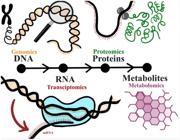
The use of high-throughput techniques has allowed not only the detection of qualitative differences but also the quantification of biomolecules that define a phenotype (Chakraborty et al., 2022; Hasin et al., 2017). Their improved sensitivity, specificity, and accuracy facilitate the rapid identification of biomarkers for biological characterization (Kim et al., 2017). The main omics approaches (Fig. 1) are discussed below.
Genomics (“what can happen”)
The development of the polymerase chain reaction (PCR) in 1986 was a major milestone in molecular biology, enabling detailed investigation of DNA structure (Mullis and Faloona, 1987). Subsequently, advances in large-scale genome sequencing and assembly enabled the publication of the first bovine reference genome in 2009, which represented a significant advance in understanding ruminant biology and evolution (Bovine Genome Sequencing and Analysis Consortium, 2009). Later developments in next-generation and long-read sequencing technologies further improved genome mapping, variant discovery, and functional genomic analyses in cattle. Single nucleotide polymorphisms (SNPs) are variations in a single base pair within the genome, typically generating two alleles at a given locus, which may be associated with specific phenotypes. Genome-wide association studies (GWAS) are used to identify significant associations between molecular markers and phenotypic traits, often using commercial panels containing thousands of SNPs available for cattle. These approaches facilitate the identification of informative SNPs and improve the precision of genetic selection by linking molecular markers to complex traits, such as daughter pregnancy rate and cow/heifer conception rate (Berry et al., 2014).
Transcriptomics (“what appears to be happening”)
The complete set of RNA transcripts, known as the transcriptome, can be characterized using transcriptomics. This high-throughput approach enables the analysis of gene expression dynamics and regulatory mechanisms involved in physiological processes and environmental adaptation (JiaYu et al., 2025). Early transcriptomic studies relied on microarray technologies, which are based on hybridisation, whereby synthetic single-stranded DNA or RNA probes bind to complementary sequences (Genomics Education Programme, 2021). However, microarray-based transcriptomic approaches are limited by their reliance on predefined probe sets designed from existing sequence knowledge, which limits their ability to detect new or unrepresented transcripts (Liu et al., 2007). RNA-sequencing (RNAseq) is currently the primary technology used in transcriptomics to map and quantify RNA molecules and is widely applied in basic research, clinical diagnostics, and drug development (Wang et al., 2009). RNAseq can detect gene polymorphisms and enable the direct measurement of isoforms or allele-specific expression, providing high-resolution transcriptome profiling. Due to its robustness and increasing accessibility, RNAseq has become the method of choice for global gene expression analysis (Corchete et al., 2020).
Proteomics (“what makes it happen”)
Proteomics focuses on the study of protein composition, structure, function, and interactions within biological systems, providing insights into the functional machinery of the cell. However, proteomic analysis is inherently complex, as protein expression is highly dynamic and influenced by temporal and environmental factors. Over the past decade, proteomics has been broadly classified into two main areas: protein expression mapping and protein interaction mapping (Al-Amrani et al., 2021). Proteome analysis is typically performed using mass spectrometry, which identifies molecules by separating positively charged ions according to their mass-to-charge ratio. Protein identification is achieved through database search algorithms that match experimental mass spectra with reference protein or peptide sequences (Al-Amrani et al., 2021; Angel et al., 2012).
Metabolomics (“what has happened”)
As the most downstream omics layer, metabolomics reflects the biochemical state most closely related to the expressed phenotype (Clish, 2015; Fiehn, 2002). It provides both quantitative and qualitative information on small-molecule metabolites and relies on high-throughput analytical techniques such as nuclear magnetic resonance, gas chromatography-mass spectrometry, and spectroscopy (Zhao et al., 2025b). These analyses reveal complex biological dynamics, enabling a better understanding of energy metabolism, redox balance, and amino acid turnover (Zhao et al., 2025a).
Application of omics technologies to improve the outcomes of assisted reproductive technologies
Each of the omics technologies described above can be applied in assisted reproduction to identify the optimal molecular characteristics of key cells and tissues involved in reproduction. The application of omics enables the study of the main biological drivers of pregnancy establishment, facilitating the identification of molecular patterns associated with successful reproductive outcomes. Therefore, these approaches represent promising tools to improve the efficiency and effectiveness of ART (Egea et al., 2014). The main omics approaches used to characterize reproductive phenotypes in both bulls and cows are discussed below. An overview of each omics is summarized in Table 1.
Overview of the main omics technologies and their applications to characterise fertility in (A) bulls and (B) cows.
Male fertility: omics-defined reproductive phenotypes
Genomics
GWAS in Holstein bulls have identified genomic regions associated with male fertility (Taylor et al., 2018). For instance, a GWAS study conducted in 1716 Irish Holstein bulls with different field fertility identified one SNP associated with pregnancy rate and 21 SNPs associated with semen quality (Abril-Parreño et al., 2023), while a GWAS in 1755 American Holstein bulls identified eight SNPs defining five quantitative trait loci that were related to sire conception rate (SCR) (Peñagaricano et al., 2012). Using a larger dataset encompassing the entire Holstein bull fertility evaluation in the United States by 2016 (44,449 SCR records), GWAS identified an additional six QTLs across multiple chromosomes, each explaining at least 0.5% of the additive genetic variance in SCR. Overall, SNPs were estimated to explain 32% of the phenotypic variance in SCR. Genes located within these regions were associated with sperm biology, including sperm development, fertilization, sperm motility, calcium channel regulation, and SNARE proteins (Han and Peñagaricano, 2016).
The combined effect of genome-wide SNP markers is captured as genomic estimated breeding value (GEBV), which is used in genomic selection. By incorporating thousands of DNA markers simultaneously, this approach aims to capture the majority of QTLs affecting a trait (Hayes et al., 2009). This allows early prediction of genetic merit in young animals without the need for progeny testing or extensive pedigree information (Habier et al., 2013; Hayes et al., 2009). Nevertheless, the prediction accuracy for SCR remains limited, which reflects the complex and polygenic nature of bull fertility (Abdollahi-Arpanahi et al., 2017).
Genomic technologies have also enabled the detection of recessive lethal haplotypes associated with embryo loss and reduced fertility (VanRaden et al., 2011; VanRaden and Miller, 2006). The combination of high-density SNP genotyping and homozygosity mapping has proven to be an efficient strategy for the rapid detection of novel genetic defects and the identification of underlying mutations, even when only a limited number of cases are available (Charlier et al., 2008; Lander and Botstein, 1987).
Transcriptomics
Analysis of the sperm transcriptome, including both coding and non-coding RNA, provides insights into spermatogenesis and the role of sperm in fertilization and early embryo development. These molecular profiles can complement outcome-based indicators such as estimated relative conception rate and non-return rate (Indriastuti et al., 2022; Selvaraju et al., 2018). Several studies have identified differentially expressed mRNA transcripts and non-coding microRNA (miRNA) between spermatozoa from high-fertility (HF) and low-fertility (LF) bulls, associated with spermatogenic competence, sperm function, fertilization potential, and early embryonic development, supporting their potential use as biomarkers for bull fertility in AI programs (Capra et al., 2017; Donnellan et al., 2022; Indriastuti et al., 2022; Selvaraju et al., 2018; Turri et al., 2021).
Proteomics
Proteomic profiling of seminal plasma and spermatozoa in bulls can help pinpoint molecular markers of sperm function, making it a valuable tool in andrological research (Wright et al., 2012). Differentially abundant proteins identified between HF and LF Holstein-Friesian bulls with divergent fertility in the field showed that proteins more abundant in HF bulls were mainly related to axoneme assembly and sperm motility, whereas those more abundant in LF bulls were related to metabolism, proteasome activity, and cell-cell recognition (Rabaglino et al., 2022). Furthermore, 34 biomarker proteins were selected using a machine learning approach, and their abundance was used to build a predictive model that classified HF and LF bulls from an external population with 94.4% accuracy.
Proteomics has also been applied to bull fertility in the context of in vitro embryo production. Bulls with similar sperm motility parameters but different fertilization outcomes exhibited significant proteomic differences in sperm and in the resulting blastocyst (Alomar et al., 2008; Ohgoda et al., 1988; Saeki et al., 1995). These differences were linked to key processes such as capacitation, the acrosome reaction, polyspermy, and the resulting blastocyst quality. This suggests that proteomics could improve selection of semen used for in vitro fertilization during IVP (Fernández-Montoro et al., 2025).
Metabolomics
Metabolites present in seminal plasma or semen play essential roles in sperm function, including energy production, motility, protection, pH regulation, and control of metabolic activity (Bieniek et al., 2016). Because metabolites are end products of biochemical pathways and closely reflect phenotypic states, metabolomics may help identify biomarkers that more accurately capture the biological mechanisms defining bull fertility (Guijas et al., 2018; Kumar et al., 2015; Velho et al., 2018). Indeed, distinct metabolic patterns were found in the seminal plasma of Holstein bulls with varying in vivo fertility, indicating that seminal metabolites may serve as predictive biomarkers (Velho et al., 2018). Hoorn et al. (2026) identified differences in semen metabolism, particularly in lipid and energy pathways, associated with bull fertility. Moreover, a supervised machine learning approach designed to detect subtle multivariable differences revealed distinct metabolic signatures in sperm from HF and LF bulls, further supporting the potential of semen metabolomics for fertility prediction.
Metabolomics has also been applied to study oxidative stress, a key factor that can damage sperm cells and impair motility, membrane integrity, and fertilization capacity (Bollwein and Bittner, 2018). For instance, bulls with a higher total progressive motile sperm count had lower reactive oxygen species levels in their semen (Pang et al., 2025), and clear metabolomic differences and oxidative stress markers have been reported in high- and low-motility sperm samples (Blanco-Prieto et al., 2023; Giaretta et al., 2025; Vigolo et al., 2022).
Female fertility: omics-defined reproductive phenotypes
Genomics
Fertility in cows is a complex, multifactorial trait encompassing oestrous cyclicity, conception, embryo survival, maintenance of pregnancy, calving, and the resumption of cyclicity postpartum. These components typically exhibit low heritability and are strongly influenced by management practices and environmental conditions (Kadarmideen and Wegmann, 2003; VanRaden et al., 2004). Results from GWAS have generally been modest and difficult to replicate across populations, reflecting the highly polygenic architecture of fertility traits. While several genomic regions have been associated with fertility, loci on BTA18 are among the most consistently reported across studies (Brand et al., 2010; Dachs et al., 2023; Freebern et al., 2020). Overall, the small effect sizes and distributed genetic control limit the power of GWAS to identify robust, reproducible associations (Ma et al., 2019). Nevertheless, genomic selection has contributed to measurable improvements in dairy cattle fertility, demonstrating that genetic progress is achievable despite the biological and statistical complexity of these traits (VanRaden et al., 2004). For example, one study showed that using genomic rather than pedigree-based relationships improved mate selection by reducing expected inbreeding in the offspring while increasing expected progeny merit (Sun et al., 2013). Another study aimed to identify gene sets associated with the number of inseminations required for heifers to achieve pregnancy after AI (n = 2754) or ET (n = 1566). The authors identified 14 genes previously reported in fertility-related GWAS, in addition to 16 genes associated with embryonic loss (Kelson et al., 2025).
Transcriptomics
Studies assessing the transcriptome in relation to female fertility must consider both the reproductive tissue and the specific stage of the cycle at which the molecular state is evaluated. Thus, the transcriptomic studies reviewed below are classified based on the cow's role within ART programs.
Cows on AI programs
During AI, sperm is deposited directly into the uterine body, bypassing the cervix. As a result, sperm first interacts with the endometrium rather than the vaginal epithelium, as occurs during natural mating. Therefore, the endometrial molecular profile plays a key role in sperm selection and capacitation (Holt and Fazeli, 2015). In a study involving 193 cows, endometrial samples collected via cytobrush immediately prior to AI revealed differences between cows that became pregnant and those that did not. A set of 57 biomarker genes, primarily involved in immune and inflammatory pathways, predicted pregnancy outcome with an average accuracy of 77% (Hoorn et al., 2024). In another study, endometrial samples collected 12 hours after AI with HF or LF bulls showed that the endometrium responded differently depending on the fertility status of the sire (Donnellan et al., 2023).
Cows on ET programs: donors
In donor cows, transcriptomics is primarily used to identify molecular markers associated with oocyte quality, fertility, and the efficiency of embryo production (Mazzoni et al., 2017; Uyar et al., 2013). Much of this research has focused on granulosa cells, which can be readily collected during oocyte aspiration and thus represent a practical, non-invasive source of biomarkers for IVP success (Mazzoni et al., 2017). Indeed, the transcriptome of cumulus cells has been shown to reflect oocyte developmental competence, as these cells maintain close paracrine communication with the oocyte (Macaulay et al., 2016, 2014; Zhang et al., 1995).
Cows on ET programs: recipients
Despite evidence supporting the benefits of uterine exposure to semen during natural mating or insemination (Chastant and Saint-Dizier, 2019), several studies indicate that the endometrium can prepare for embryo reception independently during each cycle. In virgin heifers, both the oviduct and the endometrium ipsilateral to the ovulatory ovary exhibit distinct transcriptomic profiles compared to the contralateral side as early as two days post-ovulation. Additionally, gene expression varies spatially along the reproductive tract, suggesting that intrinsic tract-specific mechanisms contribute to embryo survival (Hoorn et al., 2023). The endometrium is already responsive to embryonic signals by day 3 (Maillo et al., 2015), and the endometrial transcriptome at day 7, prior to embryo contact, differs between cows that subsequently establish pregnancy and those that do not (Mazzoni et al., 2020; Ponsuksili et al., 2012; Salilew-Wondim et al., 2010). Moreover, applying bioinformatics and machine learning to the integrated data from these studies has identified a set of 50 biomarker genes that predict endometrial receptivity with an accuracy of 96.1% (Rabaglino and Kadarmideen, 2020).
Proteomics
The identification of proteins as potential fertility biomarkers has been achieved through proteomic analyses of follicular fluid (FF), uterine fluid (UF), and oviductal fluid (OF). In FF, eight proteins related to follicular function were found to be differentially abundant in samples from less fertile cows (repeat breeder cows) compared to controls (Zachut et al., 2016). Proteomic profiling of extracellular vesicles (EV) in OF, as well as in in vitro oviduct epithelial cell models, identified 186 differentially abundant EV proteins between in vivo and in vitro systems. Notably, several proteins associated with sperm-oocyte binding, fertilization, and embryo development were absent in the in vitro system, highlighting their potential relevance for improving IVP conditions (Almiñana et al., 2017). Regarding UF, uterine histotroph collected 7 days after AI was compared between cows that retained a viable embryo and those in which the embryo degenerated or arrested. One protein, PAFAH1B3, was detected exclusively in the histotroph of cows with viable embryos, suggesting its involvement in early embryonic development (Beltman et al., 2014). In another study, the UF proteome at day 19 of pregnancy was compared between Holstein heifers with a low or high fertility index, revealing that proteins more abundant in the LF group were associated with disrupted metabolic processes (Gegenfurtner et al., 2020). Although this study reflects consequences of fertility rather than predictive markers, proteins in cervical mucus collected prior to AI have been identified as potential biomarkers of fertility in Bali heifers (Bos javanicus) (Yusuf et al., 2026).
Metabolomics
Similar to proteins, metabolites indicative of fertility traits have been measured in FF and UF. In a study conducted in Bos indicus cattle, several metabolic compounds in FF clearly discriminated donors classified according to oocyte yield: HF (>15 oocytes), medium fertility (5–15 oocytes), and LF (<5 oocytes). These metabolic markers achieved area under the curve (AUC) values ranging from 0.86 to 0.99 for distinguishing HF and LF donors (Guerreiro et al., 2018). Another study in Holstein heifers classified as Fert+ or Fert-, based on being in the top 20% or bottom 5% for calving interval genetic merit, identified differences in the abundance of several fatty acids in FF and serum, as well as amino acids in FF. These metabolites showed high predictive power for fertility genotype, as indicated by their AUC values (Moore et al., 2017). Metabolomic studies of UF have shown that its composition changes dynamically throughout the oestrous cycle, with metabolite concentrations peaking around days 5 to 7 post-ovulation (Tríbulo et al., 2019). In addition, the presence of an embryo alters the UF metabolomic profile (Sponchiado et al., 2019). These findings suggest that specific metabolites could serve as biomarkers of endometrial receptivity, particularly in the context of ET programs.
Final considerations
Fertility in cattle is a complex, polygenic trait that has long been challenging to measure accurately. In bulls, fertility has been more extensively characterized due to the large volume of data generated through AI, which maximizes the use of each ejaculate. In contrast, data collection in cows remains slower and more limited, although the use of embryo technologies has enabled a greater quantification of reproductive outcomes. Nevertheless, there is still a need to better define the biological mechanisms driving fertility differences between individuals, in order to improve our understanding of reproductive success and potentially enable its optimization.
The development and increasing accessibility of omics technologies have opened new opportunities to address this challenge. As these approaches are more widely applied, they will enhance the identification of biologically meaningful reproductive phenotypes and improve our understanding of fertility at the molecular level. It should be noted that an important omics layer not discussed in this review is epigenomics, which refers to heritable changes in gene activity or function without altering the DNA sequence. One of the best-characterized epigenetic mechanisms is DNA methylation. Previous studies have shown that the sperm DNA methylome is associated with bull fertility (Kropp et al., 2017; Zhang and Sirard, 2025), and that DNA methylation patterns in immune cells are associated with fertility in postpartum cows (Bouzeraa et al., 2025), suggesting that epigenomics may be an emerging valuable tool for improving genetic traits in cattle breeding (Wang et al., 2023).
Among the omics approaches discussed, genomics remains the most widely applied. Its development has been considered a major breakthrough in cattle breeding, and ongoing advances continue to establish genomics as a practical and widely accessible tool for farmers (Ghavi Hossein-Zadeh, 2024). This widespread adoption is largely due to the properties of DNA, which is stable and present in all tissues, allowing for easy sampling from sources such as blood or hair. As highlighted in this review, selected studies have demonstrated the successful use of genomics to identify markers associated with fertility traits in both bulls and cows. However, genomics also has important limitations. As the first layer in the molecular cascade, it reflects “what could happen” rather than “what is happening.” Although genetic variants are heritable, the polygenic nature of fertility traits and their strong environmental modulation limit the predictive accuracy of genomics alone. Consequently, genomic studies typically require large populations to detect statistically significant associations. In addition, SNP-based analyses rely on discrete, static variation, focusing on the presence or absence of genetic markers, whereas other omics approaches, such as transcriptomics, proteomics, and metabolomics, capture continuous and dynamic biological responses. These downstream omics layers may therefore provide a more accurate representation of the biological processes contributing to fertility. However, their dynamic nature also represents a limitation, as biomarker measurements may need to be repeated over time rather than assessed once in an animal’s lifetime. Future developments in these fields should aim to translate molecular discoveries into practical, user-friendly tools. For example, rapid assays based on the expression of key biomarker genes from minimally invasive samples, such as cervical swabs, could help identify cows with a higher probability of pregnancy in a given cycle following AI or ET. Finally, future research should consider integrating information from both sides of the reproductive interaction. Evaluating molecular markers in semen and the peri-ovulatory endometrium in AI, or in the embryo and the day 7 endometrium in ET, may enable the identification of optimal biological matches, thereby maximizing the likelihood of successful pregnancy establishment.
-
Financial support:
None.
-
How to cite:
Veldhuizen R, Rabaglino MB. Application of omics technologies to identify reproductive phenotypes in the context of assisted reproductive technologies for cattle. Anim Reprod. 2026;23(4):e20260084. https://doi.org/10.1590/1984-3143-AR2026-0084
Data availability statement
No research data was used.
References
-
Abdollahi-Arpanahi R, Morota G, Peñagaricano F. Predicting bull fertility using genomic data and biological information. J Dairy Sci. 2017;100(12):9656-66. https://doi.org/10.3168/jds.2017-13288 PMid:28987577.
» https://doi.org/10.3168/jds.2017-13288 -
Abril-Parreño L, Carthy TR, Keogh K, Štiavnická M, O’Meara C, Lonergan P, Kenny DA, Fair S. Genome-wide association study reveals candidate markers related to field fertility and semen quality traits in Holstein-Friesian bulls. Animal. 2023;17(6):100841. https://doi.org/10.1016/j.animal.2023.100841 PMid:37224615.
» https://doi.org/10.1016/j.animal.2023.100841 -
Al-Amrani S, Al-Jabri Z, Al-Zaabi A, Alshekaili J, Al-Khabori M. Proteomics: concepts and applications in human medicine. World J Biol Chem. 2021;12(5):57-69. https://doi.org/10.4331/wjbc.v12.i5.57 PMid:34630910.
» https://doi.org/10.4331/wjbc.v12.i5.57 -
Almiñana C, Corbin E, Tsikis G, Alcântara-Neto AS, Labas V, Reynaud K, Galio L, Uzbekov R, Garanina AS, Druart X, Mermillod P. Oviduct extracellular vesicles protein content and their role during oviduct-embryo cross-talk. Reproduction. 2017;154(3):153-68. https://doi.org/10.1530/REP-17-0054 PMid:28630101.
» https://doi.org/10.1530/REP-17-0054 -
Alomar M, Tasiaux H, Remacle S, George F, Paul D, Donnay I. Kinetics of fertilization and development, and sex ratio of bovine embryos produced using the semen of different bulls. Anim Reprod Sci. 2008;107(1-2):48-61. https://doi.org/10.1016/j.anireprosci.2007.06.009 PMid:17629423.
» https://doi.org/10.1016/j.anireprosci.2007.06.009 -
Angel TE, Aryal UK, Hengel SM, Baker ES, Kelly RT, Robinson EW, Smith RD. Mass spectrometry based proteomics: existing capabilities and future directions. Chem Soc Rev. 2012;41(10):3912-28. https://doi.org/10.1039/c2cs15331a PMid:22498958.
» https://doi.org/10.1039/c2cs15331a -
Beltman ME, Mullen MP, Elia G, Hilliard M, Diskin MG, Evans AC, Crowe MA. Global proteomic characterization of uterine histotroph recovered from beef heifers yielding good quality and degenerate day 7 embryos. Domest Anim Endocrinol. 2014;46:49-57. https://doi.org/10.1016/j.domaniend.2013.10.003 PMid:24210454.
» https://doi.org/10.1016/j.domaniend.2013.10.003 -
Berry DP, Wall E, Pryce JE. Genetics and genomics of reproductive performance in dairy and beef cattle. Animal. 2014;8(Suppl 1):105-21. https://doi.org/10.1017/S1751731114000743 PMid:24703258.
» https://doi.org/10.1017/S1751731114000743 -
Betteridge KJ. A history of farm animal embryo transfer and some associated techniques. Anim Reprod Sci. 2003;79(3-4):203-44. https://doi.org/10.1016/S0378-4320(03)00166-0 PMid:14643106.
» https://doi.org/10.1016/S0378-4320(03)00166-0 -
Bieniek JM, Drabovich AP, Lo KC. Seminal biomarkers for the evaluation of male infertility. Asian J Androl. 2016;18(3):426-33. https://doi.org/10.4103/1008-682X.175781 PMid:26975492.
» https://doi.org/10.4103/1008-682X.175781 -
Blanco-Prieto O, Mislei B, Martínez-Pastor F, Spinaci M, Mari G, Bucci D. Study of mitochondrial function in thawed bull spermatozoa using selective electron transfer chain inhibitors. Theriogenology. 2023;208:8-14. https://doi.org/10.1016/j.theriogenology.2023.05.021 PMid:37290146.
» https://doi.org/10.1016/j.theriogenology.2023.05.021 -
Bollwein H, Bittner L. Impacts of oxidative stress on bovine sperm function and subsequent in vitro embryo development. Anim Reprod. 2018;15(Suppl 1):703-10. https://doi.org/10.21451/1984-3143-AR2018-0041 PMid:36249836.
» https://doi.org/10.21451/1984-3143-AR2018-0041 -
Bouzeraa L, Martin H, Dufour P, Marques JCS, Cerri R, Sirard M-A. Epigenetic insights into fertility: involvement of immune cell methylation in dairy cows reproduction. Biol Reprod. 2025;ioaf020. https://doi.org/10.1093/biolre/ioaf020 PMid:39908274.
» https://doi.org/10.1093/biolre/ioaf020 -
Bovine Genome Sequencing and Analysis Consortium, Elsik CG, Tellam RL, Worley KC, Gibbs RA, Muzny DM, Weinstock GM, Adelson DL, Eichler EE, Elnitski L, Guigó R, Hamernik DL, Kappes SM, Lewin HA, Lynn DJ, Nicholas FW, Reymond A, Rijnkels M, Skow LC, Zdobnov EM, Schook L, Womack J, Alioto T, Antonarakis SE, Astashyn A, Chapple CE, Chen H-C, Chrast J, Câmara F, Ermolaeva O, Henrichsen CN, Hlavina W, Kapustin Y, Kiryutin B, Kitts P, Kokocinski F, Landrum M, Maglott D, Pruitt K, Sapojnikov V, Searle SM, Solovyev V, Souvorov A, Ucla C, Wyss C, Anzola JM, Gerlach D, Elhaik E, Graur D, Reese JT, Edgar RC, McEwan JC, Payne GM, Raison JM, Junier T, Kriventseva EV, Eyras E, Plass M, Donthu R, Larkin DM, Reecy J, Yang MQ, Chen L, Cheng Z, Chitko-McKown CG, Liu GE, Matukumalli LK, Song J, Zhu B, Bradley DG, Brinkman FSL, Lau LPL, Whiteside MD, Walker A, Wheeler TT, Casey T, German JB, Lemay DG, Maqbool NJ, Molenaar AJ, Seo S, Stothard P, Baldwin CL, Baxter R, Brinkmeyer-Langford CL, Brown WC, Childers CP, Connelley T, Ellis SA, Fritz K, Glass EJ, Herzig CTA, Iivanainen A, Lahmers KK, Bennett AK, Dickens CM, Gilbert JGR, Hagen DE, Salih H, Aerts J, Caetano AR, Dalrymple B, Garcia JF, Gill CA, Hiendleder SG, Memili E, Spurlock D, Williams JL, Alexander L, Brownstein MJ, Guan L, Holt RA, Jones SJM, Marra MA, Moore R, Moore SS, Roberts A, Taniguchi M, Waterman RC, Chacko J, Chandrabose MM, Cree A, Dao MD, Dinh HH, Gabisi RA, Hines S, Hume J, Jhangiani SN, Joshi V, Kovar CL, Lewis LR, Liu Y-S, Lopez J, Morgan MB, Nguyen NB, Okwuonu GO, Ruiz SJ, Santibanez J, Wright RA, Buhay C, Ding Y, Dugan-Rocha S, Herdandez J, Holder M, Sabo A, Egan A, Goodell J, Wilczek-Boney K, Fowler GR, Hitchens ME, Lozado RJ, Moen C, Steffen D, Warren JT, Zhang J, Chiu R, Schein JE, Durbin KJ, Havlak P, Jiang H, Liu Y, Qin X, Ren Y, Shen Y, Song H, Bell SN, Davis C, Johnson AJ, Lee S, Nazareth LV, Patel BM, Pu L-L, Vattathil S, Williams RL Jr, Curry S, Hamilton C, Sodergren E, Wheeler DA, Barris W, Bennett GL, Eggen A, Green RD, Harhay GP, Hobbs M, Jann O, Keele JW, Kent MP, Lien S, McKay SD, McWilliam S, Ratnakumar A, Schnabel RD, Smith T, Snelling WM, Sonstegard TS, Stone RT, Sugimoto Y, Takasuga A, Taylor JF, Van Tassell CP, Macneil MD, Abatepaulo ARR, Abbey CA, Ahola V, Almeida IG, Amadio AF, Anatriello E, Bahadue SM, Biase FH, Boldt CR, Carroll JA, Carvalho WA, Cervelatti EP, Chacko E, Chapin JE, Cheng Y, Choi J, Colley AJ, de Campos TA, De Donato M, Santos IKFM, de Oliveira CJF, Deobald H, Devinoy E, Donohue KE, Dovc P, Eberlein A, Fitzsimmons CJ, Franzin AM, Garcia GR, Genini S, Gladney CJ, Grant JR, Greaser ML, Green JA, Hadsell DL, Hakimov HA, Halgren R, Harrow JL, Hart EA, Hastings N, Hernandez M, Hu Z-L, Ingham A, Iso-Touru T, Jamis C, Jensen K, Kapetis D, Kerr T, Khalil SS, Khatib H, Kolbehdari D, Kumar CG, Kumar D, Leach R, Lee JC-M, Li C, Logan KM, Malinverni R, Marques E, Martin WF, Martins NF, Maruyama SR, Mazza R, McLean KL, Medrano JF, Moreno BT, Moré DD, Muntean CT, Nandakumar HP, Nogueira MFG, Olsaker I, Pant SD, Panzitta F, Pastor RCP, Poli MA, Poslusny N, Rachagani S, Ranganathan S, Razpet A, Riggs PK, Rincon G, Rodriguez-Osorio N, Rodriguez-Zas SL, Romero NE, Rosenwald A, Sando L, Schmutz SM, Shen L, Sherman L, Southey BR, Lutzow YS, Sweedler JV, Tammen I, Telugu BPVL, Urbanski JM, Utsunomiya YT, Verschoor CP, Waardenberg AJ, Wang Z, Ward R, Weikard R, Welsh TH Jr, White SN, Wilming LG, Wunderlich KR, Yang J, Zhao F-Q. The genome sequence of taurine cattle: a window to ruminant biology and evolution. Science. 2009;324(5926):522-8. https://doi.org/10.1126/science.1169588 PMid:19390049.
» https://doi.org/10.1126/science.1169588 -
Brand B, Baes C, Mayer M, Reinsch N, Seidenspinner T, Thaller G, Kühn C. Quantitative trait loci mapping of calving and conformation traits on Bos taurus autosome 18 in the German Holstein population. J Dairy Sci. 2010;93(3):1205-15. https://doi.org/10.3168/jds.2009-2553 PMid:20172241.
» https://doi.org/10.3168/jds.2009-2553 -
Capra E, Turri F, Lazzari B, Cremonesi P, Gliozzi TM, Fojadelli I, Stella A, Pizzi F. Small RNA sequencing of cryopreserved semen from single bull revealed altered miRNAs and piRNAs expression between High- and Low-motile sperm populations. BMC Genomics. 2017;18(1):14. https://doi.org/10.1186/s12864-016-3394-7 PMid:28052756.
» https://doi.org/10.1186/s12864-016-3394-7 -
Chakraborty D, Sharma N, Kour S, Sodhi SS, Gupta MK, Lee SJ, Son YO. Applications of Omics Technology for Livestock Selection and Improvement. Front Genet. 2022;13:774113. https://doi.org/10.3389/fgene.2022.774113 PMid:35719396.
» https://doi.org/10.3389/fgene.2022.774113 -
Charlier C, Coppieters W, Rollin F, Desmecht D, Agerholm JS, Cambisano N, Carta E, Dardano S, Dive M, Fasquelle C, Frennet J-C, Hanset R, Hubin X, Jorgensen C, Karim L, Kent M, Harvey K, Pearce BR, Simon P, Tama N, Nie H, Vandeputte S, Lien S, Longeri M, Fredholm M, Harvey RJ, Georges M. Highly effective SNP-based association mapping and management of recessive defects in livestock. Nat Genet. 2008;40(4):449-54. https://doi.org/10.1038/ng.96 PMid:18344998.
» https://doi.org/10.1038/ng.96 -
Chastant S, Saint-Dizier M. Inflammation: friend or foe of bovine reproduction? Anim Reprod. 2019;16(3):539-47. https://doi.org/10.21451/1984-3143-AR2019-0057 PMid:32435296.
» https://doi.org/10.21451/1984-3143-AR2019-0057 -
Clish CB. Metabolomics: an emerging but powerful tool for precision medicine. Cold Spring Harb Mol Case Stud. 2015;1(1):a000588. https://doi.org/10.1101/mcs.a000588 PMid:27148576.
» https://doi.org/10.1101/mcs.a000588 -
Corchete LA, Rojas EA, Alonso-López D, De Las Rivas J, Gutiérrez NC, Burguillo FJ. Systematic comparison and assessment of RNA-seq procedures for gene expression quantitative analysis. Sci Rep. 2020;10(1):19737. https://doi.org/10.1038/s41598-020-76881-x PMid:33184454.
» https://doi.org/10.1038/s41598-020-76881-x -
Dachs N, Upadhyay M, Hannemann E, Hauser A, Krebs S, Seichter D, Russ I, Gehrke LJ, Thaller G, Medugorac I. Quantitative trait locus for calving traits on Bos taurus autosome 18 in Holstein cattle is embedded in a complex genomic region. J Dairy Sci. 2023;106(3):1925-41. https://doi.org/10.3168/jds.2021-21625 PMid:36710189.
» https://doi.org/10.3168/jds.2021-21625 -
Donnellan EM, Cormican P, Reid C, Duggan G, Stiavnicka M, Meade KG, Fair S. The transcriptomic response of bovine uterine tissue is altered in response to sperm from high- and low-fertility bulls†. Biol Reprod. 2023;108(6):912-21. https://doi.org/10.1093/biolre/ioad031 PMid:36947086.
» https://doi.org/10.1093/biolre/ioad031 -
Donnellan EM, Perrier J-P, Keogh K, Štiavnická M, Collins CM, Dunleavy EM, Sellem E, Bernecic NC, Lonergan P, Kenny DA, Fair S. Identification of differentially expressed mRNAs and miRNAs in spermatozoa of bulls of varying fertility. Front Vet Sci. 2022;9:993561. https://doi.org/10.3389/fvets.2022.993561 PMid:36277068.
» https://doi.org/10.3389/fvets.2022.993561 -
Dumont, B., Ryschawy, J., Duru, M., Benoit, M., Chatellier, V., Delaby, L., Donnars, C., Dupraz, P., Lemauviel-Lavenant, S., Méda, B., Vollet, D., Sabatier, R. Review: Associations among goods, impacts and ecosystem services provided by livestock farming. Animal. 2019;13(8):1773-84. https://doi.org/10.1017/S1751731118002586
» https://doi.org/10.1017/S1751731118002586 -
Egea RR, Puchalt NG, Escrivá MM, Varghese AC. OMICS: current and future perspectives in reproductive medicine and technology. J Hum Reprod Sci. 2014;7(2):73-92. https://doi.org/10.4103/0974-1208.138857 PMid:25191020.
» https://doi.org/10.4103/0974-1208.138857 -
Fernández-Montoro A, Araftpoor E, De Coster T, Angel-Velez D, Bühler M, Hedia M, Gevaert K, Van Soom A, Pavani KC, Smits K. Decoding bull fertility in vitro: a proteomics exploration from sperm to blastocyst. Reproduction. 2025;169(4):e240296. https://doi.org/10.1530/REP-24-0296 PMid:40047448.
» https://doi.org/10.1530/REP-24-0296 -
Fiehn O. Metabolomics — the link between genotypes and phenotypes. In: Town C, editor. Functional genomics. Dordrecht: Springer Netherlands; 2002. pp. 155-71. https://doi.org/10.1007/978-94-010-0448-0_11,
» https://doi.org/10.1007/978-94-010-0448-0_11 -
Food and Agriculture Organization - FAO. Tackling Climate Change through Livestock: A global assessment of emissions and mitigation opportunities. 2013 [cited 2026 Feb 24]. Available from: https://www.fao.org/4/i3437e/i3437e00.htm
» https://www.fao.org/4/i3437e/i3437e00.htm -
Freebern E, Santos DJA, Fang L, Jiang J, Parker Gaddis KL, Liu GE, VanRaden PM, Maltecca C, Cole JB, Ma L. GWAS and fine-mapping of livability and six disease traits in Holstein cattle. BMC Genomics. 2020;21(1):41. https://doi.org/10.1186/s12864-020-6461-z PMid:31931710.
» https://doi.org/10.1186/s12864-020-6461-z -
Gegenfurtner K, Fröhlich T, Flenkenthaler F, Kösters M, Fritz S, Desnoës O, Le Bourhis D, Salvetti P, Sandra O, Charpigny G, Mermillod P, Lonergan P, Wolf E, Arnold GJ. Genetic merit for fertility alters the bovine uterine luminal fluid proteome. Biol Reprod. 2020;102(3):730-9. https://doi.org/10.1093/biolre/ioz216 PMid:31786596.
» https://doi.org/10.1093/biolre/ioz216 -
Genomics Education Programme. Hybridisation. Genomics Educ. Programme; 2021 [cited 2026 Feb 24]. Available from: https://www.genomicseducation.hee.nhs.uk/glossary/hybridisation/
» https://www.genomicseducation.hee.nhs.uk/glossary/hybridisation/ -
Ghavi Hossein-Zadeh N. An overview of recent technological developments in bovine genomics. Vet Anim Sci. 2024;25:100382. https://doi.org/10.1016/j.vas.2024.100382 PMid:39166173.
» https://doi.org/10.1016/j.vas.2024.100382 -
Giaretta E, Damato A, Zennaro L, Bonfatti V, Mislei B, Vigolo V, Falomo ME, Bertuzzo F, Gabai G, Bucci D. Metabolome and oxidative stress markers in the seminal plasma of Holstein bulls and their relationship with the characteristics of fresh and frozen/thawed sperm. Theriogenology. 2025;235:262-74. https://doi.org/10.1016/j.theriogenology.2025.01.015 PMid:39889331.
» https://doi.org/10.1016/j.theriogenology.2025.01.015 -
Guerreiro TM, Gonçalves RF, Melo CFOR, de Oliveira DN, Lima E, Visintin JA, de Achilles MA, Catharino RR. Metabolomic overview of follicular fluid in cows. Front Vet Sci. 2018;5:10. https://doi.org/10.3389/fvets.2018.00010 PMid:29473045.
» https://doi.org/10.3389/fvets.2018.00010 -
Guijas C, Montenegro-Burke JR, Warth B, Spilker ME, Siuzdak G. Metabolomics activity screening for identifying metabolites that modulate phenotype |. Nat Biotechnol. 2018;36(4):316-20. https://doi.org/10.1038/nbt.4101 PMid:29621222.
» https://doi.org/10.1038/nbt.4101 -
Habier D, Fernando RL, Garrick DJ. Genomic BLUP Decoded: A Look into the Black Box of Genomic Prediction. Genetics. 2013;194(3):597-607. https://doi.org/10.1534/genetics.113.152207 PMid:23640517.
» https://doi.org/10.1534/genetics.113.152207 -
Han Y, Peñagaricano F. Unravelling the genomic architecture of bull fertility in Holstein cattle. BMC Genet. 2016;17(1):143. https://doi.org/10.1186/s12863-016-0454-6 PMid:27842509.
» https://doi.org/10.1186/s12863-016-0454-6 -
Harindintwali JD, Zhou J, Muhoza B, Wang F, Herzberger A, Yu X. Integrated eco-strategies towards sustainable carbon and nitrogen cycling in agriculture. J Environ Manage. 2021;293:112856. https://doi.org/10.1016/j.jenvman.2021.112856 PMid:34051535.
» https://doi.org/10.1016/j.jenvman.2021.112856 -
Hasin Y, Seldin M, Lusis A. Multi-omics approaches to disease. Genome Biol. 2017;18(1):83. https://doi.org/10.1186/s13059-017-1215-1 PMid:28476144.
» https://doi.org/10.1186/s13059-017-1215-1 -
Hayes BJ, Bowman PJ, Chamberlain AJ, Goddard ME. Invited review: genomic selection in dairy cattle: progress and challenges. J Dairy Sci. 2009;92(2):433-43. https://doi.org/10.3168/jds.2008-1646 PMid:19164653.
» https://doi.org/10.3168/jds.2008-1646 -
Holt WV, Fazeli A. Do sperm possess a molecular passport? Mechanistic insights into sperm selection in the female reproductive tract. Mol Hum Reprod. 2015;21(6):491-501. https://doi.org/10.1093/molehr/gav012 PMid:25753084.
» https://doi.org/10.1093/molehr/gav012 -
Hoorn QA, Kenny DA, Fair S, Lonergan P, Simintiras CA. Metabolic profiling identifies fertility markers in bull sperm. Biol Reprod. 2026;114(3):883-92. https://doi.org/10.1093/biolre/ioaf234 PMid:41091798.
» https://doi.org/10.1093/biolre/ioaf234 -
Hoorn QA, Rabaglino MB, Amaral TF, Maia TS, Yu F, Cole JB, Hansen PJ. Machine learning to identify endometrial biomarkers predictive of pregnancy success following artificial insemination in dairy cows†. Biol Reprod. 2024;111(1):54-62. https://doi.org/10.1093/biolre/ioae052 PMid:38590174.
» https://doi.org/10.1093/biolre/ioae052 -
Hoorn QA, Rabaglino MB, Maia TS, Sagheer M, Fuego D, Jiang Z, Hansen PJ. Transcriptomic profiling of the bovine endosalpinx and endometrium to identify putative embryokines. Physiol Genomics. 2023;55(11):557-64. https://doi.org/10.1152/physiolgenomics.00064.2023 PMid:37720990.
» https://doi.org/10.1152/physiolgenomics.00064.2023 -
Indriastuti R, Pardede BP, Gunawan A, Ulum MF, Arifiantini RI, Purwantara B. Sperm transcriptome analysis accurately reveals male fertility potential in Livestock. Animals (Basel). 2022;12(21):2955. https://doi.org/10.3390/ani12212955 PMid:36359078.
» https://doi.org/10.3390/ani12212955 -
JiaYu C, Lili S, Dawei W, ZhiLin L, Xin Z, Zelin J, Yuhang Z, Huisheng X, Xueli W. Current status of transcriptome sequencing technology in ruminants. Front Vet Sci. 2025;12:1558799. https://doi.org/10.3389/fvets.2025.1558799
» https://doi.org/10.3389/fvets.2025.1558799 -
Kadarmideen HN, Wegmann S. Genetic parameters for body condition score and its relationship with type and production traits in Swiss Holsteins. J Dairy Sci. 2003;86(11):3685-93. https://doi.org/10.3168/jds.S0022-0302(03)73974-5 PMid:14672199.
» https://doi.org/10.3168/jds.S0022-0302(03)73974-5 -
Kelson V, Kiser J, Davenport K, Suarez E, Murdoch B, Neibergs H. Genomic regions associated with Holstein heifer times bred to artificial insemination and embryo transfer services. Genomics. 2025;117(1):110972. https://doi.org/10.1016/j.ygeno.2024.110972 PMid:39631552.
» https://doi.org/10.1016/j.ygeno.2024.110972 -
Kim DH, Kim YS, Son NI, Kang CK, Kim AR. Recent omics technologies and their emerging applications for personalised medicine. IET Syst Biol. 2017;11(3):87-98. https://doi.org/10.1049/iet-syb.2016.0016 PMid:28518059.
» https://doi.org/10.1049/iet-syb.2016.0016 -
Kropp J, Carrillo JA, Namous H, Daniels A, Salih SM, Song J, Khatib H. Male fertility status is associated with DNA methylation signatures in sperm and transcriptomic profiles of bovine preimplantation embryos. BMC Genomics. 2017;18(1):280. https://doi.org/10.1186/s12864-017-3673-y PMid:28381255.
» https://doi.org/10.1186/s12864-017-3673-y -
Kumar A, Kroetsch T, Blondin P, Anzar M. Fertility-associated metabolites in bull seminal plasma and blood serum: 1H nuclear magnetic resonance analysis. Mol Reprod Dev. 2015;82(2):123-31. https://doi.org/10.1002/mrd.22450 PMid:25640164.
» https://doi.org/10.1002/mrd.22450 -
Lander ES, Botstein D. Homozygosity mapping: a way to map human recessive traits with the DNA of inbred children. Science. 1987;236(4808):1567-70. https://doi.org/10.1126/science.2884728 PMid:2884728.
» https://doi.org/10.1126/science.2884728 -
Liu F, Jenssen T-K, Trimarchi J, Punzo C, Cepko CL, Ohno-Machado L, Hovig E, Patrick Kuo W. Comparison of hybridization-based and sequencing-based gene expression technologies on biological replicates. BMC Genomics. 2007;8(1):153. https://doi.org/10.1186/1471-2164-8-153 PMid:17555589.
» https://doi.org/10.1186/1471-2164-8-153 -
Ma L, Cole JB, Da Y, VanRaden PM. Symposium review: genetics, genome-wide association study, and genetic improvement of dairy fertility traits. J Dairy Sci. 2019;102(4):3735-43. https://doi.org/10.3168/jds.2018-15269 PMid:30268602.
» https://doi.org/10.3168/jds.2018-15269 -
Macaulay AD, Gilbert I, Caballero J, Barreto R, Fournier E, Tossou P, Sirard M-A, Clarke HJ, Khandjian ÉW, Richard FJ, Hyttel P, Robert C. The Gametic Synapse: RNA Transfer to the Bovine Oocyte1. Biol Reprod. 2014;91(4):90. https://doi.org/10.1095/biolreprod.114.119867 PMid:25143353.
» https://doi.org/10.1095/biolreprod.114.119867 -
Macaulay AD, Gilbert I, Scantland S, Fournier E, Ashkar F, Bastien A, Saadi HAS, Gagné D, Sirard M-A, Khandjian ÉW, Richard FJ, Hyttel P, Robert C. Cumulus cell transcripts transit to the bovine oocyte in preparation for maturation. Biol Reprod. 2016;94(1):16. https://doi.org/10.1095/biolreprod.114.127571 PMid:26586844.
» https://doi.org/10.1095/biolreprod.114.127571 -
Maillo V, Gaora PÓ, Forde N, Besenfelder U, Havlicek V, Burns GW, Spencer TE, Gutierrez-Adan A, Lonergan P, Rizos D. Oviduct-embryo interactions in cattle: two-way traffic or a one-way street? Biol Reprod. 2015;92(6):144. https://doi.org/10.1095/biolreprod.115.127969 PMid:25926440.
» https://doi.org/10.1095/biolreprod.115.127969 -
Mapletoft RJ, Hasler JF. Assisted reproductive technologies in cattle: a review. Rev Sci Tech. 2005;24(1):393-403. https://doi.org/10.20506/rst.24.1.1582 PMid:16110904.
» https://doi.org/10.20506/rst.24.1.1582 -
Mazzoni G, Pedersen HS, Rabaglino MB, Hyttel P, Callesen H, Kadarmideen HN. Characterization of the endometrial transcriptome in early diestrus influencing pregnancy status in dairy cattle after transfer of in vitro-produced embryos. Physiol Genomics. 2020;52(7):269-79. https://doi.org/10.1152/physiolgenomics.00027.2020 PMid:32508252.
» https://doi.org/10.1152/physiolgenomics.00027.2020 -
Mazzoni G, Salleh SM, Freude K, Pedersen HS, Stroebech L, Callesen H, Hyttel P, Kadarmideen HN. Identification of potential biomarkers in donor cows for in vitro embryo production by granulosa cell transcriptomics. PLoS One. 2017;12(4):e0175464. https://doi.org/10.1371/journal.pone.0175464 PMid:28403200.
» https://doi.org/10.1371/journal.pone.0175464 -
Moore SG, O’Gorman A, Brennan L, Fair T, Butler ST. Follicular fluid and serum metabolites in Holstein cows are predictive of genetic merit for fertility. Reprod Fertil Dev. 2017;29(4):658-69. https://doi.org/10.1071/RD15182 PMid:26511586.
» https://doi.org/10.1071/RD15182 -
Mullis KB, Faloona FA. Specific synthesis of DNA in vitro via a polymerase-catalyzed chain reaction. Methods Enzymol. 1987;155:335-50. https://doi.org/10.1016/0076-6879(87)55023-6 PMid:3431465.
» https://doi.org/10.1016/0076-6879(87)55023-6 -
Ohgoda O, Niwa K, Yuhara M, Takahashi S, Kanoya K. Variations in penetration rates in vitro of bovine follicular oocytes do not reflect conception rates after artificial insemination using frozen semen from different bulls. Theriogenology. 1988;29(6):1375-81. https://doi.org/10.1016/0093-691X(88)90018-0
» https://doi.org/10.1016/0093-691X(88)90018-0 -
Pang F, Sheng Y, Gao L, Rushdi HE, Loor JJ, Tian Q, Liu S. Seminal plasma metabolomics and sperm lipidomics profiles of bull semen with different total progressive motile sperm count. J Anim Sci. 2025;103:skaf012. https://doi.org/10.1093/jas/skaf012 PMid:39887007.
» https://doi.org/10.1093/jas/skaf012 -
Peñagaricano F, Weigel KA, Khatib H. Genome-wide association study identifies candidate markers for bull fertility in Holstein dairy cattle. Anim Genet. 2012;43(Suppl 1):65-71. https://doi.org/10.1111/j.1365-2052.2012.02350.x PMid:22742504.
» https://doi.org/10.1111/j.1365-2052.2012.02350.x -
Ponsuksili S, Murani E, Schwerin M, Schellander K, Tesfaye D, Wimmers K. Gene expression and DNA-methylation of bovine pretransfer endometrium depending on its receptivity after in vitro-produced embryo transfer. PLoS One. 2012;7(8):e42402. https://doi.org/10.1371/journal.pone.0042402 PMid:22952593.
» https://doi.org/10.1371/journal.pone.0042402 -
Rabaglino MB, Kadarmideen HN. Machine learning approach to integrated endometrial transcriptomic datasets reveals biomarkers predicting uterine receptivity in cattle at seven days after estrous. Sci Rep. 2020;10(1):16981. https://doi.org/10.1038/s41598-020-72988-3 PMid:33046742.
» https://doi.org/10.1038/s41598-020-72988-3 -
Rabaglino MB, Le Danvic C, Schibler L, Kupisiewicz K, Perrier JP, O’Meara CM, Kenny DA, Fair S, Lonergan P. Identification of sperm proteins as biomarkers of field fertility in Holstein-Friesian bulls used for artificial insemination. J Dairy Sci. 2022;105(12):10033-46. https://doi.org/10.3168/jds.2022-22273 PMid:36307245.
» https://doi.org/10.3168/jds.2022-22273 -
Saeki K, Nagao Y, Hoshi M, Nagai M. Effects of heparin, sperm concentration and bull variation on in vitro fertilization of bovine oocytes in a protein-free medium. Theriogenology. 1995;43(4):751-9. https://doi.org/10.1016/0093-691X(95)00017-3 PMid:16727665.
» https://doi.org/10.1016/0093-691X(95)00017-3 -
Salilew-Wondim D, Hölker M, Rings F, Ghanem N, Ulas-Cinar M, Peippo J, Tholen E, Looft C, Schellander K, Tesfaye D. Bovine pretransfer endometrium and embryo transcriptome fingerprints as predictors of pregnancy success after embryo transfer. Physiol Genomics. 2010;42(2):201-18. https://doi.org/10.1152/physiolgenomics.00047.2010 PMid:20388838.
» https://doi.org/10.1152/physiolgenomics.00047.2010 - Selvaraju, S., Parthipan, S., Somashekar, L., Binsila, B.K., Kolte, A.P., Aragsamy, A., Ravindra, J.P., Krawetz, S.A. Current status of sperm functional genomics and its diagnostic potential of fertility in bovine (Bos taurus). Syst Biol Reprod Med. 2018;64(6):484-501.
-
Sponchiado M, Gonella-Diaza AM, Rocha CC, Turco EGL, Pugliesi G, Leroy JLMR, Binelli M. The pre-hatching bovine embryo transforms the uterine luminal metabolite composition in vivo. Sci Rep. 2019;9(1):8354. https://doi.org/10.1038/s41598-019-44590-9 PMid:31175317.
» https://doi.org/10.1038/s41598-019-44590-9 -
Sun C, VanRaden PM, O’Connell JR, Weigel KA, Gianola D. Mating programs including genomic relationships and dominance effects1. J Dairy Sci. 2013;96(12):8014-23. https://doi.org/10.3168/jds.2013-6969 PMid:24119810.
» https://doi.org/10.3168/jds.2013-6969 -
Taylor JF, Schnabel RD, Sutovsky P. Genomics of bull fertility. Animal. 2018;12(s1):s172-83. https://doi.org/10.1017/S1751731118000599 PMid:29618393.
» https://doi.org/10.1017/S1751731118000599 -
Tríbulo P, Balzano-Nogueira L, Conesa A, Siqueira LG, Hansen PJ. Changes in the uterine metabolome of the cow during the first 7 days after estrus. Mol Reprod Dev. 2019;86(1):75-87. https://doi.org/10.1002/mrd.23082 PMid:30383328.
» https://doi.org/10.1002/mrd.23082 -
Turri F, Capra E, Lazzari B, Cremonesi P, Stella A, Pizzi F. A combined flow cytometric semen analysis and miRNA profiling as a tool to discriminate between high- and low-fertility bulls. Front Vet Sci. 2021;8:703101. https://doi.org/10.3389/fvets.2021.703101 PMid:34355036.
» https://doi.org/10.3389/fvets.2021.703101 -
Uyar A, Torrealday S, Seli E. Cumulus and granulosa cell markers of oocyte and embryo quality. Fertil Steril. 2013;99(4):979-97. https://doi.org/10.1016/j.fertnstert.2013.01.129 PMid:23498999.
» https://doi.org/10.1016/j.fertnstert.2013.01.129 -
VanRaden PM, Miller RH. Effects of Nonadditive Genetic Interactions, Inbreeding, and Recessive Defects on Embryo and Fetal Loss by Seventy Days. J Dairy Sci. 2006;89(7):2716-21. https://doi.org/10.3168/jds.S0022-0302(06)72347-5 PMid:16772590.
» https://doi.org/10.3168/jds.S0022-0302(06)72347-5 -
VanRaden PM, Olson KM, Null DJ, Hutchison JL. Harmful recessive effects on fertility detected by absence of homozygous haplotypes. J Dairy Sci. 2011;94(12):6153-61. https://doi.org/10.3168/jds.2011-4624 PMid:22118103.
» https://doi.org/10.3168/jds.2011-4624 -
VanRaden PM, Sanders AH, Tooker ME, Miller RH, Norman HD, Kuhn MT, Wiggans GR. Development of a national genetic evaluation for cow fertility. J Dairy Sci. 2004;87(7):2285-92. https://doi.org/10.3168/jds.S0022-0302(04)70049-1 PMid:15328243.
» https://doi.org/10.3168/jds.S0022-0302(04)70049-1 -
Velho ALC, Menezes E, Dinh T, Kaya A, Topper E, Moura AA, Memili E. Metabolomic markers of fertility in bull seminal plasma. PLoS One. 2018;13(4):e0195279. https://doi.org/10.1371/journal.pone.0195279 PMid:29634739.
» https://doi.org/10.1371/journal.pone.0195279 - Viana, J.H. 2024 Statistics of embryo production and transfer in domestic farm animals. Embryo Technology Newsletter. 2025;43(4):1-15.
-
Vigolo V, Giaretta E, Da Dalt L, Damiani J, Gabai G, Bertuzzo F, Falomo ME. Relationships between biomarkers of oxidative stress in seminal plasma and sperm motility in bulls before and after cryopreservation. Animals (Basel). 2022;12(19):2534. https://doi.org/10.3390/ani12192534 PMid:36230273.
» https://doi.org/10.3390/ani12192534 -
Wang X, Li W, Feng X, Li J, Liu GE, Fang L, Yu Y. Harnessing male germline epigenomics for the genetic improvement in cattle. J Anim Sci Biotechnol. 2023;14(1):76. https://doi.org/10.1186/s40104-023-00874-9 PMid:37277852.
» https://doi.org/10.1186/s40104-023-00874-9 -
Wang Y, Cui X, Chen Z. Innovations in cattle breeding technology: prospects in the era of gene editing. Animals (Basel). 2025;15(10):1364. https://doi.org/10.3390/ani15101364 PMid:40427242.
» https://doi.org/10.3390/ani15101364 -
Wang Z, Gerstein M, Snyder M. RNA-Seq: a revolutionary tool for transcriptomics. Nat Rev Genet. 2009;10(1):57-63. https://doi.org/10.1038/nrg2484 PMid:19015660.
» https://doi.org/10.1038/nrg2484 -
Wright PC, Noirel J, Ow S-Y, Fazeli A. A review of current proteomics technologies with a survey on their widespread use in reproductive biology investigations. Theriogenology. 2012;77(4):738-765.e52. https://doi.org/10.1016/j.theriogenology.2011.11.012 PMid:22325247.
» https://doi.org/10.1016/j.theriogenology.2011.11.012 -
Yusuf M, Toleng AL, Hasrin H, Baharun A, Diansyah AM, Santoso S, Rahmat R, Alfian AM, Masturi M, Sahiruddin S, Amrullah MF, Adam AAS, Jannah M. Proteomic signatures of cervical mucus associated with fertility in Bali heifers (Bos javanicus): implications for biomarker-based selection in artificial insemination programs. Vet World. 2026;19(1):135-48. https://doi.org/10.14202/vetworld.2026.135-148 PMid:41822590.
» https://doi.org/10.14202/vetworld.2026.135-148 -
Zachut M, Sood P, Livshitz L, Kra G, Levin Y, Moallem U. Proteome dataset of pre-ovulatory follicular fluids from less fertile dairy cows. Data Brief. 2016;7:1515-8. https://doi.org/10.1016/j.dib.2016.04.051 PMid:27182550.
» https://doi.org/10.1016/j.dib.2016.04.051 -
Zhang L, Jiang S, Wozniak PJ, Yang X, Godke RA. Cumulus cell function during bovine oocyte maturation, fertilization, and embryo development in vitro. Mol Reprod Dev. 1995;40(3):338-44. https://doi.org/10.1002/mrd.1080400310 PMid:7772344.
» https://doi.org/10.1002/mrd.1080400310 -
Zhang Y, Sirard MA. From sperm to offspring: epigenetic markers for dairy herd fertility. Biol Reprod. 2025;ioaf056. https://doi.org/10.1093/biolre/ioaf056 PMid:40156927.
» https://doi.org/10.1093/biolre/ioaf056 -
Zhao W, Gu N, Liu X, Qing N, Sheng J, Lin X, Huang H. D-Mannose-Mediated metabolic pathways sustain the molecular signatures of sperm function and fertilization. J Adv Res. 2025a;76:251-69. https://doi.org/10.1016/j.jare.2024.12.035 PMid:39733858.
» https://doi.org/10.1016/j.jare.2024.12.035 -
Zhao X, Tsermoula P, Khakimov B. Applications of metabolomics in cow health assessment. Metabolomics. 2025b;21(6):159. https://doi.org/10.1007/s11306-025-02364-7 PMid:41206408.
» https://doi.org/10.1007/s11306-025-02364-7
Edited by
-
Editor-in-Chief:
Carlos Eduardo Ambrósio, Felipe Perecin.