In vivo versus in vitro embryo production in small ruminants: strengths, limitations, and practical outcomes
Abstract
Embryo transfer technologies have become essential tools for accelerating genetic gain, disseminating superior genetics, and preserving valuable germplasm in livestock species. In small ruminants, embryo production has historically relied on multiple ovulation and embryo transfer (MOET), whereas in vitro embryo production (IVEP) has expanded substantially over recent decades. Despite this technological progress, the global embryo industry in sheep and goats remains dominated by MOET systems, in contrast to cattle, where IVEP has rapidly become the prevailing technology. This review critically examines the biological, technical, and logistical factors underlying the current balance between MOET and IVEP in small ruminants. We discuss the main biological determinants influencing embryo production efficiency, including oocyte developmental competence, species-specific reproductive traits, and the intrinsic sensitivity of small ruminant gametes and embryos to in vitro environments. A comparative analysis with bovine systems highlights that several operational advantages that have driven the expansion of IVEP in cattle, such as the possibility of repeated ovum pick-up without hormonal superstimulation, minimally invasive oocyte recovery, and efficient use of sex-sorted semen, are not readily transferable to sheep and goats. Conversely, recent advances in nonsurgical embryo recovery have reinforced the practical advantages of MOET in small ruminants by reducing procedural invasiveness and improving animal welfare. Emerging perspectives for IVEP are also discussed, including innovations in embryo culture systems and the integration of IVEP with genomic selection and sexed semen technologies. Overall, current evidence indicates that the predominance of MOET in small ruminants reflects not a technological delay but rather a complex interplay of biological robustness and operational simplicity. We believe that IVEP is more likely to expand as a complementary tool rather than to replace MOET as the dominant embryo production strategy in sheep and goats.
Keywords:
IVEP; IVF; MOET; goat; sheep
Introduction
Small ruminants play an important role in global livestock systems, contributing to food security, rural livelihoods, and the sustainability of agricultural production in diverse ecological contexts. Sheep and goats are particularly relevant in low-input production systems, where their adaptability to harsh environments and efficiency in converting marginal resources into animal products make them essential for producing meat, milk, wool, and fiber. In this context, reproductive biotechnologies have become increasingly important tools for accelerating genetic progress, disseminating superior genetics, and preserving valuable germplasm. Among those, embryo transfer programs have long relied on multiple ovulation and embryo transfer (MOET) as the principal strategy for embryo production in sheep and goats (Souza-Fabjan et al., 2023a). Over the past decades, however, substantial efforts have been devoted to developing in vitro embryo production (IVEP) systems in small ruminants (Souza-Fabjan et al., 2023b), which led to a considerable increase in the use of this technology (Viana, 2025).
The annual statistics compiled by the International Embryo Technology Society (IETS) Data Retrieval Committee represent the most comprehensive and widely recognized source of information on global embryo production and transfer in domestic animals. However, interpretation of these data in small ruminants requires caution, as reporting is voluntary and the number of participating countries varies among years. Consequently, fluctuations in reported embryo numbers may partly reflect differences in reporting coverage rather than actual changes in global production trends. In the most recent report, embryo transfer activity was recorded in 41 countries for cattle, compared with only 11 and seven countries reporting activity in sheep and goats, respectively. In the previous newsletter, however, ovine embryo activity had been reported by 16 countries (Viana, 2024), meaning that five countries were no longer represented in the most recent dataset, which can substantially affect global totals. For example, in that report, the United Kingdom alone recorded 31,449 MOET-derived ovine embryos produced (Viana, 2024), yet no data from this country were included in the most recent IETS newsletter (Viana, 2025) (Table 1). Given that similar levels of commercial activity likely persisted, the absence of data from major contributors can markedly influence global statistics. Therefore, IETS reports should be interpreted primarily as indicators of industry trends rather than as an exact quantification of worldwide embryo production.
Global trends in ovine and caprine embryo production over the last decade based on International Embryo Technology Society (IETS) Data Retrieval Committee newsletters, including in vivo and in vitro embryos, leading reporting countries, and the number of countries contributing data each year.
The increase in IVEP in sheep and goats in the most recent report, and the “apparent” reduction (due to unreported data and thus underestimation), reduced the numerical gap between IVEP and MOET compared with previous years. However, interpretation of IVEP activity requires consideration of the geographic distribution of reported data. In the most recent IETS reports, all goat IVEP embryos originated from only two reporting countries, China and the United States, indicating that current IVEP activity in goats is highly concentrated geographically rather than broadly adopted across the global small-ruminant embryo industry. Despite significant technological advances in assisted reproductive technologies, MOET remains the dominant system in small ruminants, whereas adoption of IVEP remains comparatively limited. In contrast, IVEP has expanded rapidly in cattle over the last decade and now dominates the global embryo industry, with more than two million embryos reported worldwide in 2024 alone. This raises an important question: why has the technological trajectory observed in cattle not yet been replicated in sheep and goats?
A simplistic interpretation might suggest that IVEP in small ruminants remains technologically immature. Rather than representing a simple technological competition, the relationship between MOET and IVEP in small ruminants should be understood as a complex interplay among biological efficiency, operational/logistical feasibility, infrastructure requirements, and economic constraints. Thus, the objective of this review is to critically examine MOET and IVEP in small ruminants with respect to biological performance, technical requirements, logistical feasibility, and practical outcomes. We argue that the continued predominance of MOET in sheep and goats reflects the combined influence of biological consistency, operational simplicity, and economic considerations rather than merely a technological delay in IVEP development.
MOET versus IVEP in small ruminants: insights from bovine systems
A comparison between MOET and IVEP in small ruminants should consider not only embryo output but also biological and logistical factors, including donor physiology, oocyte recovery procedures, and laboratory infrastructure (Table 2; Figure 1). Importantly, many assisted reproductive technologies and culture systems currently used in sheep and goats were initially developed and optimized in cattle. However, small ruminants cannot be considered simply “small cows”, as significant species-specific differences exist in ovarian physiology, follicular dynamics, and reproductive management. In cattle, IVEP efficiency benefits from the possibility of repeated ovum pick-up (OPU) without hormonal superstimulation, because the large antral follicle population supports a continuous pool of recruitable follicles (Bó et al., 2019). In contrast, sheep and goats exhibit smaller follicular populations and consequently lower oocyte yield per collection. While cattle typically present 20–40 recruitable follicles per follicular wave, small ruminants generally recruit only 5–20 follicles, reflecting important differences in ovarian reserve and follicular dynamics (Ginther et al., 1997; Evans et al., 2000; Ireland et al., 2007; Cushman et al., 2009). As a consequence, gonadotropin stimulation is usually required to obtain adequate numbers of oocytes for IVEP in these species (Baldassarre and Karatzas, 2004; Souza-Fabjan et al., 2014a; Baldassarre, 2021).
Comparative overview of in vivo embryo production through MOET (by non-surgical embryo recovery) and in vitro embryo production (IVEP) in small ruminants.

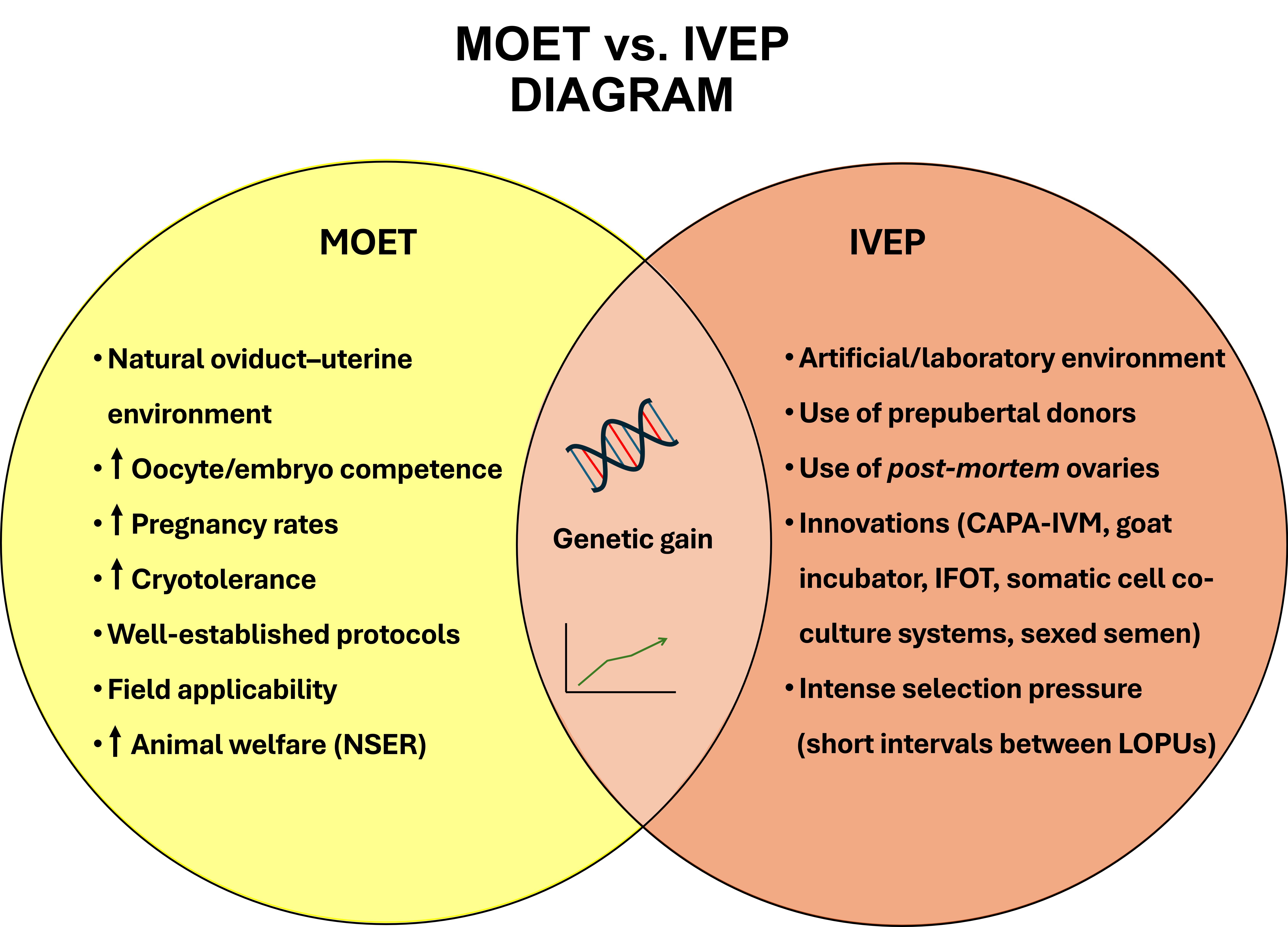
Conceptual comparison between in vivo and in vitro embryo production strategies in small ruminants. The Venn diagram summarizes key procedures and technological components specific to the MOET and IVEP systems, as well as shared elements between the approaches, illustrating their complementary roles in modern breeding programs. MOET: multiple ovulation and embryo transfer; IVEP: in vitro embryo production; LOPU: laparoscopic ovum pick-up; NSER: non-surgical embryo recovery; CAPA-IVM: capacitation in vitro maturation; IFOT: intrafollicular oocyte transfer.
Procedural aspects further influence the relative advantages of these technologies. In cattle, OPU can be performed transvaginally under epidural anesthesia, allowing frequent and minimally invasive oocyte collections. In small ruminants, however, oocyte recovery generally requires laparoscopic ovum pick-up (LOPU), which increases technical complexity and requires specialized equipment (Baldassarre and Karatzas, 2004; Souza-Fabjan et al., 2014a; Baldassarre, 2021). Embryo recovery in MOET programs may also involve invasive procedures, as laparotomy has historically been the most reliable method in sheep, although non-surgical embryo recovery approaches are increasingly being explored (Table 3). Despite these limitations, IVEP offers opportunities that are not readily achievable with MOET. Because oocyte recovery does not depend on ovulation or pregnancy establishment, IVEP can be applied to a broader range of donors, including prepubertal females, aged animals, pregnant donors, and post-mortem ovaries, expanding the range of exploitable genetic resources. IVEP also enables the use of laboratory-based technologies such as sex-sorted semen and experimental manipulation of gametes and embryos, although the availability of sexed semen in small ruminants remains limited (Souza-Fabjan et al., 2023a). Conversely, MOET benefits from embryo development in vivo within the maternal reproductive tract, which generally supports higher developmental competence and embryo quality compared with in vitro systems (Sirard, 2019; Lonergan and Fair, 2016; Coy et al., 2012). Thus, while MOET offers biological advantages through in vivo embryo development, IVEP provides greater operational and technological flexibility. Rather than representing competing approaches, both technologies should be viewed as complementary strategies that can be integrated in modern small ruminant breeding programs.
Comparison between surgical (laparotomy) and nonsurgical (NSER, transcervical) embryo recovery in small ruminants: efficiency, technical requirements, and biological constraints.
Multiple ovulation and embryo transfer (MOET) in small ruminants
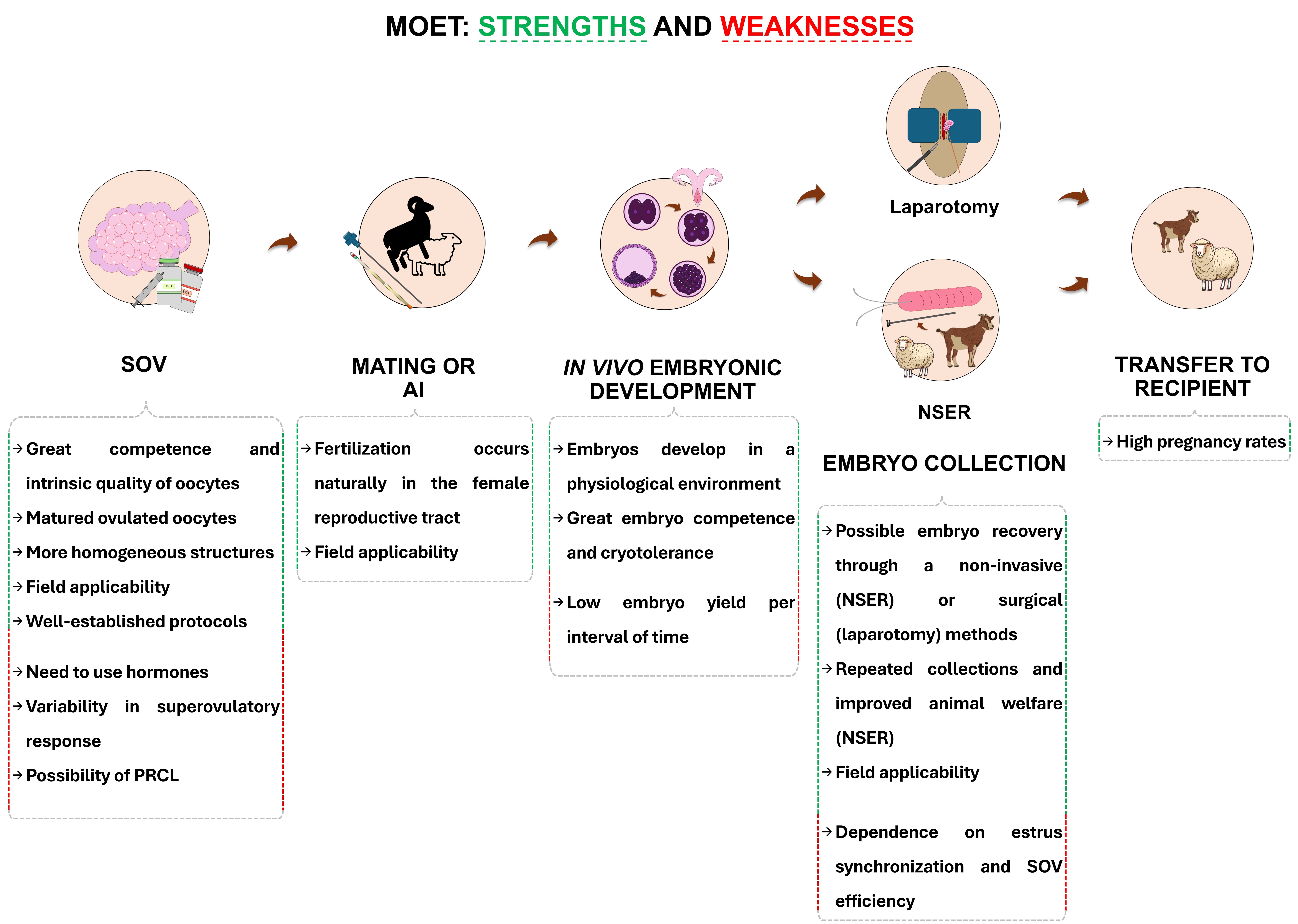
In vivo embryo production in small ruminants is primarily achieved through MOET. In this technique, donor females are hormonally stimulated to induce the development and ovulation of multiple follicles within a single estrous cycle. Following natural mating or artificial insemination, fertilization and early embryo development occur within the physiological environment of the reproductive tract (Figure 2). Embryos are subsequently recovered from the uterus six to seven days after fertilization and either transferred to synchronized recipient females or cryopreserved for later use (Souza-Fabjan et al., 2023a).
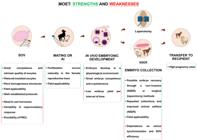
Workflow of in vivo embryo production in small ruminants. The scheme illustrates the main procedural steps—from ovarian superstimulation to embryo recovery—and highlights biological and technical strengths and weaknesses that may influence embryo yield and quality at each stage. MOET: multiple ovulation and embryo transfer; SOV: superovulation; PRCL: premature regression of the corpora lutea; AI: artificial insemination; NSER: non-surgical embryo recovery.
Biological strengths
In vivo embryo production through MOET has long been the predominant method for generating embryos in small ruminants (Paramio and Izquierdo, 2016; Souza-Fabjan et al., 2023a). One of the main biological advantages of this approach is that oocyte maturation, fertilization, and early embryonic development occur within the natural reproductive tract, allowing embryos to develop under physiological conditions in the ovary, oviduct, and uterus. Follicular growth and oocyte maturation occur under tightly regulated endocrine and paracrine signaling pathways, including interactions with granulosa and cumulus cells that support both nuclear and cytoplasmic maturation and the acquisition of developmental competence (Sirard et al., 2006).
After fertilization, the embryo continues its development within the maternal reproductive tract, where it is exposed to a highly regulated microenvironment characterized by dynamic changes in hormones, metabolites, growth factors, extracellular vesicles, and maternal transcripts (Saint-Dizier et al., 2020; Bastos et al., 2022). These components actively modulate embryo gene expression, metabolism, and epigenetic regulation, mediating embryo–maternal communication during the earliest stages of development (Coy et al., 2012; Lonergan and Fair, 2016; Mazzarella et al., 2024). As a result, embryos produced in vivo typically display high developmental competence and balanced cellular organization, reflecting the tightly regulated physiological environment in which early development occurs.
Another frequently reported feature of embryos produced through MOET is their superior cryopreservation tolerance and more consistent pregnancy outcomes following embryo transfer. In cattle, pregnancy rates obtained after transfer of in vitro-produced embryos are commonly reported to be approximately 20% lower than those achieved with in vivo–derived embryos under comparable conditions (Ealy et al., 2019; Lonergan and Fair, 2016; Hansen, 2020). Similar trends have been reported in small ruminants from experimental and field studies. In sheep, comparative studies evaluating different cryopreservation methods demonstrated markedly higher survival rates after transfer for MOET-derived embryos (53.3%) than for IVEP (20.8%) (Dos Santos-Neto et al., 2017). Likewise, in the same study, survival following conventional freezing or vitrification was substantially higher for embryos recovered from superovulated donors than for those produced in vitro. Consistent with these findings, pregnancy establishment after embryo transfer has generally been reported to be higher with in vivo embryos than with IVP embryos in small ruminants, even though improvements in cryopreservation techniques have progressively reduced these differences (Menchaca et al., 2016; Falchi et al., 2022).
The higher cryotolerance of in vivo embryos has been linked to several structural and metabolic characteristics, including lower intracellular lipid accumulation, more stable membrane composition, and more balanced cellular metabolism (Rizos et al., 2008; Banliat et al., 2022). Although substantial progress in culture systems, media formulation, and vitrification techniques has improved the survival of in vitro–produced embryos, these features contribute to improved resistance to osmotic and thermal stress during cryopreservation in MOET-embryos. These characteristics help explain why in vivo–derived embryos are frequently preferred in programs focused on cryobanking and long-term germplasm preservation (Arrais et al., 2021; Fonseca et al., 2022).
Beyond these aspects, MOET could theoretically offer advantages in sanitary safety and germplasm exchange. According to IETS guidelines, the risk of disease transmission through embryo transfer, regardless of the technique, is very low when standardized sanitary procedures are applied. Most infectious agents do not penetrate the zona pellucida but may attach to its outer surface, which is why sanitary handling protocols focus primarily on decontaminating it before transfer (IETS Manual, Chapter 2). However, structural differences in the zona pellucida between the two types of embryos may also influence pathogen interactions (Vanroose et al., 2000). During early development within the reproductive tract, the zona pellucida undergoes modifications through exposure to oviductal glycoproteins and other maternal secretions, which alter its biochemical composition and surface properties (de la Fuente et al., 2025), whereas embryos produced through IVEP are not exposed to this environment. Experimental studies suggest that such modifications may influence the adhesion of certain pathogens to the IVEP-derived embryo surface, although the biological significance of these observations under field conditions remains uncertain (IETS Manual, Chapter 3).
Technical and logistical advantages
In small ruminants, embryo recovery can be performed using three main approaches: laparotomy, laparoscopy, or nonsurgical embryo recovery (NSER) (Table 3). Historically, laparotomy was the most widely used technique in these species, largely due to the anatomical complexity of their cervix, which impairs transcervical access to the uterus. Although surgical approaches can provide reliable embryo recovery rates, they are invasive procedures associated with anesthesia, postoperative recovery, and potential surgical complications, which may restrict the frequency with which valuable donors can be used. Over the past decade, however, several research groups, including ours, have focused on developing and refining transcervical embryo recovery techniques. Advances in catheter design, cervical manipulation methods, and hormonal protocols to induce cervical relaxation have progressively improved the feasibility of NSER in both sheep and goats when performed by trained operators (Fonseca et al., 2019; Souza-Fabjan et al., 2023a).
In this aspect, MOET benefits from decades of technical refinement and field application. A practical advantage of MOET systems in small ruminants is the possibility of performing embryo recovery through NSER, a minimally invasive technique that allows repeated collections without the need for surgical intervention. Compared with traditional surgical approaches, NSER reduces animal stress, eliminates the risks associated with abdominal surgery and general anesthesia, and shortens recovery time, thereby improving animal welfare and facilitating the repeated use of valuable donors within breeding programs (Santos et al., 2020; Souza-Fabjan et al., 2022).
Furthermore, NSER can be conducted under field conditions with relatively simple equipment and short procedural times, typically requiring less than 30 minutes per donor when performed by experienced operators. Because the procedure does not require complex surgical infrastructure, it represents a logistically advantageous approach for commercial embryo transfer programs (Figueira et al., 2020; Souza-Fabjan et al., 2023a). The possibility of performing repeated embryo collections at relatively short intervals without major surgical intervention contributes to the operational flexibility and scalability of MOET programs in small ruminants, with no relevant species-specific differences reported between sheep and goats under comparable conditions.
Taken together, these technical and logistical characteristics contribute to the operational simplicity of MOET systems. When efficient NSER protocols are available, in vivo embryo production can be implemented with relatively limited laboratory infrastructure and fewer technical steps than in vitro embryo production systems.
Limitations of MOET
One important biological characteristic of MOET programs is the substantial variability observed among donor females in their response to superovulatory treatments. Differences in ovarian follicular dynamics, follicular reserve, endocrine status, genetic background, and physiological conditions can lead to marked variation in the number of ovulations and embryos recovered per treatment cycle (Cognié et al., 2003; Gonzalez-Bulnes et al., 2004; Bartlewski et al., 2016). In sheep and goats, the stage of the follicular wave at the onset of gonadotropin treatment, as well as individual differences in follicle recruitment and dominance patterns, are important determinants of superovulatory response (Bartlewski et al., 2016). As a result, the number of embryos recovered per donor may vary substantially between individuals (0-12; Morais et al., 2025) and across treatment cycles, representing a major biological limitation of MOET programs. Several strategies have therefore been proposed to reduce this variability, including protocols designed to synchronize the emergence of the follicular wave before the initiation of gonadotropin treatment (Gonzalez-Bulnes et al., 2004; Bartlewski et al., 2016). In this context, approaches such as the Day-0 protocol, in which superovulatory treatment is initiated at the time of a newly emerged follicular wave, have been proposed to improve follicular recruitment and enhance the consistency of superovulatory responses in small ruminants (Menchaca et al., 2009; Souza-Fabjan et al., 2017).
In addition to intrinsic biological variability, extrinsic factors associated with hormonal protocols are major determinants of superovulatory response. These include the type and origin of gonadotropins (e.g., pituitary-derived FSH, recombinant FSH, eCG, or their combinations), dosing regimens and administration schedules, as well as synchronization strategies based on progestogens (e.g., CIDR or intravaginal sponges) and luteolytic agents such as prostaglandin F2α or its analogs. Variations in these protocols can markedly influence follicular dynamics, ovulatory response, and embryo yield (Bartlewski et al., 2016; Aybazov et al., 2023; Souza-Fabjan et al., 2023a).
A further limitation affecting the efficiency of MOET programs in small ruminants is the occurrence of premature regression of the corpus luteum (PRCL). This luteal dysfunction results in an early decline in progesterone concentrations, compromising embryo development and frequently reducing embryo recovery and the number of transferable embryos. Studies in both goats and sheep have shown that PRCL may occur in a substantial proportion of superovulated females, with reported incidences ranging from approximately 20% to over 60% depending on the protocol and experimental conditions (Bartlewski et al., 2016; Souza-Fabjan et al., 2017; Rocha et al., 2022). This phenomenon has been associated with endocrine disturbances during superovulation, including elevated estradiol concentrations and the presence of anovulatory follicles, which may trigger premature prostaglandin release and luteolysis. Consequently, PRCL is considered one of the main factors reducing embryo recovery rates and overall efficiency of MOET programs in sheep (Forcada et al., 2011; Rocha et al., 2022). To mitigate PRCL, strategies such as the use of flunixin meglumine (a non-steroidal anti-inflammatory drug inhibiting prostaglandin synthesis) during the early luteal phase have been proposed, leading to improved embryo recovery and production in superovulated ewes (Maia et al., 2024).
Another operational limitation of MOET programs is that embryos recovered from a given superovulatory cycle typically originate from a single male when artificial insemination or natural mating is used. In contrast, IVEP systems allow oocytes to be fertilized with semen from multiple sires within the same session using separate fertilization droplets, increasing flexibility for genetic combinations and enabling more efficient use of semen from elite sires.
In vitro embryo production (IVEP) in small ruminants
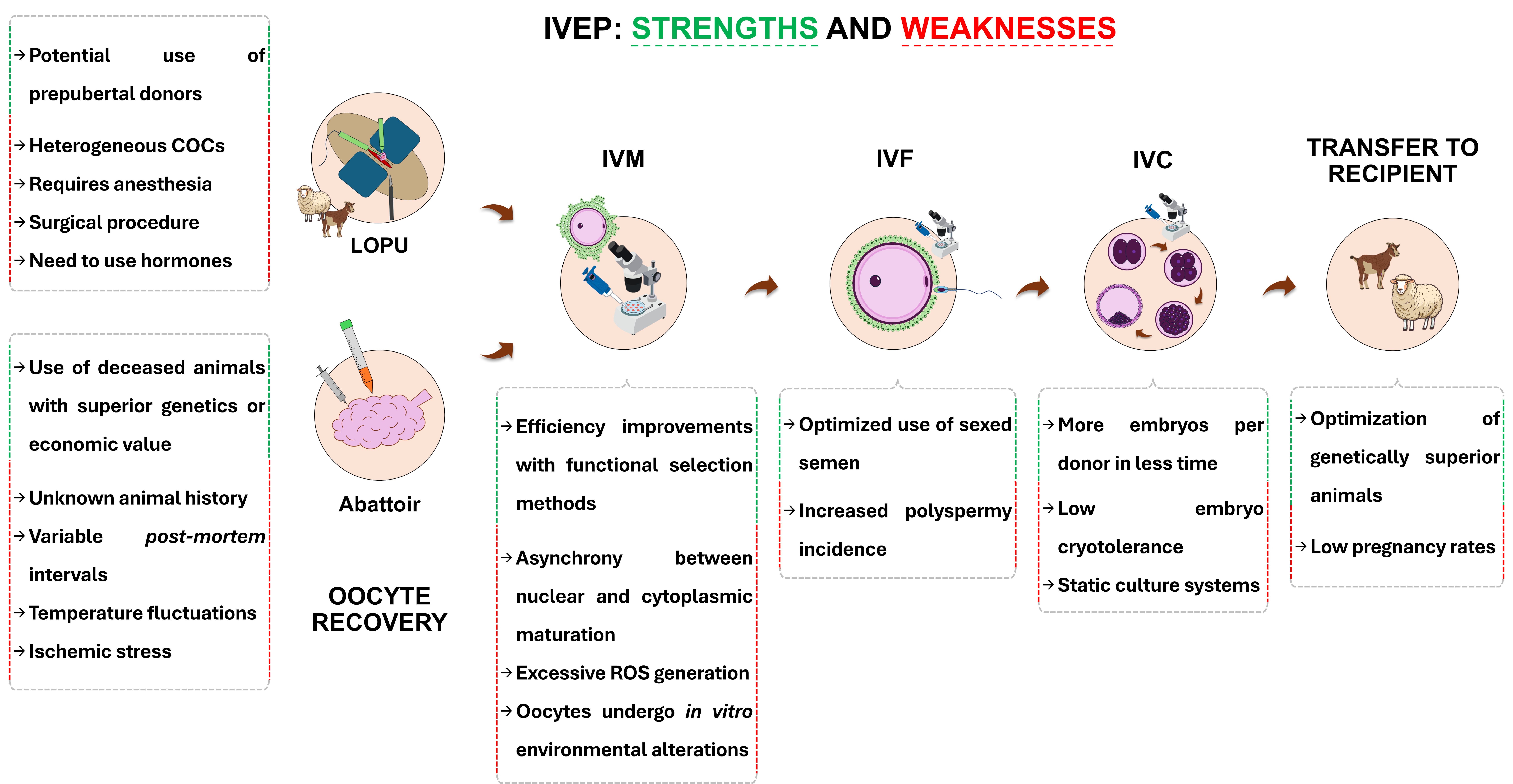
In vitro embryo production, comprising oocyte recovery, in vitro maturation (IVM), fertilization (IVF), and embryo culture (IVC), is an important tool for genetic improvement, germplasm conservation, and experimental research in small ruminants (Figure 3). Despite substantial progress, IVEP efficiency in sheep and goats remains lower than that achieved in cattle. This limitation arises from the interaction between species-specific biological characteristics and technical constraints affecting not only oocyte competence, but also sperm selection and quality, fertilization success, and subsequent embryo development. These constraints can be conceptually organized into three interconnected levels: biological, technical, and logistical limitations. Biological factors define the physiological boundaries of follicular development and gamete competence, whereas technical and operational factors influence the efficiency and reproducibility of IVEP systems. Understanding these layers is essential for identifying the main bottlenecks limiting IVEP performance in small ruminants.
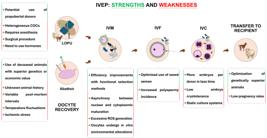
Main steps involved in in vitro embryo production in small ruminants, from oocyte recovery to embryo culture. Potential biological and technical strengths and weaknesses affecting developmental competence and embryo quality are indicated along the IVEP pipeline. IVEP: in vitro embryo production; LOPU: laparoscopic ovum pick-up; COCs: cumulus–oocyte complexes; IVM: in vitro maturation; IVF: in vitro fertilization; IVC: in vitro culture; ROS: reactive oxygen species.
Biological limitations
Biological limitations represent intrinsic constraints affecting IVEP efficiency in small ruminants and arise mainly from species-specific characteristics of follicular dynamics, oocyte developmental competence, and early embryonic physiology. A central challenge is the high heterogeneity of oocytes recovered during routine procedures, largely influenced by follicle size, donor age, physiological status, and environmental conditions. This variability directly affects the ability of oocytes to complete maturation, sustain fertilization, and support early embryonic development, thereby defining the biological baseline upon which technical IVEP procedures operate.Oocyte developmental competence
Oocyte developmental competence is widely recognized as the primary biological bottleneck limiting IVEP efficiency in small ruminants. Competence refers to the ability of the oocyte to undergo coordinated nuclear and cytoplasmic maturation and subsequently sustain fertilization and early embryonic development (Sirard, 2019). In sheep and goats, oocytes are frequently recovered from follicles at heterogeneous developmental stages, particularly when follicular wave synchronization is not performed, resulting in a large proportion of oocytes with reduced developmental potential (Crozet et al., 1995; Mermillod et al., 1999; Gandolfi et al., 2005; Falchi et al., 2022; Souza-Fabjan et al., 2023b). Oocytes derived from small and medium follicles often exhibit incomplete cytoplasmic maturation, which affects key determinants of competence such as cytoskeletal organization, cortical granule redistribution, and mitochondrial activity (Velilla et al., 2004, 2005, 2006; Anguita et al., 2007, 2008). In contrast, oocytes obtained from larger follicles generally display higher developmental competence, although they represent only a small fraction of recovered oocytes.
In superstimulated ewes, extending the coasting period (the interval between gonadotropin withdrawal and oocyte collection) to 60 h increased the proportion of large follicles and produced oocytes with larger diameters, more advanced germinal vesicle chromatin condensation, and higher expression of maternal-effect and cumulus–oocyte communication genes, despite no detectable differences in conventional morphological or metabolic selection criteria (Pinheiro et al., 2023). These observations highlight that the developmental potential of oocytes entering IVEP systems is strongly conditioned by their follicular history, emphasizing the importance of physiological factors influencing follicular development.
Physiological modulators of IVEP efficiency
Beyond intrinsic oocyte competence, several physiological factors influence IVEP efficiency in small ruminants by affecting follicular dynamics and the follicular microenvironment. Reproductive seasonality is a major determinant, as photoperiod-driven changes in ovarian activity influence follicular development and endocrine support, resulting in higher embryo development during the breeding season and reduced outcomes during seasonal anestrus (Tibary et al., 2005; Souza-Fabjan et al., 2021). However, in lower-latitude regions, such as the tropical and subtropical areas of South America, including Brazil, seasonal effects may be less pronounced due to reduced photoperiod variation and the use of management strategies that mitigate reproductive seasonality. Donor age also affects developmental competence, as oocytes from prepubertal females show reduced potential due to incomplete nuclear and cytoplasmic maturation (Paramio and Izquierdo, 2014, 2016; Souza-Fabjan et al., 2023b). Nutritional and metabolic status further modulate oocyte quality through their effects on endocrine balance and follicular fluid composition (Diskin et al., 2003; Leroy et al., 2004). These physiological factors (including season, age, and nutrition) also influence the response to ovarian superstimulation and, consequently, affect the outcomes of MOET. However, their impact may be more pronounced in IVEP systems because oocytes must undergo IVM, making their developmental competence more dependent on their initial physiological condition. In addition, genetic and breed-related variability influence ovarian reserve, follicular dynamics, and reproductive patterns, ultimately affecting oocyte yield and developmental competence in IVEP systems (Cognié et al., 2004; Mermillod et al., 2008). Together, these physiological factors contribute to the variability observed in embryo production outcomes in small ruminants.
Sensitivity to in vitro environments
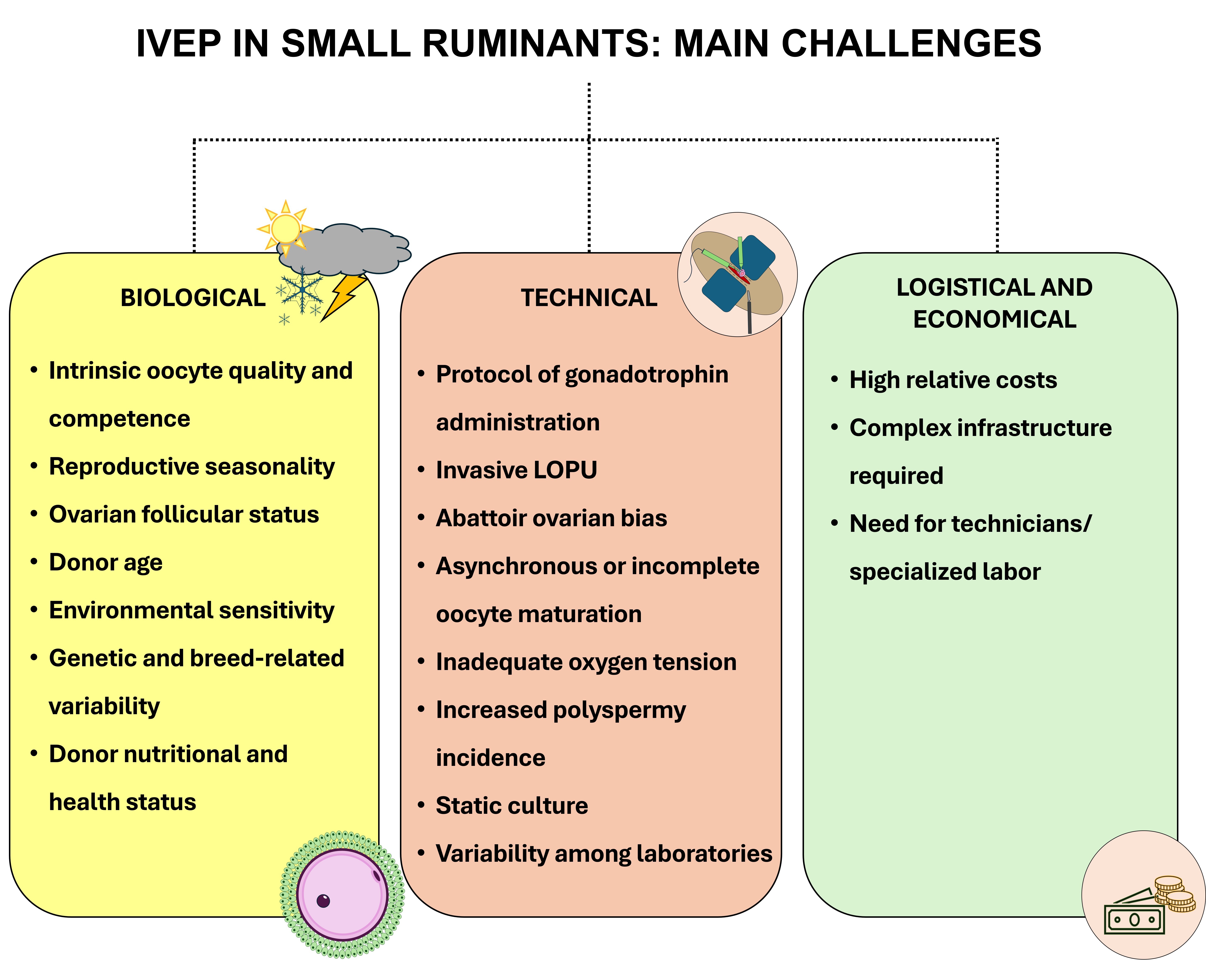
In addition to intrinsic competence and physiological background, gametes and embryos from ovine and caprine species exhibit increased sensitivity to in vitro environments compared with bovine embryos and with embryos derived in vivo. However, this apparent sensitivity may not solely reflect intrinsic biological differences, but also the widespread use of in vitro culture systems originally optimized for bovine embryos, which may not fully meet the specific physiological and metabolic requirements of small ruminants (Souza-Fabjan et al., 2023a). Ovine and caprine oocytes and embryos show limited tolerance to variations in oxygen tension, redox balance, and metabolic conditions (Rodríguez-Dorta et al., 2007; Natarajan et al., 2010; Amiridis and Cseh, 2012). This vulnerability is partly associated with limited antioxidant defenses and metabolic buffering capacity, increasing susceptibility to reactive oxygen species (ROS), mitochondrial dysfunction, and redox imbalance (Rizos et al., 2002; Mauchart et al., 2023). As a consequence, developmental arrest frequently occurs during critical stages of early embryogenesis, particularly around embryonic genome activation and during the transition from the 8-cell stage to the morula. Importantly, environmental sensitivity interacts with intrinsic oocyte competence and follicular background. Oocytes with incomplete cytoplasmic maturation exhibit reduced tolerance to oxidative and metabolic stress, thereby amplifying developmental losses during IVEP procedures. Together, intrinsic competence, physiological modulation of follicular development, and sensitivity to in vitro environments (both biological and system-derived) define the framework that limits IVEP efficiency in small ruminants and ultimately shapes the outcomes of subsequent technical procedures (Figure 4).
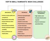
Overview of the major constraints limiting in vitro embryo production efficiency in small ruminants. Challenges are organized into distinct interacting domains—biological, technical, logistical, and economical—that illustrate the multifactorial nature of the limitations affecting the scalability and field application of IVEP systems. IVEP: in vitro embryo production; LOPU: laparoscopic ovum pick-up.
Technical limitations in IVEP
Beyond intrinsic biological constraints, the efficiency and reproducibility of IVEP in small ruminants are also influenced by technical limitations inherent to current laboratory systems. Importantly, many of these limitations reflect incomplete knowledge of the biological dynamics regulating gamete maturation, fertilization, and early embryonic development in these species. As a result, IVM, IVF, and IVC systems remain unable to fully reproduce the dynamic ovarian, oviductal, and uterine environments that coordinate these processes in vivo. This limitation is particularly evident for the oviduct, whose microenvironment plays a critical role in sperm capacitation, gamete interaction, and early embryo development. Despite its importance, detailed information on the biochemical composition and temporal dynamics of the oviductal environment in sheep and goats remains scarce, making it difficult to accurately mimic these conditions in vitro. Consequently, several aspects of current IVEP systems are simplified approximations of complex physiological processes, contributing to reduced efficiency and variability in embryo production in small ruminants.
Oocyte recovery: LOPU and abattoir-derived ovaries
Oocyte recovery represents a major technical bottleneck in small ruminant IVEP, as collection methods directly influence oocyte yield, integrity, and developmental competence. Laparoscopic ovum pick-up is indispensable for recovering oocytes from live donors but is invasive and technically demanding, requiring specialized equipment, trained personnel, and careful animal management, which limits collection frequency and scalability (Souza-Fabjan et al., 2014a; Baldassarre, 2021). Procedural parameters such as aspiration pressure, needle gauge, and operator proficiency can affect cumulus–oocyte complex (COC) integrity, with suboptimal settings causing mechanical damage and reduced in vitro performance (Wieczorek et al., 2020; Souza-Fabjan et al., 2014a; Baldassarre et al., 2014). In addition, repeated ovarian manipulation during frequent LOPU sessions may induce local tissue stress and inflammatory responses, compromising recovery efficiency and embryo development (Teixeira et al., 2011).
Abattoir-derived ovaries are widely used for methodological optimization due to their availability, low cost, and high oocyte throughput (Ebrahimi et al., 2025). This approach has supported the development of IVM, IVF, and IVC systems under controlled laboratory conditions (Souza-Fabjan et al., 2023b), but introduces technical bias because oocytes originate from donors of unknown age, metabolic and reproductive status and are exposed to variable post-mortem conditions before processing (Paramio and Izquierdo, 2014). Such factors can impair oocyte quality, mitochondrial function, and redox balance (Lonergan and Fair, 2014; Reader et al., 2017). Consequently, IVEP systems optimized with slaughterhouse-derived oocytes may overestimate robustness and perform inconsistently when applied to LOPU-derived oocytes from live donors, reinforcing oocyte recovery as a key technical limitation affecting IVEP reproducibility in sheep and goats (Souza-Fabjan et al., 2014b).
Oocyte selection and in vitro maturation (IVM)
Oocyte selection represents the first technical filter in small ruminant IVEP, as the developmental competence of the oocyte entering IVM largely determines subsequent embryonic outcomes. Morphological evaluation based on cumulus investment and cytoplasmic appearance remains the most commonly used approach; however, it is subjective and only weakly predictive of true developmental competence (Ebrahimi et al., 2025). Functional selection methods, such as brilliant cresyl blue (BCB) staining, which reflects glucose-6-phosphate dehydrogenase (G6PDH) activity and cytoplasmic maturity, have therefore been proposed as more biologically informative indicators (Bragança et al., 2021a; Pinheiro et al., 2023). The BCB test is typically applied to immature COCs immediately after recovery and prior to IVM. However, the proportion of BCB-positive oocytes varies markedly according to donor characteristics. For instance, approximately 29% of oocytes were classified as BCB-positive in prepubertal goats (Rodríguez-González et al., 2002), whereas 19–47% were reported in prepubertal sheep depending on the BCB concentration used (Catalá et al., 2011). In contrast, higher proportions have been reported in adult donor populations, including 66–69% BCB-positive oocytes in slaughterhouse-derived sheep ovaries (Wang et al., 2012) and up to 85% among morphologically normal oocytes from FSH-primed adult goats (Paramio and Izquierdo, 2014). Consistent with this metabolic selection, BCB-positive oocytes generally show higher maturation, cleavage, and blastocyst rates than BCB-negative counterparts (Rodríguez-González et al., 2002; Catalá et al., 2011; Wang et al., 2012). However, although BCB staining enriches the oocyte population for developmental competence, its effect on the total number of embryos produced is less consistent because the procedure reduces the number of oocytes entering the IVEP pipeline.
Despite improvements in selection strategies, IVM remains a major technical bottleneck in small ruminant IVEP. Conventional maturation systems are inherently static and fail to reproduce the dynamic regulatory mechanisms present in vivo, including meiotic arrest control, cyclic nucleotide signaling, and cumulus–oocyte metabolic coupling (Paramio and Izquierdo, 2016). As a consequence, nuclear maturation frequently occurs without coordinated cytoplasmic and metabolic remodeling, limiting developmental competence. In addition, culture under supraphysiological oxygen tension can exacerbate oxidative stress, particularly in oocytes with incomplete cytoplasmic maturation (Luvoni et al., 1996; Agarwal et al., 2006).
Recent transcriptomic studies provide molecular evidence of these limitations, demonstrating that oocytes matured in vitro exhibit altered signaling pathways, endoplasmic reticulum stress, and disrupted redox regulation compared with in vivo-matured counterparts (Cui et al., 2025). Historically, attempts to overcome these deficiencies have relied on increasingly complex IVM supplementation strategies using serum, follicular fluid, antioxidants, or growth factors (Paramio and Izquierdo, 2016; Souza-Fabjan et al., 2023b). Although these approaches may partially improve embryo development, they often reduce reproducibility across laboratories. More recently, alternative strategies have focused on restoring physiological control of meiotic progression. Biphasic maturation systems based on cyclic AMP modulation, such as Capacitation-IMV (CAPA-IVM), delay meiotic resumption and improve coordination between nuclear and cytoplasmic maturation, resulting in increased blastocyst production (Rose et al., 2013; Medina-Chávez et al., 2021; Ferrer-Roda et al., 2024a,b). Taken together, these findings indicate that improving IVEP efficiency in small ruminants will depend less on refining morphological selection criteria and more on developing maturation systems capable of restoring the dynamic regulatory environment that governs oocyte competence in vivo.
In vitro fertilization (IVF)
IVF is another critical step that influences embryo yield and quality in small ruminant IVEP systems. Even when nuclear maturation is achieved during IVM, oocyte populations remain heterogeneous in their ability to regulate fertilization events such as cortical granule exocytosis and the establishment of an effective polyspermy block. In this context, sperm preparation and capacitation procedures are major sources of variability affecting fertilization outcomes. In ovine and caprine IVF systems, spermatozoa are typically prepared using swim-up or density-gradient centrifugation (e.g., Percoll) to enrich for motile sperm. The swim-up method is more commonly applied to fresh semen, whereas density-gradient centrifugation is frequently preferred for frozen–thawed samples due to its superior capacity to recover functionally competent spermatozoa. Density-gradient centrifugation acts as a selective washing technique that separates sperm based on motility and density, yielding highly enriched populations of motile and morphologically normal spermatozoa while effectively removing cellular debris, leukocytes, epithelial cells, bacteria, and immature or abnormal sperm, including those with compromised DNA integrity (Mortimer, 2000; Henkel and Schill, 2003; Souza-Fabjan et al., 2023a). This method allows further selection of sperm according to their kinetic properties and head density, thereby enhancing the overall quality of the recovered fraction (Mortimer, 2000; Henkel and Schill, 2003).
Following sperm selection, fertilization is generally performed in specialized media. Synthetic Oviductal Fluid (SOF) is the medium most commonly used for ovine IVF, whereas Tyrode’s Albumin Lactate Pyruvate (TALP) is widely used as a fertilization medium in caprine IVF systems (Paramio and Izquierdo, 2014; Souza-Fabjan et al., 2023b). These media are typically supplemented with bovine serum albumin and bicarbonate, which support sperm capacitation, together with heparin or related glycosaminoglycans as capacitating agents, similar to fertilization systems commonly used in bovine IVF. In some protocols, additional capacitation-promoting compounds such as penicillamine, hypotaurine, and epinephrine or estrous serum are included to enhance sperm functionality and fertilization rates (Souza-Fabjan et al., 2023b).
Despite these strategies, ovine and caprine spermatozoa remain highly sensitive to handling, cryopreservation, and oxidative stress, which can compromise motility, membrane integrity, and fertilization capacity (Peris et al., 2004; Barbas et al., 2018; Jurado-Campos et al., 2023). Moreover, conventional static IVF systems fail to fully reproduce the dynamic regulatory environment of the oviduct, increasing the incidence of polyspermy commonly reported (Palomo et al., 2010; Romar et al., 2019; Anzalone et al., 2021). Although polyspermy is particularly pronounced in porcine IVF systems, it also represents a recurrent challenge in ovine and caprine IVF, reflecting the difficulty of reproducing the physiological mechanisms that regulate sperm–oocyte interaction within the oviduct. Supplementation with oviductal fluid proteins has been shown to improve monospermic fertilization and IVF efficiency, highlighting the limitations of conventional systems in reproducing physiological gamete interactions (Bragança et al., 2021b).
The use of sex-sorted semen introduces additional technical challenges in small ruminant reproduction. Flow cytometric sorting exposes spermatozoa to mechanical and oxidative stress, which can reduce motility, membrane integrity, and fertilization potential (Hollinshead et al., 2004; de Graaf et al., 2007a,b; Ngcobo et al., 2024). Embryos generated from sorted sperm may also display altered expression of genes involved in DNA methylation, chromatin remodeling, and metabolic regulation (Beilby et al., 2011). Although commercial availability has recently expanded, particularly in dairy goat breeding systems, the practical use of sex-sorted semen in small ruminants remains geographically limited. Importantly, the impact of sex-sorted semen differs between reproductive technologies. In MOET programs relying on artificial insemination, the reduced fertility associated with sorted sperm can directly compromise fertilization rates and embryo yield. In contrast, IVF systems offer greater flexibility in sperm concentration and capacitation conditions, partially compensating for reduced sperm quality. Consequently, while sex-sorted semen introduces biological and technical constraints, its integration may be more compatible with IVEP systems than with MOET-based embryo production.
In vitro embryo culture (IVC)
IVC represents the final stage of IVEP and plays a central role in determining embryo quality. Conventional culture systems remain largely static and only partially reproduce the dynamic physicochemical environment of the oviduct and uterus (Souza-Fabjan et al., 2023b; Ebrahimi et al., 2025). The development of SOF, formulated to mimic to composition of the OF in energy substrates (lactate-pyruvate-glucose) and salts, enabled ovine embryos to overcome the classical developmental block at the 8–16-cell stage (Tervit et al., 1972) and is now widely used. However, SOF-based media do not reproduce the complex composition of the native OF and rely on fixed nutrient concentrations despite clear stage-specific metabolic requirements during development (Gardner and Lane, 1997; Thompson et al., 2007). Early embryos primarily utilize pyruvate and lactate, whereas glucose uptake increases during compaction and blastulation, and failure to accommodate these shifts can compromise blastocyst quality.
Culture conditions also influence oxidative balance. Atmospheric oxygen tension increases ROS production, whereas reduced oxygen levels (approximately 5–7%) improve blastocyst development, mitochondrial function, and cryotolerance (Thompson, 2000; Leoni et al., 2007; Souza-Fabjan et al., 2023b). Strategies such as somatic cell co-culture or fetal calf serum supplementation may increase embryo yield but introduce variability and may impair embryo quality or postnatal outcomes (Young et al., 1998; Souza-Fabjan et al., 2023b). In ruminants, exposure to serum during early embryo culture has also been associated with abnormal fetal growth and developmental disorders collectively referred to as large offspring syndrome, which was initially described in sheep and later extensively reported in cattle produced by assisted reproductive technologies (Young et al., 1998; Chen et al., 2013). These abnormalities are thought to arise from epigenetic dysregulation and altered metabolic conditions during early embryogenesis.
Consequently, most IVEP systems currently rely on semi-defined SOF-based media supplemented with bovine serum albumin and amino acids to reduce variability and improve reproducibility. Despite these improvements, developmental efficiency remains limited. In small ruminants, blastocyst rates following IVEP typically range from approximately 20–40% of fertilized oocytes, depending on donor characteristics and laboratory conditions (Cognié et al., 2003; Paramio and Izquierdo, 2016; Souza-Fabjan et al., 2023b). Moreover, embryos produced in vitro frequently exhibit altered transcriptomic and epigenetic profiles compared with in vivo-derived embryos, as demonstrated in ovine models showing differential gene expression and impaired embryo–endometrium interactions, highlighting that current IVC systems still fail to fully replicate the physiological conditions of the reproductive tract (Wei et al., 2016; Zhu et al., 2021).
Together, these limitations suggest that contemporary IVC systems impose a technical ceiling on embryo developmental potential in small ruminant IVEP. Future improvements in embryo culture efficiency will likely depend on the development of dynamic, physiologically relevant culture systems, including sequential media formulations, microfluidic platforms, and oviduct-on-a-chip technologies that better replicate the in vivo reproductive environment (Ferraz and Ferronato, 2023). Overall, these emerging approaches highlight the need for next-generation embryo culture systems that integrate dynamic nutrient regulation, physiological oxygen tension, and reproductive tract-derived signals to more accurately recapitulate early embryonic development in vivo.
Logistical and economic challenges
The implementation of IVEP in small ruminants is constrained by logistical and economic barriers that limit its scalability and commercial feasibility. Compared with cattle, IVEP in sheep and goats requires greater technical specialization per embryo produced, resulting in higher operational costs and strong dependence on centralized infrastructure. From a logistical perspective, the technique relies on highly trained personnel for LOPU, anesthesia, gamete handling, and embryo culture. A single LOPU session often requires multiple specialized operators and veterinary anesthesia support, increasing labor requirements compared with MOET (Souza-Fabjan et al., 2014a; Baldassarre, 2021). In addition, IVEP depends on advanced laboratory infrastructure, including surgical facilities, controlled-atmosphere incubators, and dedicated embryo culture laboratories, which restricts its use to specialized centers and limits on-farm application (Paramio and Izquierdo, 2014).
Another economic constraint is the routine use of hormonal stimulation to promote follicular development before LOPU (Souza-Fabjan et al., 2023b; Baldassarre, 2021). In sheep and goats, IVEP typically relies on FSH- or eCG-based protocols to obtain adequate oocyte yields, generating recurring donor-level costs (Baldassarre and Karatzas, 2004). Variability in ovarian response may also require protocol adjustments or repeated stimulation cycles, further increasing hormone use and overall costs (Baldassarre, 2021). As a result, the combined expenses associated with surgical procedures, anesthesia, consumables, hormonal treatments, and specialized labor lead to a relatively high cost per transferable embryo, which is often higher than that of MOET-derived embryos (Baldassarre and Karatzas, 2004). Scalability represents an additional limitation. The invasive nature of LOPU restricts the frequency of oocyte collection from individual donors, while the reliance on laboratory-based workflows limits large-scale application under field conditions. Consequently, IVEP in small ruminants remains largely confined to high-value genetic programs, germplasm conservation, and research applications.
Emerging perspectives for IVEP in small ruminants
Despite decades of methodological refinement, IVEP in small ruminants remains constrained by biological inefficiencies, logistical complexity, and relatively high operational costs, which together limit its scalability and field applicability. These challenges have shifted the research focus from incremental optimization of conventional protocols toward innovative strategies that integrate advances in reproductive biology, cellular metabolism, and bioengineering. Current perspectives increasingly emphasize improving oocyte developmental competence, reducing procedural invasiveness and hormonal dependency, and enhancing system reliability under less restrictive laboratory conditions. The next phase of technological progress will likely depend on translating mechanistic insights into simplified, cost-effective platforms capable of expanding IVF beyond specialized centers (Figure 5).
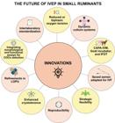
Emerging innovations and research directions aimed at improving in vitro embryo production (IVEP) efficiency in small ruminants. Strategies include advances in oocyte competence, fertilization systems, and embryo culture technologies that may enhance embryo yield, cryotolerance, and scalability of IVEP platforms. COCs: cumulus–oocyte complexes; CAPA-IVM: biphasic in vitro maturation; IFOT: intrafollicular oocyte transfer; LOPU: laparoscopic ovum pick-up; IVF: in vitro fertilization.
What could change the balance?
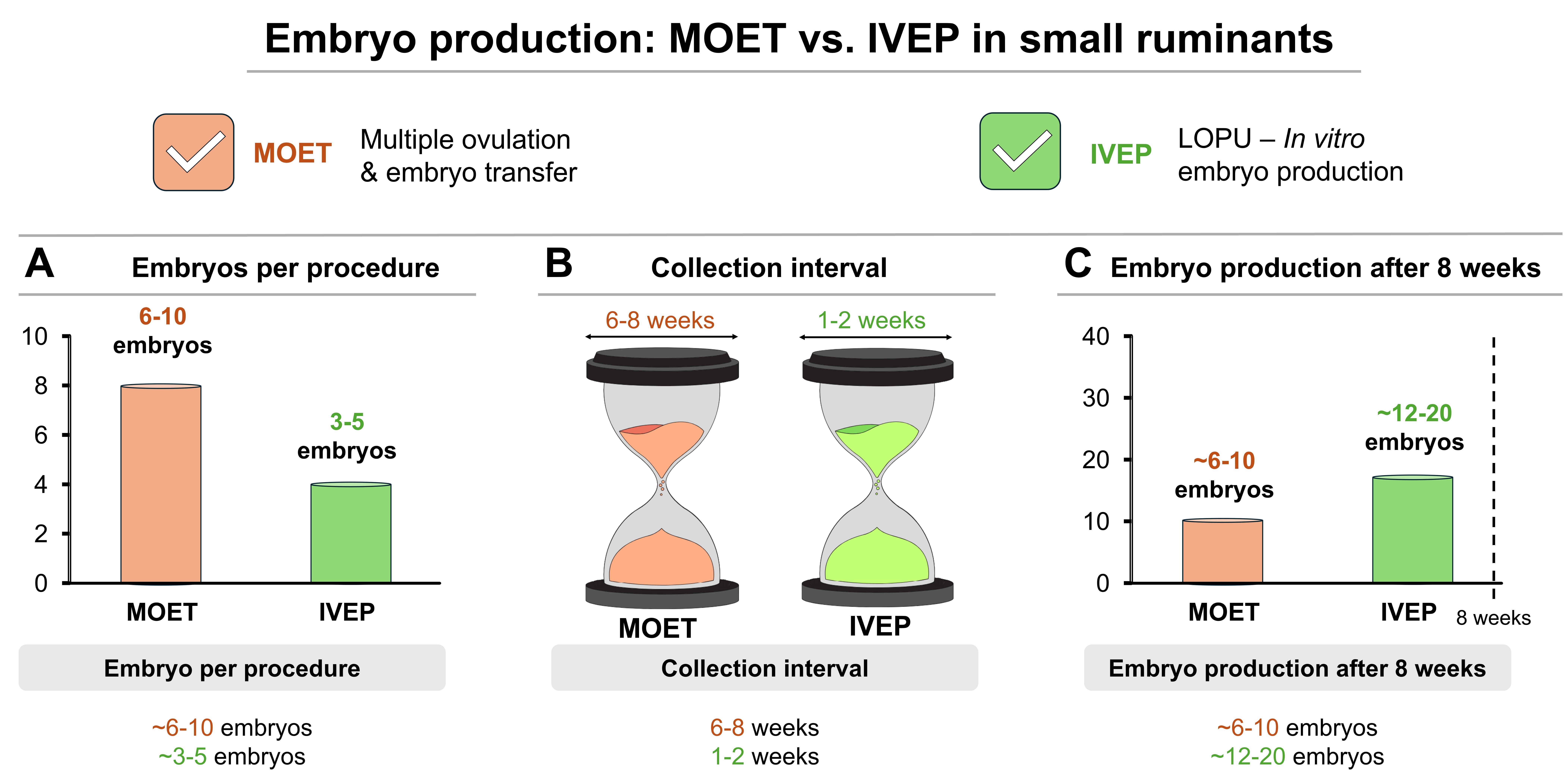
Historically, the balance between MOET and IVEP in small ruminants has been determined not only by biological efficiency but also by operational simplicity, cost–benefit relationships, and robustness under field conditions. Although MOET remains the dominant approach due to its reproducibility and logistical feasibility, advances across the IVEP pipeline suggest that this balance could shift if improvements translate into scalable and economically viable outcomes. Current LOPU–IVEP systems typically recover approximately 10 and 14 viable oocytes per session in sheep and goats, respectively, resulting in 3–5 transferable embryos and pregnancy rates exceeding 50% after embryo transfer (Baldassarre, 2021). Although this embryo yield per session is generally lower than that achieved through MOET, which commonly produces 6–10 transferable embryos per superovulatory cycle, IVEP offers the possibility of shorter intervals between collections, often every 1–2 weeks, compared with the 6–8 week recovery interval typically required for MOET. Consequently, when embryo output is evaluated over a defined period, IVEP may achieve comparable or even greater cumulative embryo production (Gibbons et al., 2007; Souza-Fabjan et al., 2023a) (Figure 6). When pregnancy rates are also considered, the overall number of pregnancies generated per donor over time may approach similar levels between systems under optimized conditions.

Conceptual comparison between in vivo embryo production (MOET) and in vitro embryo production (IVEP) systems in small ruminants based on embryo production dynamics. (A) Typical number of embryos obtained per procedure, (B) interval between consecutive collections, and (C) estimated cumulative embryo production over an eight-week period. Although MOET generally produces more embryos per session, the shorter collection interval of IVEP may result in comparable cumulative embryo output over time.
One factor contributing to the potential strategic value of IVEP is the increasing availability of sex-sorted semen adapted for IVF. Historically, the use of sexed sperm in sheep and goats has been limited by reduced fertilization rates, increased DNA fragmentation, and altered capacitation dynamics associated with sorting and cryopreservation (Hollinshead et al., 2004; de Graaf et al., 2007b; Bathgate, 2008). However, recent refinements in sperm handling and IVF-specific capacitation protocols have improved fertilization outcomes and blastocyst development (Bai et al., 2025), increasing the strategic value of IVEP in breeding programs where control of offspring sex may be advantageous. For example, the production of female offspring may be preferred in dairy-oriented systems to accelerate genetic gain through replacement females, whereas male offspring may be desirable in specific breeding or terminal-cross programs. Nevertheless, in many sheep and goat breeding schemes, both sexes are economically valuable, and in some programs, sex-sorted semen is not routinely used because both male and female offspring contribute to genetic progress and herd structure. On the female side, refinements in LOPU, including improved aspiration control and follicular synchronization, have increased oocyte recovery and procedural consistency (Teixeira et al., 2011; Baldassarre, 2021). Nevertheless, oocyte quality remains a major limitation, and integrating morphological and functional selection methods, such as BCB staining, can enrich oocyte populations with greater developmental potential (Rodríguez-González et al., 2003; Catala et al., 2011; Alm et al., 2020).
Advances in embryo culture systems may also improve IVEP outcomes. Studies in bovine and human models indicate that reduced oxygen tension, dynamic culture systems, and supplementation with bioactive components can enhance blastocyst quality and post-cryopreservation survival (Chen et al., 2023; Alegretti et al., 2024; Báez et al., 2024). Similarly, extracellular vesicles derived from reproductive fluids have been shown to improve embryo cryosurvival by modulating lipid metabolism and redox balance (Leal et al., 2022). These findings highlight embryo cryotolerance as a critical determinant of IVEP competitiveness, a domain in which MOET-derived embryos continue to hold a practical advantage. However, translating these strategies to ovine and caprine systems remains challenging due to species-specific differences in embryo metabolism and oxidative sensitivity. Without rigorous species-specific validation and cost–benefit evaluation, improvements in laboratory performance may not substantially alter the economic balance between MOET and IVEP.
Global embryo production data reported by the IETS in 2024 clearly reflect these constraints, where MOET-based production still accounted for more than 95% of ovine embryos worldwide. Although ovine IVEP increased substantially between 2022 and 2023 (+62%), in vivo production also expanded (+79%), suggesting overall market growth rather than technological replacement. In goats, IVEP activity also increased but remains geographically concentrated. Importantly, this pattern is not consistently observed in the 2025 dataset, largely due to a reduction in the number of reporting countries, which limits direct comparisons and likely underestimates global embryo production trends. Integration with genomic selection may represent one of the most transformative applications of advanced reproductive technologies in small ruminants. By enabling earlier and more precise identification of superior donors and sires, genomic information can improve the efficiency of both MOET and IVEP programs. In this context, IVEP may provide additional advantages by allowing repeated oocyte recovery and rapid multiplication of genetically superior females, thereby increasing selection intensity and shortening generation intervals (Kasinathan et al., 2015; Meuwissen et al., 2016). Ultimately, a shift in the balance between MOET and IVEP will depend on the convergence of improved biological robustness, embryo cryotolerance, economic scalability, and alignment with breeding objectives.
Realistic expectations
Despite significant technological progress, the expansion of IVEP in small ruminants must be interpreted within realistic biological, logistical, and economic constraints. Many advances that could theoretically shift the balance between IVEP and MOET remain limited by challenges related to cryotolerance, reproducibility, and scalability. Consequently, current evidence suggests that IVEP is more likely to expand as a strategic complementary tool rather than replace MOET as the dominant reproductive technology in sheep and goat production systems. MOET remains technically simpler, more convincing under field conditions, and economically predictable for large-scale application, particularly in extensive and semi-intensive systems. In contrast, IVEP depends on specialized infrastructure, skilled personnel, and tightly controlled laboratory environments, restricting its routine use primarily to nucleus herds, research institutions, and high-value breeding programs. Under these circumstances, IVEP is best positioned to complement MOET in situations where in vivo embryo production is limited. Such scenarios include the use of prepubertal donors, animals with reduced reproductive performance, or individuals under intense genetic selection. IVEP also offers strategic advantages when integrated with sexed semen, genomic selection, and intensive breeding schemes designed to accelerate genetic gain, even when total embryo output remains lower than that achievable with MOET. In this context, IVEP functions as a precision-oriented rather than volume-oriented reproductive technology.
Future progress will likely depend on approaches that simultaneously improve oocyte competence, embryo cryotolerance, and the robustness of simplified culture systems. Additional knowledge on peri-conception in small ruminants and derived potential innovations, including non-invasive embryo assessment, dynamic culture environments, and improved metabolic support during early development, may further expand the applicability of IVEP. However, the long-term impact of these technologies will ultimately depend on their economic feasibility and compatibility with production systems. When strategically integrated, IVEP can complement MOET and enhance the flexibility and genetic efficiency of advanced breeding programs without fundamentally replacing existing reproductive frameworks.
Conclusions
Multiple ovulation and embryo transfer remains the backbone of embryo production in small ruminants, largely due to its technical simplicity (when NSER is applied), robustness under field conditions, and predictable cost–benefit ratio across diverse production systems. Despite significant technological advances, IVEP has not yet achieved the operational scalability required to replace MOET as the dominant reproductive technology. Nevertheless, IVEP has undergone substantial biological and technical refinement over the past decade. Improvements in oocyte recovery, fertilization protocols, embryo culture systems, and the integration of sexed semen and genomic tools have increased its strategic value, particularly in high-value breeding contexts. These advances position IVEP as a precision-oriented technology capable of addressing specific limitations of MOET rather than functioning as a direct substitute.
Looking forward, the most realistic trajectory for IVEP lies in its targeted integration within advanced breeding programs. Future progress will depend less on maximizing blastocyst yield and more on improving embryo cryotolerance, system reproducibility, interlaboratory standardization, and economic viability under field conditions. When aligned with clearly defined breeding objectives—such as accelerating genetic gain or disseminating elite female genetics—IVEP has the potential to complement MOET and enhance the flexibility of reproductive management systems in small ruminants. Ultimately, the future of assisted reproduction in these species will likely depend not on replacing existing technologies but on strategically integrating complementary approaches that combine biological efficiency with practical applicability across diverse production environments.
Acknowledgements
JMGS-F is both a FAPERJ and a CNPq fellow. The authors thank both Post-Graduation Programs at Fluminense Federal University: Science and Biotechnology (PPBI) and Veterinary Medicine (PPGMedVet). We also thank all the students who participated in this work.
-
Financial support:
This research received financial support from Coordenação de Aperfeiçoamento de Pessoal de Nível Superior (CAPES, code 001), CAPES–COFECUB Program (88887.711957/2022-00), Conselho Nacional de Desenvolvimento Científico e Tecnológico (CNPq), and Fundação Carlos Chagas Filho de Amparo à Pesquisa do Estado do Rio de Janeiro (FAPERJ).
-
How to cite:
Souza-Fabjan JMG, Cupello APS, Rocha NO, Gomes MSA, Saint-Dizier M, Batista RITP. In vivo versus in vitro embryo production in small ruminants: strengths, limitations, and practical outcomes. Anim Reprod. 2026;23(4):e20260048. https://doi.org/10.1590/1984-3143-AR2026-0048
Data availability statement
No research data was used.
References
-
Agarwal A, Said TM, Bedaiwy MA, Banerjee J, Alvarez JG. Oxidative stress in assisted reproductive techniques setting. Fertil Steril. 2006;86(3):503-12. https://doi.org/10.1016/j.fertnstert.2006.02.088 PMid:16860798.
» https://doi.org/10.1016/j.fertnstert.2006.02.088 -
Alegretti JR, Rocha AMD, Nogueira-de-Souza NC, Kato N, Barros BC, Motta ELA, Serafini PC, Takayama S, Smith GD. Controlled dynamic microfluidic culture of murine, bovine, and human embryos improves development: proof-of-concept studies. Cells. 2024;13(24):2080. https://doi.org/10.3390/cells13242080 PMid:39768173.
» https://doi.org/10.3390/cells13242080 - Alm H, Torner H, Löhrke B, Viergutz T, Kanitz W. BCB staining as a tool for oocyte selection in farm animals: relationship with mitochondrial activity and embryo development. Theriogenology. 2020;150:119-28.
-
Amiridis GS, Cseh S. Assisted reproductive technologies in the reproductive management of small ruminants. Anim Reprod Sci. 2012;130(3-4):152-61. https://doi.org/10.1016/j.anireprosci.2012.01.009 PMid:22381207.
» https://doi.org/10.1016/j.anireprosci.2012.01.009 -
Anguita B, Jimenez-Macedo A, Izquierdo D, Mogas T, Paramio MT. Effect of oocyte diameter on meiotic competence, embryo development, p34(cdc2) expression and MPF activity in prepubertal goat oocytes. Theriogenology. 2007;67(3):526-36. https://doi.org/10.1016/j.theriogenology.2006.09.003 PMid:17014901.
» https://doi.org/10.1016/j.theriogenology.2006.09.003 -
Anguita B, Paramio MT, Jimenez-Macedo AR, Morato R, Mogas T, Izquierdo D. Total RNA and protein content, cyclin B1 expression and developmental competence of prepubertal goat oocytes. Anim Reprod Sci. 2008;103(3-4):290-303. https://doi.org/10.1016/j.anireprosci.2006.12.018 PMid:17250980.
» https://doi.org/10.1016/j.anireprosci.2006.12.018 -
Anzalone DA, Palazzese L, Czernik M, Sabatucci A, Valbonetti L, Capra E, Loi P. Controlled spermatozoa-oocyte interaction improves embryo quality in sheep. Sci Rep. 2021;11(1):22629. https://doi.org/10.1038/s41598-021-02000-z PMid:34799642.
» https://doi.org/10.1038/s41598-021-02000-z -
Arrais AM, Mello MRB, Vergani GB, Figueira LM, Esteves SN, Pereira VSA, Bartlewski PM, Oliveira MEF, Souza-Fabjan JMG, Fonseca JF. Nonsurgical embryo recovery from estrus-synchronized or superovulated Morada Nova ewes: a feasible strategy for sheep embryo banking. Biopreserv Biobank. 2021;19(5):360-8. https://doi.org/10.1089/bio.2020.0125 PMid:33769085.
» https://doi.org/10.1089/bio.2020.0125 -
Aybazov M, Selionova M, Trukhachev V, Malorodov V, Yuldashbayev Y, Easa AA. Embryo production and transplantation in non-breeding season of meat sheep breeds by stimulating superovulation with different follicle-stimulating hormone preparations. Reprod Domest Anim. 2023;58(2):230-7. https://doi.org/10.1111/rda.14279 PMid:36222218.
» https://doi.org/10.1111/rda.14279 -
Báez F, de Brun V, Rodriguez-Osorio N, Vinoles C. Low oxygen tension during in vitro embryo production improves the yield, quality, and cryotolerance of bovine blastocysts. Anim Sci J. 2024;95(1):e13941. https://doi.org/10.1111/asj.13941 PMid:38549361.
» https://doi.org/10.1111/asj.13941 -
Bai L, Zhao Y, Zhou Y, Song Y, Xiao H, Zhao G, Wang Z, Li X. Advances in immunological sorting of X and Y chromosome-bearing sperm: from proteome to sex-specific proteins. Front Vet Sci. 2025;12:1523491. https://doi.org/10.3389/fvets.2025.1523491 PMid:40144522.
» https://doi.org/10.3389/fvets.2025.1523491 -
Baldassarre H, Karatzas CN. Advanced assisted reproduction technologies (ART) in goats. Anim Reprod Sci. 2004;82-83:255-66. https://doi.org/10.1016/j.anireprosci.2004.04.027 PMid:15271458.
» https://doi.org/10.1016/j.anireprosci.2004.04.027 -
Baldassarre H, Rao KM, Neveu N, Brochu E, Begin I, Karatzas CN. Laparoscopic ovum pick-up followed by in vitro embryo production in sheep and goats. Theriogenology. 2014;82(2):213-21. https://doi.org/10.1016/j.theriogenology.2014.03.010 PMid:24768058.
» https://doi.org/10.1016/j.theriogenology.2014.03.010 -
Baldassarre H. Laparoscopic ovum pick-up followed by in vitro embryo production and transfer in assisted breeding programs for ruminants. Animals (Basel). 2021;11(1):216. https://doi.org/10.3390/ani11010216 PMid:33477298.
» https://doi.org/10.3390/ani11010216 -
Banliat C, Mahe C, Lavigne R, Com E, Pineau C, Labas V, Guyonnet B, Mermillod P, Saint-Dizier M. The proteomic analysis of bovine embryos developed in vivo or in vitro reveals the contribution of the maternal environment to early embryo. BMC Genomics. 2022;23(1):839. https://doi.org/10.1186/s12864-022-09076-5 PMid:36536309.
» https://doi.org/10.1186/s12864-022-09076-5 -
Barbas JP, Leahy T, Horta AE, Garcia-Herreros M. Sperm kinematics and subpopulational responses during the cryopreservation process in caprine ejaculates. Cryobiology. 2018;82:137-47. https://doi.org/10.1016/j.cryobiol.2018.03.005 PMid:29571632.
» https://doi.org/10.1016/j.cryobiol.2018.03.005 - Bartlewski PM, Baby TE, Giffin JL. Reproductive cycles in sheep. Theriogenology. 2016;86:229-40.
-
Bastos NM, Ferst JG, Goulart RS, Silveira JC. The role of the oviduct and extracellular vesicles during early embryo development in bovine. Anim Reprod. 2022;19(1):e20220015. https://doi.org/10.1590/1984-3143-ar2022-0015 PMid:35493787.
» https://doi.org/10.1590/1984-3143-ar2022-0015 -
Bathgate R. Functional integrity of sex-sorted, frozen-thawed boar sperm and its potential for artificial insemination. Theriogenology. 2008;70(8):1234-41. https://doi.org/10.1016/j.theriogenology.2008.06.009 PMid:18640713.
» https://doi.org/10.1016/j.theriogenology.2008.06.009 -
Beilby KH, de Graaf SP, Evans G, Maxwell WMC, Wilkening S, Wrenzycki C, Grupen CG. Quantitative mRNA expression in ovine blastocysts produced from X- and Y-chromosome bearing sperm, both in vitro and in vivo. Theriogenology. 2011;76(3):471-81. https://doi.org/10.1016/j.theriogenology.2011.02.024 PMid:21497386.
» https://doi.org/10.1016/j.theriogenology.2011.02.024 -
Bó GA, Baruselli PS, Mapletoft RJ. Strategies to increment in vivo and in vitro embryo production and transfer in cattle. Anim Reprod. 2019;16(3):411-22. https://doi.org/10.21451/1984-3143-AR2019-0042 PMid:32435285.
» https://doi.org/10.21451/1984-3143-AR2019-0042 -
Bragança GM, Batista RITP, Souza-Fabjan JMG, Alfradique VAP, Arashiro EKN, Pinto PHN, Santos JDR, Camargo LSA, Menchaca A, da Fonseca JF, Brandao FZ. Exogenous progestogens differentially alter gene expression of immature cumulus-oocyte complexes in sheep. Domest Anim Endocrinol. 2021a;74:106518. https://doi.org/10.1016/j.domaniend.2020.106518 PMid:32711283.
» https://doi.org/10.1016/j.domaniend.2020.106518 -
Bragança GM, Alcantara-Neto AS, Batista RITP, Brandao FZ, Freitas VJF, Mermillod P, Souza-Fabjan JMG. Oviduct fluid during IVF moderately modulates polyspermy in in vitro-produced goat embryos during the non-breeding season. Theriogenology. 2021b;168:59-65. https://doi.org/10.1016/j.theriogenology.2021.03.022 PMid:33857909.
» https://doi.org/10.1016/j.theriogenology.2021.03.022 -
Catalá MG, Izquierdo D, Uzbekova S, Morato R, Roura M, Romaguera R, Papillier P, Paramio MT. Brilliant cresyl blue stain selects largest oocytes with highest mitochondrial activity, maturation-promoting factor activity and embryo developmental competence in prepubertal sheep. Reproduction. 2011;142(4):517-27. https://doi.org/10.1530/REP-10-0528 PMid:21757473.
» https://doi.org/10.1530/REP-10-0528 -
Chen HH, Lee CI, Huang CC, Cheng EH, Lee TH, Lin PY, Chen CH, Lee MS. Biphasic oxygen tension promotes the formation of transferable blastocysts in patients without euploid embryos in previous monophasic oxygen cycles. Sci Rep. 2023;13(1):4330. https://doi.org/10.1038/s41598-023-31472-4 PMid:36922540.
» https://doi.org/10.1038/s41598-023-31472-4 -
Chen Z, Robbins KM, Wells KD, Rivera RM. Large offspring syndrome: a bovine model for the human loss-of-imprinting overgrowth syndrome Beckwith-Wiedemann. Epigenetics. 2013;8(6):591-601. https://doi.org/10.4161/epi.24655 PMid:23751783.
» https://doi.org/10.4161/epi.24655 -
Cognié Y, Baril G, Poulin N, Mermillod P. Current status of embryo technologies in sheep and goat. Theriogenology. 2003;59(1):171-88. https://doi.org/10.1016/S0093-691X(02)01270-0 PMid:12499027.
» https://doi.org/10.1016/S0093-691X(02)01270-0 -
Cognié Y, Poulin N, Locatelli Y, Mermillod P. State-of-the-art production, conservation and transfer of in-vitro-produced embryos in small ruminants. Reprod Fertil Dev. 2004;16(4):437-45. https://doi.org/10.1071/RD04029 PMid:15315742.
» https://doi.org/10.1071/RD04029 -
Coy P, Garcia-Vazquez FA, Visconti PE, Aviles M. Roles of the oviduct in mammalian fertilization. Reproduction. 2012;144(6):649-60. https://doi.org/10.1530/REP-12-0279 PMid:23028122.
» https://doi.org/10.1530/REP-12-0279 -
Crozet N, Ahmed-Ali M, Dubos MP. Developmental competence of goat oocytes from follicles of different size categories following maturation, fertilization and culture in vitro. J Reprod Fertil. 1995;103(2):293-8. https://doi.org/10.1530/jrf.0.1030293 PMid:7616502.
» https://doi.org/10.1530/jrf.0.1030293 -
Cui J, Zhao X, Hao J, Liu X, Wang W, He L, Wang Y, Rong J, Qiu C, Chen D, Cheng L, Tian J, Zhang J, Xi G. Cross-species insights into in vitro maturation defects of the oocyte and identification of crucial regulators for sheep oocyte maturation. Antioxidants. 2025;14(12):1499. https://doi.org/10.3390/antiox14121499 PMid:41462698.
» https://doi.org/10.3390/antiox14121499 -
Cushman RA, Allan MF, Kuehn LA, Snelling WM, Cupp AS, Freetly HC. Evaluation of antral follicle count and ovarian morphology in crossbred beef cows: investigation of influence of stage of the estrous cycle, age, and birth weight. J Anim Sci. 2009;87(6):1971-80. https://doi.org/10.2527/jas.2008-1728 PMid:19286826.
» https://doi.org/10.2527/jas.2008-1728 -
de Graaf SP, Evans G, Gillan L, Guerra MM, Maxwell WMC, O’Brien JK. The influence of antioxidant, cholesterol and seminal plasma on the in vitro quality of sorted and non-sorted ram spermatozoa. Theriogenology. 2007a;67(2):217-27. https://doi.org/10.1016/j.theriogenology.2006.07.008 PMid:16934321.
» https://doi.org/10.1016/j.theriogenology.2006.07.008 -
de Graaf SP, Evans G, Maxwell WMC, Cran DG, O’Brien JK. Birth of offspring of pre-determined sex after artificial insemination of frozen-thawed, sex-sorted and refrozen-thawed ram spermatozoa. Theriogenology. 2007b;67(2):391-8. https://doi.org/10.1016/j.theriogenology.2006.08.005 PMid:17034842.
» https://doi.org/10.1016/j.theriogenology.2006.08.005 -
de la Fuente D, Maroto M, Cajas YN, Canon-Beltran K, Fernandez-Gonzalez R, Munoz-Maceda A, Sanchez-Puig JM, Blasco R, Cots P, Aviles M, Rizos D, Gutierrez-Adan A. Oviductin sets the species-specificity of the mammalian zona pellucida. eLife. 2025;14:e101338. https://doi.org/10.7554/eLife.101338.4 PMid:40488743.
» https://doi.org/10.7554/eLife.101338.4 -
Diskin MG, Mackey DR, Roche JF, Sreenan JM. Effects of nutrition and metabolic status on circulating hormones and ovarian follicle development in cattle. Anim Reprod Sci. 2003;78(3-4):345-70. https://doi.org/10.1016/S0378-4320(03)00099-X PMid:12818653.
» https://doi.org/10.1016/S0378-4320(03)00099-X -
Dos Santos-Neto PC, Cuadro F, Barrera N, Crispo M, Menchaca A. Embryo survival and birth rate after minimum volume vitrification or slow freezing of in vivo and in vitro produced ovine embryos. Cryobiology. 2017;78:8-14. https://doi.org/10.1016/j.cryobiol.2017.08.002 PMid:28803846.
» https://doi.org/10.1016/j.cryobiol.2017.08.002 -
Ealy AD, Wooldridge LK, McCoski SR. Board invited review: post-transfer consequences of in vitro-produced embryos in cattle. J Anim Sci. 2019;97(6):2555-68. https://doi.org/10.1093/jas/skz116 PMid:30968113.
» https://doi.org/10.1093/jas/skz116 -
Ebrahimi M, Mara L, Chessa F, Angioi AG, Berlinguer F, Dattena M. Protocol for in vitro embryo production in sheep using slaughterhouse-derived ovaries. STAR Protoc. 2025;6(3):104027. https://doi.org/10.1016/j.xpro.2025.104027 PMid:40815567.
» https://doi.org/10.1016/j.xpro.2025.104027 -
Evans AC, Duffy P, Hynes N, Boland MP. Waves of follicle development during the estrous cycle in sheep. Theriogenology. 2000;53(3):699-715. https://doi.org/10.1016/S0093-691X(99)00268-X PMid:10735037.
» https://doi.org/10.1016/S0093-691X(99)00268-X -
Falchi L, Ledda S, Zedda MT. Embryo biotechnologies in sheep: achievements and new improvements. Reprod Domest Anim. 2022;57(5, Suppl 5):22-33. https://doi.org/10.1111/rda.14127 PMid:35437835.
» https://doi.org/10.1111/rda.14127 -
Ferraz MAMM, Ferronato GA. Opportunities involving microfluidics and 3D culture systems to the in vitro embryo production. Anim Reprod. 2023;20(2):e20230058. https://doi.org/10.1590/1984-3143-ar2023-0058 PMid:37638255.
» https://doi.org/10.1590/1984-3143-ar2023-0058 -
Ferrer-Roda M, Gil A, Paramio MT, Izquierdo D. Effect of biphasic in vitro maturation (CAPA-IVM) on EGF receptor and embryo development of prepubertal goat oocytes according to follicle size. Anim Biotechnol. 2024a;35(1):2422316. https://doi.org/10.1080/10495398.2024.2422316 PMid:39522054.
» https://doi.org/10.1080/10495398.2024.2422316 -
Ferrer-Roda M, Izquierdo D, Gil A, Oliveira MEF, Paramio MT. Oocyte competence of prepubertal sheep and goat oocytes: an assessment of large-scale chromatin configuration and epidermal growth factor receptor expression in oocytes and cumulus cells. Int J Mol Sci. 2024b;25(8):4474. https://doi.org/10.3390/ijms25084474 PMid:38674059.
» https://doi.org/10.3390/ijms25084474 -
Figueira LM, Alves NG, Souza-Fabjan JMG, Vergani GB, Oliveira MEF, Lima RR, Fonseca JF. Ultrasonographic cervical evaluation: a tool to select ewes for non-surgical embryo recovery. Reprod Domest Anim. 2020;55(11):1638-45. https://doi.org/10.1111/rda.13825 PMid:32959440.
» https://doi.org/10.1111/rda.13825 -
Fonseca JF, Oliveira MEF, Brandao FZ, Batista RITP, Garcia AR, Bartlewski PM, Souza-Fabjan JMG. Non-surgical embryo transfer in goats and sheep: the Brazilian experience. Reprod Fertil Dev. 2019;31(1):17-26. https://doi.org/10.1071/RD18324 PMid:32188539.
» https://doi.org/10.1071/RD18324 -
Fonseca JF, Vergani GB, Lima MSD, Silva KM, Monteiro AWU, Ramos AF, Alves BRC, Souza-Fabjan JMG, Oliveira MEF, Batista RITP. Nonsurgical embryo recovery as a feasible tool for supporting embryo biobanks of locally adapted Brazilian sheep and goats. Biopreserv Biobank. 2022;20(6):493-501. https://doi.org/10.1089/bio.2021.0066 PMid:34747654.
» https://doi.org/10.1089/bio.2021.0066 -
Forcada F, Ait Amer-Meziane M, Abecia JA, Maurel MC, Cebrian-Perez JA, Muino-Blanco T, Asenjo B, Vazquez MI, Casao A. Repeated superovulation using a simplified FSH/eCG treatment for in vivo embryo production in sheep. Theriogenology. 2011;75(4):769-76. https://doi.org/10.1016/j.theriogenology.2010.10.019 PMid:21144569.
» https://doi.org/10.1016/j.theriogenology.2010.10.019 -
Gandolfi F, Brevini TA, Cillo F, Antonini S. Cellular and molecular mechanisms regulating oocyte quality and the relevance for farm animal reproductive efficiency. Rev Sci Tech. 2005;24(1):413-23. https://doi.org/10.20506/rst.24.1.1580 PMid:16110906.
» https://doi.org/10.20506/rst.24.1.1580 -
Gardner DK, Lane M. Culture and selection of viable blastocysts: a feasible proposition for human IVF? Hum Reprod Update. 1997;3(4):367-82. https://doi.org/10.1093/humupd/3.4.367 PMid:9459282.
» https://doi.org/10.1093/humupd/3.4.367 -
Gibbons A, Pereyra Bonnet F, Cueto MI, Catala M, Salamone DF, Gonzalez-Bulnes A. Procedure for maximizing oocyte harvest for in vitro embryo production in small ruminants. Reprod Domest Anim. 2007;42(4):423-6. https://doi.org/10.1111/j.1439-0531.2006.00802.x PMid:17635781.
» https://doi.org/10.1111/j.1439-0531.2006.00802.x -
Ginther OJ, Kot K, Kulick LJ, Wiltbank MC. Emergence and deviation of follicles during the development of follicular waves in cattle. Theriogenology. 1997;48(1):75-87. https://doi.org/10.1016/S0093-691X(97)00192-1 PMid:16728109.
» https://doi.org/10.1016/S0093-691X(97)00192-1 -
Gonzalez-Bulnes A, Baird DT, Campbell BK, Cocero MJ, Garcia-Garcia RM, Inskeep EK, Lopez-Sebastian A, McNeilly AS, Santiago-Moreno J, Souza CJH, Veiga-Lopez A. Multiple factors affecting the efficiency of multiple ovulation and embryo transfer in sheep and goats. Reprod Fertil Dev. 2004;16(4):421-35. https://doi.org/10.1071/RD04033 PMid:15315741.
» https://doi.org/10.1071/RD04033 -
Hansen PJ. The incompletely fulfilled promise of embryo transfer in cattle: why aren’t pregnancy rates greater and what can we do about it? J Anim Sci. 2020;98(11):skaa288. https://doi.org/10.1093/jas/skaa288 PMid:33141879.
» https://doi.org/10.1093/jas/skaa288 -
Henkel RR, Schill WB. Sperm preparation for ART. Reprod Biol Endocrinol. 2003;1(1):108. https://doi.org/10.1186/1477-7827-1-108 PMid:14617368.
» https://doi.org/10.1186/1477-7827-1-108 -
Hollinshead FK, Evans G, Evans KM, Catt SL, Maxwell WMC, O’Brien JK. Birth of lambs of a pre-determined sex after in vitro production of embryos using frozen-thawed sex-sorted and refrozen-thawed ram spermatozoa. Reproduction. 2004;127(5):557-68. https://doi.org/10.1530/rep.1.00049 PMid:15129011.
» https://doi.org/10.1530/rep.1.00049 -
Ireland JJ, Ward F, Jimenez-Krassel F, Ireland JL, Smith GW, Lonergan P, Evans AC. Follicle numbers are highly repeatable within individual animals but are inversely correlated with FSH concentrations and the proportion of good-quality embryos after ovarian stimulation in cattle. Hum Reprod. 2007;22(6):1687-95. https://doi.org/10.1093/humrep/dem071 PMid:17468258.
» https://doi.org/10.1093/humrep/dem071 -
Jurado-Campos A, Soria-Meneses PJ, Arenas-Moreira M, Alonso-Moreno C, Rodriguez-Robledo V, Soler AJ, Garde JJ, Del Rocio Fernandez-Santos M. Minimizing sperm oxidative stress using nanotechnology for breeding programs in rams. J Anim Sci Biotechnol. 2023;14(1):106. https://doi.org/10.1186/s40104-023-00907-3 PMid:37559077.
» https://doi.org/10.1186/s40104-023-00907-3 -
Kasinathan P, Wei H, Xiang T, Molina JA, Metzger J, Broek D, Kasinathan S, Faber DC, Allan MF. Acceleration of genetic gain in cattle by reduction of generation interval. Sci Rep. 2015;5(1):8674. https://doi.org/10.1038/srep08674 PMid:25728468.
» https://doi.org/10.1038/srep08674 -
Leal CLV, Canon-Beltran K, Cajas YN, Hamdi M, Yaryes A, Millan de la Blanca MG, Beltran-Brena P, Mazzarella R, da Silveira JC, Gutierrez-Adan A, Gonzalez EM, Rizos D. Extracellular vesicles from oviductal and uterine fluids supplementation in sequential in vitro culture improves bovine embryo quality. J Anim Sci Biotechnol. 2022;13(1):116. https://doi.org/10.1186/s40104-022-00763-7 PMid:36280872.
» https://doi.org/10.1186/s40104-022-00763-7 -
Leoni GG, Rosati I, Succu S, Bogliolo L, Bebbere D, Berlinguer F, Ledda S, Naitana S. A low oxygen atmosphere during IVF accelerates the kinetic of formation of in vitro produced ovine blastocysts. Reprod Domest Anim. 2007;42(3):299-304. https://doi.org/10.1111/j.1439-0531.2006.00783.x PMid:17506809.
» https://doi.org/10.1111/j.1439-0531.2006.00783.x -
Leroy JL, Vanholder T, Delanghe JR, Opsomer G, Van Soom A, Bols PE, Dewulf J, de Kruif A. Metabolic changes in follicular fluid of the dominant follicle in high-yielding dairy cows early post partum. Theriogenology. 2004;62(6):1131-43. https://doi.org/10.1016/j.theriogenology.2003.12.017 PMid:15289052.
» https://doi.org/10.1016/j.theriogenology.2003.12.017 -
Lonergan P, Fair T. Maturation of oocytes in vitro. Annu Rev Anim Biosci. 2016;4(1):255-68. https://doi.org/10.1146/annurev-animal-022114-110822 PMid:26566159.
» https://doi.org/10.1146/annurev-animal-022114-110822 -
Lonergan P, Fair T. The ART of studying early embryo development: progress and challenges in ruminant embryo culture. Theriogenology. 2014;81(1):49-55. https://doi.org/10.1016/j.theriogenology.2013.09.021 PMid:24274409.
» https://doi.org/10.1016/j.theriogenology.2013.09.021 -
Luvoni GC, Keskintepe L, Brackett BG. Improvement in bovine embryo production in vitro by glutathione-containing culture media. Mol Reprod Dev. 1996;43(4):437-43. https://doi.org/10.1002/(SICI)1098-2795(199604)43:4<437::AID-MRD5>3.0.CO;2-Q PMid:9052934.
» https://doi.org/10.1002/(SICI)1098-2795(199604)43:4<437::AID-MRD5>3.0.CO;2-Q -
Maia ALRS, Figueira LM, Rocha MS, Pinheiro JBS, Oliveira TA, Brandão FZ, Fonseca JF, Oliveira MEF, Oliveira CA, Souza-Fabjan JMG. The effect of flunixin meglumine on the premature regression of corpus luteum, recovery rate, and embryo production in superovulated Dorper ewes. Anim Reprod Sci. 2024;270:107595. https://doi.org/10.1016/j.anireprosci.2024.107595 PMid:39236591.
» https://doi.org/10.1016/j.anireprosci.2024.107595 -
Mauchart P, Vass RA, Nagy B, Sulyok E, Bódis J, Kovács K. Oxidative stress in assisted reproductive techniques, with a focus on an underestimated risk factor. Curr Issues Mol Biol. 2023;45(2):1272-86. https://doi.org/10.3390/cimb45020083 PMid:36826028.
» https://doi.org/10.3390/cimb45020083 -
Mazzarella R, Cajas YN, Gonzalez-Martínez ME, Rizos D. Extracellular vesicles: emerging paradigms in bovine embryo-maternal communication. Anim Reprod. 2024;21(3):e20240065. https://doi.org/10.1590/1984-3143-ar2024-0065 PMid:39286362.
» https://doi.org/10.1590/1984-3143-ar2024-0065 -
Medina-Chávez DA, Sánchez-Ajofrín I, Peris-Frau P, Maside C, Montoro V, Fernández-Santos R, Garde JJ, Soler AJ. cAMP modulators before in vitro maturation decrease DNA damage and boost developmental potential of sheep oocytes. Animals (Basel). 2021;11(9):2512. https://doi.org/10.3390/ani11092512 PMid:34573478.
» https://doi.org/10.3390/ani11092512 -
Menchaca A, Barrera N, dos Santos Neto PC, Cuadro F, Crispo M. Advances and limitations of in vitro embryo production in sheep and goats. Anim Reprod. 2016;13(3):273-8. https://doi.org/10.21451/1984-3143-AR871
» https://doi.org/10.21451/1984-3143-AR871 - Menchaca A, Hunton JR. SOP for small ruminant embryo technology. Part 1: In vivo embryo production. In: Leelawardana S, Perry G, Fiéni F, editors. Manual of the International Embryo Technology Society. 5th ed. Champaign: International Embryo Technology Society; 2025. Chap. 24, p. 290-296.
-
Menchaca A, Rubianes E, et al. Superovulation in sheep: follicular wave synchronization and embryo production. Theriogenology. 2009;72:477-83. https://doi.org/10.1016/j.theriogenology.2009.04.002 PMid:19515409.
» https://doi.org/10.1016/j.theriogenology.2009.04.002 -
Mermillod P, Dalbiès-Tran R, Uzbekova S, Thélie A, Traverso JM, Perreau C, Papillier P, Monget P. Factors affecting oocyte quality: who is driving the follicle? Reprod Domest Anim. 2008;43(2, Suppl 2):393-400. https://doi.org/10.1111/j.1439-0531.2008.01190.x PMid:18638152.
» https://doi.org/10.1111/j.1439-0531.2008.01190.x - Mermillod P, Oussaid B, Cognié Y. Aspects of follicular and oocyte maturation that affect the developmental potential of embryos. J Reprod Fertil Suppl. 1999;54:449-60. PMid:10692875.
-
Meuwissen T, Hayes B, Goddard M. Genomic selection: a paradigm shift in animal breeding. Anim Front. 2016;6(1):6-14. https://doi.org/10.2527/af.2016-0002
» https://doi.org/10.2527/af.2016-0002 -
Morais MCC, Alcântara-Neto AS, Xavier-Getirana BR, Schmidt APP, Brandão FZ, Da Silveira JC, Renovato-Martins M, Nogueira FCS, Bustamante-Filho IC, Souza-Fabjan JMG. Extracellular vesicles in ewe blood plasma as biomarkers of hormonal treatment response and superovulation outcomes. Biol Reprod. 2025;113(6):1616-30. https://doi.org/10.1093/biolre/ioaf228 PMid:41182237.
» https://doi.org/10.1093/biolre/ioaf228 -
Mortimer D. Sperm preparation methods. J Androl. 2000;21(3):357-66. https://doi.org/10.1002/j.1939-4640.2000.tb03390.x PMid:10819443.
» https://doi.org/10.1002/j.1939-4640.2000.tb03390.x -
Natarajan R, Shankar MB, Munuswamy D. Effect of α-tocopherol supplementation on in vitro maturation of sheep oocytes and in vitro development of preimplantation sheep embryos to the blastocyst stage. J Assist Reprod Genet. 2010;27(8):483-90. https://doi.org/10.1007/s10815-010-9430-7 PMid:20454845.
» https://doi.org/10.1007/s10815-010-9430-7 -
Ngcobo JN, Nedambale TL, Sithole SM, Mtileni B, Mpofu TJ, Ramukhithi FV, Chokoe TC, Nephawe KA. A systematic review on the prospects of X- and Y-sexed semen in ruminant livestock: implications for conservation, a South African perspective. Front Vet Sci. 2024;11:1384768. https://doi.org/10.3389/fvets.2024.1384768 PMid:38655533.
» https://doi.org/10.3389/fvets.2024.1384768 -
Palomo MJ, Mogas T, Izquierdo D, Paramio MT. The influence of sperm concentration, length of the gamete co-culture and the evolution of different sperm parameters on the in vitro fertilization of prepubertal goat oocytes. Zygote. 2010;18(4):345-55. https://doi.org/10.1017/S0967199410000055 PMid:20334721.
» https://doi.org/10.1017/S0967199410000055 -
Paramio MT, Izquierdo D. Current status of in vitro embryo production in sheep and goats. Reprod Domest Anim. 2014;49(4, Suppl 4):37-48. https://doi.org/10.1111/rda.12334 PMid:25277431.
» https://doi.org/10.1111/rda.12334 -
Paramio MT, Izquierdo D. Recent advances in in vitro embryo production in small ruminants. Theriogenology. 2016;86(1):152-9. https://doi.org/10.1016/j.theriogenology.2016.04.027 PMid:27157391.
» https://doi.org/10.1016/j.theriogenology.2016.04.027 -
Peris SI, Morrier A, Dufour M, Bailey JL. Cryopreservation of ram semen facilitates sperm DNA damage: relationship between sperm andrological parameters and the sperm chromatin structure assay. J Androl. 2004;25(2):224-33. https://doi.org/10.1002/j.1939-4640.2004.tb02782.x PMid:14760008.
» https://doi.org/10.1002/j.1939-4640.2004.tb02782.x -
Pinheiro JBS, Figueira LM, Correia LFL, Oliveira TA, Brair VL, Barros FFPC, Ascoli FO, Batista RITP, Brandão FZ, Souza-Fabjan JMG. The coasting time affects the quality of cumulus-oocyte complexes in superstimulated ewes. Theriogenology. 2023;196:236-43. https://doi.org/10.1016/j.theriogenology.2022.11.026 PMid:36434845.
» https://doi.org/10.1016/j.theriogenology.2022.11.026 -
Reader KL, Stanton JL, Juengel JL. The role of oocyte organelles in determining developmental competence. Biology (Basel). 2017;6(3):35. https://doi.org/10.3390/biology6030035 PMid:28927010.
» https://doi.org/10.3390/biology6030035 -
Rizos D, Clemente M, Bermejo-Alvarez P, De La Fuente J, Lonergan P, Gutiérrez-Adán A. Consequences of in vitro culture conditions on embryo development and quality. Reprod Domest Anim. 2008;43(2, Suppl 4):44-52. https://doi.org/10.1111/j.1439-0531.2008.01230.x PMid:18803756.
» https://doi.org/10.1111/j.1439-0531.2008.01230.x -
Rizos D, Ward F, Duffy P, Boland MP, Lonergan P. Consequences of bovine oocyte maturation, fertilization or early embryo development in vitro versus in vivo: implications for blastocyst yield and blastocyst quality. Mol Reprod Dev. 2002;61(2):234-48. https://doi.org/10.1002/mrd.1153 PMid:11803560.
» https://doi.org/10.1002/mrd.1153 -
Rocha MS, Maia ALRS, Rangel PSC, Oliveira MEF, Fonseca JF, Oliveira CA, Souza-Fabjan JMG. Occurrence of premature regression of corpus luteum in MOET programs in Dorper ewes under subtropical climate. Livest Sci. 2022;255:104808. https://doi.org/10.1016/j.livsci.2021.104808
» https://doi.org/10.1016/j.livsci.2021.104808 -
Rodríguez-Dorta N, Cognié Y, González F, Poulin N, Guignot F, Touzé JL, Baril G, Cabrera F, Alamo D, Batista M, Gracia A, Mermillod P. Effect of coculture with oviduct epithelial cells on viability after transfer of vitrified in vitro produced goat embryos. Theriogenology. 2007;68(6):908-13. https://doi.org/10.1016/j.theriogenology.2007.07.004 PMid:17719625.
» https://doi.org/10.1016/j.theriogenology.2007.07.004 -
Rodríguez-González E, López-Béjar M, Velilla E, Paramio MT. Selection of prepubertal goat oocytes using the brilliant cresyl blue test. Theriogenology. 2002;57(5):1397-409. https://doi.org/10.1016/S0093-691X(02)00645-3 PMid:12054199.
» https://doi.org/10.1016/S0093-691X(02)00645-3 -
Rodríguez-González E, López-Bejar M, Izquierdo D, Paramio MT. Developmental competence of prepubertal goat oocytes selected with brilliant cresyl blue and matured with cysteamine supplementation. Reprod Nutr Dev. 2003;43(2):179-87. https://doi.org/10.1051/rnd:2003012 PMid:12956317.
» https://doi.org/10.1051/rnd:2003012 - Romar R, Funahashi H, Coy P. In vitro fertilization in pigs: new molecules and protocols to consider in sperm capacitation. Theriogenology. 2019;137:169-77.
-
Rose RD, Gilchrist RB, Kelly JM, Thompson JG, Sutton-McDowall ML. Regulation of sheep oocyte maturation using cAMP modulators. Theriogenology. 2013;79(1):142-8. https://doi.org/10.1016/j.theriogenology.2012.09.020 PMid:23102843.
» https://doi.org/10.1016/j.theriogenology.2012.09.020 -
Saint-Dizier M, Schoen J, Chen S, Banliat C, Mermillod P. Composing the early embryonic microenvironment: physiology and regulation of oviductal secretions. Int J Mol Sci. 2020;21(1):223. https://doi.org/10.3390/ijms21010223 PMid:31905654.
» https://doi.org/10.3390/ijms21010223 -
Santos JDR, Ungerfeld R, Balaro MFA, Souza-Fabjan JMG, Cosentino IO, Brair VL, Souza CV, Pinto PHN, Bade ALC, Fonseca JF, Brandão FZ. Transcervical vs. laparotomy embryo collection in ewes: the effectiveness and welfare implications of each technique. Theriogenology. 2020;153:112-21. https://doi.org/10.1016/j.theriogenology.2020.05.004 PMid:32450469.
» https://doi.org/10.1016/j.theriogenology.2020.05.004 -
Sirard MA. Folliculogenesis and acquisition of oocyte competence in cows. Anim Reprod. 2019;16(3):449-54. https://doi.org/10.21451/1984-3143-AR2019-0038 PMid:32435288.
» https://doi.org/10.21451/1984-3143-AR2019-0038 -
Sirard MA, Richard F, Blondin P, Robert C. Contribution of the oocyte to embryo quality. Theriogenology. 2006;65(1):126-36. https://doi.org/10.1016/j.theriogenology.2005.09.020 PMid:16256189.
» https://doi.org/10.1016/j.theriogenology.2005.09.020 -
Souza-Fabjan JMG, Locatelli Y, Freitas VJF, Mermillod P. Laparoscopic ovum pick up (LOPU) in goats: from hormonal treatment to oocyte possible destinations. Rev Bras Cienc Vet. 2014a;21(1):3-11. https://doi.org/10.4322/rbcv.2014.021
» https://doi.org/10.4322/rbcv.2014.021 -
Souza-Fabjan JM, Locatelli Y, Duffard N, Corbin E, Touzé JL, Perreau C, Beckers JF, Freitas VJ, Mermillod P. In vitro embryo production in goats: slaughterhouse and laparoscopic ovum pick up-derived oocytes have different kinetics and requirements regarding maturation media. Theriogenology. 2014b;81(8):1021-31. https://doi.org/10.1016/j.theriogenology.2014.01.023 PMid:24582267.
» https://doi.org/10.1016/j.theriogenology.2014.01.023 -
Souza-Fabjan JMG, Rosa RM, Balaro MFA, Pinto PHN, Santos GB, Arashiro EKN, Fonseca JF, Ungerfeld R, Brandão FZ. Effect of different hormonal combinations on follicular wave emergence and superovulatory response in sheep. Theriogenology. 2017;103:24-9. https://doi.org/10.1016/j.theriogenology.2017.07.036 PMid:28763726.
» https://doi.org/10.1016/j.theriogenology.2017.07.036 -
Souza-Fabjan JMG, Correia LFL, Batista RITP, Locatelli Y, Freitas VJF, Mermillod P. Reproductive seasonality affects in vitro embryo production outcomes in adult goats. Animals (Basel). 2021;11(3):873-81. https://doi.org/10.3390/ani11030873 PMid:33803854.
» https://doi.org/10.3390/ani11030873 -
Souza-Fabjan JMG, Batista RITP, Melo LM, Oliveira MAL, Chaves MS, Fonseca JF, Freitas VJF. Transcervical versus laparotomy embryo recovery: what strategy is best for embryo bank formation in the Canindé goat conservation program? Biopreserv Biobank. 2022;20(2):204-7. https://doi.org/10.1089/bio.2021.0041 PMid:34491078.
» https://doi.org/10.1089/bio.2021.0041 -
Souza-Fabjan JMG, Oliveira MEF, Guimarães MPP, Brandão FZ, Bartlewski PM, Fonseca JF. Non-surgical artificial insemination and embryo recovery as safe tools for genetic preservation in small ruminants. Animal. 2023a;17(Suppl 1):100787. https://doi.org/10.1016/j.animal.2023.100787 PMid:37567658.
» https://doi.org/10.1016/j.animal.2023.100787 -
Souza-Fabjan JMG, Leal GR, Monteiro CAS, Batista RITP, Barbosa NO, Freitas VJF. In vitro embryo production in small ruminants: what is still missing? Anim Reprod. 2023b;20(3):e20230055. https://doi.org/10.1590/1984-3143-ar2023-0055 PMid:38025995.
» https://doi.org/10.1590/1984-3143-ar2023-0055 -
Teixeira PP, Padilha LC, Oliveira ME, Motheo TF, Silva AS, Barros FF, Coutinho LN, Flôres FN, Lopes MC, Bandarra MB, Silva MA, Vasconcelos RO, Rodrigues LF, Vicente WR. Laparoscopic ovum collection in sheep: gross and microscopic evaluation of the ovary and influence on oocyte production. Anim Reprod Sci. 2011;127(3-4):169-75. https://doi.org/10.1016/j.anireprosci.2011.08.001 PMid:21907507.
» https://doi.org/10.1016/j.anireprosci.2011.08.001 -
Tervit HR, Whittingham DG, Rowson LE. Successful culture in vitro of sheep and cattle ova. J Reprod Fertil. 1972;30(3):493-7. https://doi.org/10.1530/jrf.0.0300493 PMid:4672493.
» https://doi.org/10.1530/jrf.0.0300493 -
Thompson JG. In vitro culture and embryo metabolism of cattle and sheep embryos: a decade of achievement. Anim Reprod Sci. 2000;60-61:263-75. https://doi.org/10.1016/S0378-4320(00)00096-8 PMid:10844200.
» https://doi.org/10.1016/S0378-4320(00)00096-8 -
Thompson JG, Lane M, Gilchrist RB. Metabolism of the bovine cumulus-oocyte complex and influence on subsequent developmental competence. Soc Reprod Fertil Suppl. 2007;64(1):179-90. https://doi.org/10.5661/RDR-VI-179 PMid:17491147.
» https://doi.org/10.5661/RDR-VI-179 -
Tibary A, Anouassi A, Khatir H. Update on reproductive biotechnologies in small ruminants and camelids. Theriogenology. 2005;64(3):618-38. https://doi.org/10.1016/j.theriogenology.2005.05.016 PMid:15964064.
» https://doi.org/10.1016/j.theriogenology.2005.05.016 -
Vanroose G, Nauwynck H, Van Soom A, Ysebaert MT, Charlier G, Van Oostveldt P, de Kruif A. Structural aspects of the zona pellucida of in vitro-produced bovine embryos: a scanning electron and confocal laser scanning microscopic study. Biol Reprod. 2000;62(2):463-9. https://doi.org/10.1095/biolreprod62.2.463 PMid:10642588.
» https://doi.org/10.1095/biolreprod62.2.463 -
Velilla E, Izquierdo D, Rodríguez-González E, López-Béjar M, Vidal F, Paramio MT. Distribution of prepubertal and adult goat oocyte cortical granules during meiotic maturation and fertilization: ultrastructural and cytochemical study. Mol Reprod Dev. 2004;68(4):507-14. https://doi.org/10.1002/mrd.20111 PMid:15236337.
» https://doi.org/10.1002/mrd.20111 -
Velilla E, Rodríguez-González E, Vidal F, Paramio MT. Microtubule and microfilament organization in immature, in vitro matured and in vitro fertilized prepubertal goat oocytes. Zygote. 2005;13(2):155-65. https://doi.org/10.1017/S0967199405003229 PMid:16128411.
» https://doi.org/10.1017/S0967199405003229 -
Velilla E, Rodríguez-González E, Vidal F, Izquierdo D, Paramio MT. Mitochondrial organization in prepubertal goat oocytes during in vitro maturation and fertilization. Mol Reprod Dev. 2006;73(5):617-26. https://doi.org/10.1002/mrd.20426 PMid:16450409.
» https://doi.org/10.1002/mrd.20426 - Viana JHM. 2023 statistics of embryo production and transfer in domestic farm animals. Embryo Technol Newsl. 2024;42(4):1-15.
- Viana JHM. 2024 statistics of embryo production and transfer in domestic farm animals. Embryo Technol Newsl. 2025;43(4):1-15.
-
Wang L, Lin J, Huang J, Wang J, Zhao Y, Chen T. Selection of ovine oocytes by brilliant cresyl blue staining. J Biomed Biotechnol. 2012;2012:161372. https://doi.org/10.1155/2012/161372 PMid:22675245.
» https://doi.org/10.1155/2012/161372 -
Wei X, Xiaoling Z, Kai M, Rui W, Jing X, Min G, Zhonghong W, Jianhui T, Xinyu Z, Lei A. Characterization and comparative analyses of transcriptomes for in vivo and in vitro produced peri-implantation conceptuses and endometria from sheep. J Reprod Dev. 2016;62(3):279-87. https://doi.org/10.1262/jrd.2015-064 PMid:26946921.
» https://doi.org/10.1262/jrd.2015-064 -
Wieczorek J, Koseniuk J, Skrzyszowska M, Cegła M. L-OPU in goat and sheep: different variants of the oocyte recovery method. Animals (Basel). 2020;10(4):658. https://doi.org/10.3390/ani10040658 PMid:32290243.
» https://doi.org/10.3390/ani10040658 -
Young LE, Sinclair KD, Wilmut I. Large offspring syndrome in cattle and sheep. Rev Reprod. 1998;3(3):155-63. https://doi.org/10.1530/ror.0.0030155 PMid:9829550.
» https://doi.org/10.1530/ror.0.0030155 -
Zhu L, Marjani SL, Jiang Z. The epigenetics of gametes and early embryos and potential long-range consequences in livestock species-filling in the picture with epigenomic analyses. Front Genet. 2021;12:557934. https://doi.org/10.3389/fgene.2021.557934 PMid:33747031.
» https://doi.org/10.3389/fgene.2021.557934
Edited by
-
Academic Editors:
Carlos Eduardo Ambrósio, Felipe Perecin.