 Zinc Nanoparticles Mitigated Anemia Induced by High Fat/Sucrose Diet Via Regulation of Hepcidin Gene and Combating Iron-Overload Disease.
Abstract
Nanomaterials are used in numerous fields due to rapid nanotechnology advancement. Zinc oxide nanoparticles (ZnONPs) are used in medicine, diagnosis, and treatment. This research examined zinc nanoparticles' ability to treat anaemia in obese rats by preserving iron homeostasis and reducing oxidative stress. A group of rats were assigned as non-obese and over of 16 weeks, a total of 24 Wistar adult male rats were subjected to a diet high in and sucrose. Then rats were assigned to 3 groups; obese, 5 or 10 mg/Kg ZnONPs IP that were treated for the next 8 weeks. Concerning blood cells, the treatment of obese rats with ZnONPs significantly alleviated the increase in WBCs and platelets count, ESR, and plasma fibrinogen level. Also, ZnONPs attenuated the decrease in bleeding time, RBCs count, MCV, lymphocyte %, and Hb level of obese rats. Otherwise, obese rats exhibited a significant rise in plasma cholesterol, triglycerides, malondialdehyde (MDA), superoxide dismutase (SOD), TNF-α (Tumor necrosis factor alpha), IL6 (Interleukin 6), and CRP (C-reactive protein) levels, while the level of reduced glutathione decreased. The high-fat diet also disrupted adipocyte secretion, resulting in a significant drop in adiponectin and a rise in leptin. ZnONPs also altered earlier parameters. Also, the handling of obese rats with ZnONPs led to a significant decrease in body mass index (BMI) and body weight gain. It appears that the virtue of ZnONPs in maintaining iron homeostasis and reducing inflammation and oxidative stress in obese rats led to improvement in blood cells and lessening of anemia.
Keywords:
BMI; Nanoparticle; Obesity; ROS; Trace Elements; Hepcidin; Iron Overload
HIGHLIGHTS
ZnONPs prevented anemia in obese rats regulating iron homeostasis.
ZnONPs regulated hepcidin expression, oxidative stress and inflammatory indicators.
Anemia-linked iron-dysregulated metabolism may be treatable with hepcidin.
By reducing BMI and adipose tissue iron, ZnONPs decreased anemia in obese rats.

INTRODUCTION
Obesity is defined as too much fat accumulation that is considered a source of various diseases such as type 2 diabetes mellitus, hypertension, myocardial infarction, stroke, and osteoarthritis. Obesity is linked with an increase in mortality. The prevalence of obesity has increased worldwide, exceeding the expected levels [1]. In the human obese, the increase in BMI led to a fall in plasma iron concentration and rising in CRP level [2]. The relationship among obesity and iron deficiency may be due to increased hepcidin levels mediated by chronic inflammation. Hepcidin is a small peptide hormone that functions as a negative regulator of intestinal iron absorption [3]. High blood hepcidin levels and iron homeostasis may be caused by the inflammatory consequences of being overweight or obese. Iron excess, which may be linked to metabolic dysfunction and increased risk of metabolic illness, is exacerbated by dysregulation of iron metabolism due to enhanced hepcidin expression and increased ferritin [4]. Increased levels of circulating leptin have been associated with decreased intestinal absorption and a considerable drop in m iron as a result of hepcidin-induced decreased intestinal absorption. Iron Retention in Macrophages [5]. Susceptibility to oxidative damage is even greater in obese subjects because of depleted endogenous antioxidants [6].
Nanotechnology involves the use of materials that have nanoscale dimensions in the range of 1-100 nm. Nanoparticles gained attention for their therapeutic purposes and diagnostic applications and are widely used in a number of processes that include material science, agriculture, the food industry, and cosmetics [7-9]. Zinc oxide nanoparticles have received much attention recently as they act as a potent antioxidant agent with the least side effects and exhibit anti-inflammatory activity [10,11]. Zinc is a crucial mineral for human nutrition [12]. An inadequate zinc intake has been linked to serious health consequences and clinical symptoms in humans [13]. Red blood cells (RBCs) in mammals are constantly renewed by erythropoiesis. Anemia is characterized by a decrease in red blood cell count. Zinc, in addition to iron, folate, and vitamin B12, is regarded as a crucial component for erythropoiesis [14]. Studies have indicated that anemia is significantly associated with zinc deficiency [15]. As a component of zinc finger proteins, which play a crucial role in regulating erythroid cell development by modifying erythroid-specific gene expression, zinc plays a catalytic role in heme metabolism and engages in transcriptional control during erythropoiesis [16].
The aim of this study was to evaluate the role of zinc nanoparticles as a therapeutic agent in combating anemia via maintaining iron homeostasis and lessening oxidative stress in obese rats.
MATERIAL AND METHODS
Reagents
The ZnO nanoparticles were synthesized by sol-the gel method. The synthesis and characterization of ZnONPs were described deeply in our previous published work [17]. The size of ZnONPs used in this study was 32 nm.
Experimental Protocol
Thirty-two Wistar adult male rats at the age of 10 weeks weighing 138-155 g “(Animal house of National Research Center, Egypt) were stayed at stable room temperature (25 Co) with 12 h light/dark cycles and free access to food and water” [18]. “The animals were left a week for adaptation. Eight rats were kept as control, and others were given a high-fat diet and water from the tap with 25% sucrose for 16 weeks to develop obesity” [19]. “The high-fat diet consists of carbohydrate 42.3%, protein 17%, fat 22.50%, fiber 3,2%, minerals 5%, and moisture 10%”. Non-obese rats were fed free standard chow pellets [20].
The rats were separated into 4 groups:
-
Group 1: Non-obese rats served as control
-
Group 2: Obese rats injected with the same volume of vehicle
-
Group 3: Obese rats treated with 5mg/Kg ZnO NPs (IP injection) for 8 weeks
-
Group 4: Obese rats treated with 10mg/Kg ZnO NPs (IP injection) for 8 weeks
The doses of ZnO NPs used were according to previous work [21]. ZnO NPs were suspended in distilled water. Animal handling was carried out in according to recommendations and under the regulations of Animal Care and Use of the National Research Centre in Egypt with ethical approval No.19218. All surgery was performed under anesthesia and all efforts were made to reduce suffering.
Anthropometric measures
Body mass index (BMI) can be calculated concerning the height and weight of a rat according to the formula: “BMI = body weight (g)/height (cm2)”. The circumference of the waist was measured. BMI was recorded at basal and at an interval of two weeks till the sixth week.
Samples
Following the end of the treatment phase the animals were anesthetized by ketamine/ xylazine, “the rats of all groups were fasted about 10 hours. Blood samples were collected from a retro-orbital vein and separated plasma was stored in Eppendorf tubes at -30˚ for biochemical analysis” [21,22].
Blood cell count
Blood cell count was determined by using a hematology analyzer, Scil Vet ABC, and operations manual, USA. ESR was determined by filling a Westergren tube with blood mixed with an anticoagulant with an adjusted level of blood. The blood was left for one hour, after which the height of clear plasma in the upper limit of the column was read off to be the nearest millimeter.
Bleeding time
The rat's tail was boiled for 1 minute at 40 degrees Celsius to kill any bacteria, then dried. A tiny incision was made down the center of the tail using a sterile lancet. The clock was begun ticking right away. We used filter paper to blot the blood every 30 seconds [23].
Biochemical parameters
An immunoassay method was used to determine the levels of plasma leptin, adiponectin-α, IL6, CRP (ELISA, Sunlong Biotech Co. Kit, China), fibrinogen (antibodies-online GmbHSchloss-Rahe-Str. Aachen, Germany) and ferritin (Abcam, UK). Cholesterol, triglycerides, and iron were colorimetrically determined in blood plasma using kits from the Salucea Company, Netherlands. Hb was evaluated colorimetrically by using a kit produced by Abcam. MDA, SOD, and GSH were determined colorimetrically using kits produced by Elabscience biotechnology Inc [24,25].
Determination of iron in adipose tissue
After euthanasia by cervical dislocation under anesthesia after blood sampling, iron was determined in adipose tissue according to the method described by Imeryuz and coauthors (2007). Briefly, the adipose tissue (1 g) was treated with 65% of nitric acid for 30 minutes and then with 60% perchloric acid [26]. After centrifugation (3000 g for 12 minutes), the supernatant was diluted with deionized water, and non-heme iron was determined by graphite furnace atomic absorption spectrophotometer and expressed as mg non-heme iron/g of tissue weight [26].
Hepcidin gene expression
The gene expression was evaluated in adipose tissue by Real-Time Quantitative PCR Analysis. “RNA was extracted from the adipose tissue by QIAamp RNA Mini kit (CAT#52304 QIAGEN), and cDNA was generated by the Quantitect reverse transcription kit (CAT# 205311 QIAGEN)”. RT-qPCR reactions were performed on 96-well plates (Applied Biosystems qPCR). “The fluorescence signals were evaluated with the Applied Biosystems Sequence Detection Software and CT (cycle threshold) and fold change in gene expression was calculated using glyceraldehyde 3-phosphate dehydrogenase (GAPDH) gene as the reference gene-housekeeping gene” [27].
Statistical analysis
“Before the statistical analysis, data values were checked for normality using the Shapiro test. The data are presented as means ± S.E. Data were processed by one-way ANOVA followed by the Tukey-Kramer Post hoc test. GraphPad Prism software (version 9, California, USA) was employed to perform the statistical analysis and establish the represented graphs. The significance level was set top< 0.05 for all statistical tests” as per Elbaset and coauthors [28].
RESULTS
Anthropometric measures
The results showed a decrease in waist circumference and BMI of obese rats treated with 5 or 10 mg/Kg ZnONPs as compared to the obese group in all time intervals of the experiment (Table 1). The body gain percentages at 6 weeks post-treatment relative to initial values of control, obese and obese + ZnONPs (5 or 10 mg/Kg) groups were 84%, 191%, 150%, and 140%, respectively.
Biochemical parameters
The plasma levels of leptin, CPR, IL6, TNF-α, cholesterol, triglycerides, and ferritin or iron level in adipose tissue elevated significantly (P≤0.05) in obese rats (Table 2, Figure 1), while blood Hb level or plasma adiponectin and iron levels declined significantly (P≤0.05). On the other hand, the previous parameters were alleviated by the treatment of obese rats with ZnONPs. The results indicated a positive correlation between iron content in adipose tissue and BMI, ESR, bleeding time, MDA, and CRP levels. Otherwise, a negative correlation was recorded between adipose tissue iron content and adiponectin or GSH levels (Figure 2).

Effect of ZnONPs on (a) iron in plasma, (b) adipose tissue, (c) HB, and (d) plasma ferritin of obese rats.Each value represents the mean of 8 animals ±SE. Statistical analysis was performed using one-way ANOVA followed by Tukey-Kramer multiple comparisons test. (* vs control group, @ vs obese group and # vs ZnONPs 5mg/kg) at p<0.05. O: Obese.

Correlation between iron in adipose tissue vs (a) ESR, (b) MDA, (c) bleeding time, (d) CRP, (e) BMI, (f) GSH and (g) adiponectin.
Oxidative stress parameters in plasma
The treatment of obese rats with ZnONPs led to a significant decrease (P≤0.05) in plasma MDA or SOD levels as compared to the obese group (Table 3). On the other hand, the GSH level increased significantly in the O+ ZnONPs group relative to the obese group. The results are represented in Figure 3 illustrated a positive correlation between MDA and CRP, ESR, and cholesterol. On the other hand, there is a negative correlation between MDA and Hb or bleeding time.

Correlation between MDA vs (a) hemoglobin, (b) CRP, (c) total cholesterol, (d) ESR and (e) bleeding time.
Blood cell count
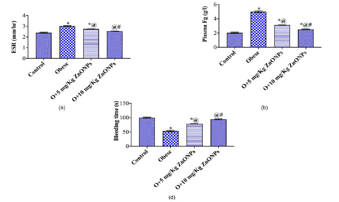
The treatment of obese rats with ZnONPs relieved the significant increase in WBCs count, ESR, and platelets count. On the other hand, ZnONPs combated the decrease in RBCs count and lymphocyte % (Figure 4).

Effect of ZnONPs on blood (a) RBCs count, (b) MCV, (c) lymphocytes count, (d) WBCs count and platelets count of obese rats. Each value represents the mean of 8 animals ±SE. Statistical analysis was performed using one-way ANOVA followed by Tukey-Kramer multiple comparisons test. (* vs control group, @ vs obese group and # vs ZnONPs 5mg/kg) at p<0.05. O: Obese.
Bleeding time, ESR, Fibrinogen
The bleeding time decreased significantly in obese rats, while ESR and fibrinogen levels increased significantly. The treatment of obese rats with ZnONPs mitigated the previous parameters (Figure 5). We observed a positive correlation between ESR and inflammatory markers, BMI, and Fg (Figure 6). On the other hand, a negative correlation was recorded between ESR and bleeding time.

Effect of ZnONPs on (a) ESR, (b) Fg, and (d) bleeding time of obese rats. Each value represents the mean of 8 animals ±SE. Statistical analysis was performed using one-way ANOVA followed by Tukey-Kramer multiple comparisons test. (* vs control group, @ vs obese group and # vs ZnONPs 5mg/kg) at p<0.05. O: Obese.
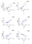
Correlation between ESR vs (a) TNF-alpha, (b) CRP, (c) IL-6, (d) BMI, (e) Fibrinogen and bleeding time.
Hepcidin gene expression
Figure 7 illustrates the means ±SE of fold change for hepcidin gene (HAMP) expression in the group sample. The results indicated a significant upregulation (P<0.05) of the gene in the obese samples compared with the control. On the other hand, a significant downregulation (P<0.05) of the gene was noticed in the samples concerned with obese rats treated with ZnONPs ( 5 or 10 mg/Kg ) compared to the obese group. Moreover, the hepcidin gene of obese rats treated with 10 mg/Kg ZnONPs was downregulated (P<0.05) relative to those given 5mg/Kg of ZnONPs.

Comparison of hepcidin gene (HAMP) expression fold change in adipose tissue of different studied groups. Each value represents the mean±SE. Statistical analysis was performed using one-way ANOVA followed by Tukey-Kramer multiple comparisons test. (* vs control group, @ vs obese group and # vs ZnONPs 5mg/kg) at p<0.05. O: Obese.
DISCUSSION
Being overweight and obesity are the most widespread metabolic disorders in the world. The waist circumference and BMI are important predictors of the metabolic abnormalities associated with metabolic syndrome [29]. It appeared that the decrease of BMI, waist circumference, and body weight gain in obese rats by ZnONPs can be referred to as controlling adipocyte hormones. Studies have shown that adiponectin also helps to manage weight by controlling the levels of glucose within the body and by breaking down fatty acids [30]. Total adiponectin levels increased, and insulin resistance decreased significantly in those who followed a low-calorie diet-based weight-loss program for 16 weeks [31]. Our results indicate a significant decrease in plasma cholesterol and triglycerides in O+ ZnONPs (5mg or 10mg/kg) as compared to the obese group. El-Bahr and coauthors (2020) found that the supplementation of ZnO-NPs at doses of 30 or 60 mg/kg in the diet of Japanese quails led to a significant decrease in triglycerides [32]. Zinc's presence in the structure of metalloenzymes involved in lipid metabolism may account for the hypolipidemic impact of ZnO-NPs [33] or decreasing lipid absorption from food [34]. Moreover, The hypolipidemic effect of ZnONPs in obese rats can be attributed to the enhancement of adiponectin levels since it increases fatty acid oxidation in muscle and reduces free fatty acids and triglycerides [35]. Since elevated lipid concentrations affect the lipid composition of the erythrocyte membrane, leading to increased erythrocyte fragility, with consequent leaking of cellular material like hemoglobin, ZnONPs appear to have enhanced blood cells indices via its hypolipidemic function [36].
The bleeding time increased significantly in the obese rats given ZnONPs when compared with the obese group. It was suggested that visceral obesity is related to accelerated blood coagulation in addition to disrupted metabolism of lipids [37]. The bleeding time decreased significantly in obese groups associated with a significant increase in the platelet count. It was reported that feeding animals with a high-fat, high-carbohydrate diet led to a rise in the platelets count in the blood with change in their membrane lipids and phosphorylation of platelet contractile proteins associated with an increase of blood clotting [38].
The present study showed an increase in free radical damage in obese rats as evidenced by an increase in MDA level and a decrease in GSH level. MDA is a product indicative of lipid peroxidation. Increased lipid peroxidation is thought to be a consequence of oxidative stress, which occurs when the dynamic balance between the prooxidant and antioxidant mechanism is impaired. It is known that hyperlipidemic states are associated with altered physical properties of cellular membranes [39] which may facilitate the escape of free radicals from the mitochondrial electron transport chain. Regarding GSH properties, it plays a vital role as an antioxidant and as a scavenger of oxidant species [40]. In this case, ZnONPs acted as antioxidants by decreasing MDA and raising GSH levels. Inducing metallothioneins, a potent scavenger of free radicals, protecting sulfhydryl groups from oxidation, maintaining intracellular levels of reduced glutathione, and reducing the production of lipid peroxidation products are only some of the ways in which zinc exerts its antioxidant function [41-43]. It was reported that antioxidants inhibit the accumulation of lipid peroxidation products during oxidative stress and protect the RBCs and hemoglobin from alteration [44]. The maintenance of Hb and RBCs by ZnONPs in obese rats can be attributed to their antioxidant and anti-inflammatory properties. A negative correlation was found between MDA level in blood and Hb, which may indicate an inverse relationship between MDA and Hb, or MDA had an adverse effect on Hb. On the other hand, a positive correlation between MDA and both adipose iron, CRP, ESR, and cholesterol was observed.
Obesity is associated with increased concentrations of circulating inflammatory cytokines, which are produced by the adipose tissue as TNF-α and IL6 that contribute to the inflammatory process [45].TNF -α and IL6 estimate the liver to produce fibrinogen and CRP, which in turn increases ESR level [46]. The erythrocyte sedimentation rate is a simple inflammatory marker [47]. Oxidative stress is strongly associated with CRP levels [48]. TNF-α is one of the proinflammatory cytokines that reportedly inhibit the generation of glycophorin cells, and decreased differentiation of erythroid cells exacerbates ineffective erythropoiesis [49]. It appears that the treatment of obese rats with ZnONPs maintains RBCs count and hemoglobin level via its anti-inflammatory properties since they lowered inflammatory markers, TNF-α, IL6, and CPR. Here, the correlation analysis showed a positive correlation between ESR and inflammatory markers (CRP, IL6, TNF-α), BMI, and fibrinogen. Moreover, ZnONPs mitigate the increase of WBCs and platelets observed in obese rats. Both elevation of WBCs and platelets are considered biomarkers of inflammation and they are associated with several chronic conditions [50,51].
Iron dysregulation is a potential contributor to the pathology of obesity-related metabolic complications [52,53]. The connotation amid increased iron overburden and inflammation or oxidative stress had been reported. Hepcidin production is highly prompted by inflammation and iron excess [54]. Here, the obese rats showed a significant increase in adipose tissue iron content and expression of the hepcidin gene, while plasma iron and blood Hb levels lowered significantly, which may indicate the obese rats suffer from anemia and oxidative stress due to iron overload. Hepcidin is the central regulator of systemic iron homeostasis. Iron-restricted erythropoiesis and anemia result from high hepcidin levels, which prevent iron absorption in the intestines and iron recycling in macrophages [55]. Inflammatory or chronic illness anemia is characterized by low blood iron, increased iron storage as seen by raised ferritin levels, and low transferrin levels. What's more, we discovered that hepcidin and IL-6 levels are positively correlated with one another [56]. Numerous inflammatory and degenerative disorders are associated with elevated levels of serum ferritin, which is thought to be a leaky indicator from damaged cells [57]. It has been suggested that ferritin functions as a local cytokine and that activation of MAPK-triggered NF-κB increases the production of proinflammatory mediators, including inducible nitric oxide synthase [58]. It has been established that IL-6 is both a required and sufficient cytokine for the induction of hepcidin during inflammation and that the IL-6-hepcidin axis is responsible for the hypoferremia seen in inflammatory diseases [59]. The results of the correlation demonstrated an inverse relationship between iron content in adipose tissue and both adiponectin, bleeding time, and GSH. Moreover, a proportional relationship between iron content in adipose tissue and MDA, BMI, CRP, and ESR was observed.
Disturbances in lipid metabolism and oxidative stress due to iron excess have been seen in rats, with endogenous antioxidant levels being significantly lowered and mild lipid peroxidation damage occurring [60]. Our results showed that the treatment of obese rats with ZnONPs affects iron homeostasis, as evidenced by a significant decrease in the iron content of adipose tissue and an increase in blood iron and hemoglobin levels. It was discovered that zinc level strongly affects intestinal absorption and systemic use of iron via regulation of iron transporter expression [61]. Insufficient zinc can cause abnormalities in proteins that transport, store, and regulate iron, leading to an increase in an iron buildup [62]. It is possible to suggest that supplementation with ZnONPs would mitigate the anemia and other associated consequences observed in the experimental protocol.
Limitations
The study you provided has a comprehensive analysis of the effects of ZnONPs (Zinc Oxide Nanoparticles) treatment on various parameters in obese rats. While the study seems to provide valuable insights, there are some limitations and considerations to keep in mind; (1) the study was conducted on obese rats, which may not perfectly represent human physiology and response to interventions. While animal models are useful for initial studies, the results might not directly translate to humans. (2)The study examines two different doses of ZnONPs (5 mg/kg and 10 mg/kg). While the study discusses the effects of these doses, it doesn't elaborate on why these specific doses were chosen or the potential dose-response relationship. (3) While the study observes correlations between parameters, it doesn't delve into the underlying mechanisms behind these correlations. Mechanistic insights are crucial for a comprehensive understanding of the observed effects. (4) While correlations provide insights into potential relationships between variables, they do not imply causation. Further experimentation, such as intervention studies, is needed to establish causative links. (5) some related clotting parameters was not measured as D-dimer, Arachidonic acid, Thromboxane A2, collagen and thrombin.
In summary, while the study provides valuable information about the effects of ZnONPs treatment on various parameters in obese rats, further research, including human clinical trials, mechanistic studies, and consideration of potential long-term effects, is necessary before drawing definitive conclusions about the therapeutic potential of ZnONPs for obesity-related anemia in humans.
CONCLUSION
In conclusion, the treatment of obese rats with ZnONPs combated anemia via regulation of iron homeostasis or hepcidin gene expression, maintaining hemoglobin concentration, and decreasing oxidative stress or inflammatory markers. Hepcidin may provide a promising therapeutic target in anemia-linked iron-dysregulated metabolism. Moreover, the positive role of ZnONPs in controlling anemia in obese rats can be attributed to a reduction in BMI since BMI was correlated positively with iron content in adipose tissue.
We can conclude that the integrated of nanoparticles in the therapeutic strategies would provide new pathway to increase their potency against chronic disorders like anemia. To the best of our knowledge, we appear to be the pioneers in discovering that zinc nanoparticles effectively alleviated anemia triggered by a high-fat/sucrose diet by modulating the hepcidin gene and addressing iron-overload conditions.
Acknowledgments
The authors would like to thank Science Shake Inc. for conducting proofreading and English language editing (https://science-shake.com).
REFERENCES
- 1 Blüher M. Obesity: global epidemiology and pathogenesis. Nat Rev Endocrinol. 2019 May 27;15(5):288-98.
- 2 Eftekhari MH, Mozaffari-Khosravi H, Shidfar F. The relationship between BMI and iron status in iron-deficient adolescent Iranian girls. Public Health Nutr. 2009 Dec 12;12(12):2377-81.
- 3 Alshwaiyat N, Ahmad A, Wan Hassan WMR, Al jamal H. Association between obesity and iron deficiency (Review). Exp Ther Med. 2021 Sep 7;22(5):1268.
- 4 Moore Heslin A, O’Donnell A, Buffini M, Nugent AP, Walton J, Flynn A, et al. Risk of Iron Overload in Obesity and Implications in Metabolic Health. Nutrients. 2021 May 2;13(5):1539.
- 5 Chung B, Matak P, McKie AT, Sharp P. Leptin increases the expression of the iron regulatory hormone hepcidin in HuH7 human hepatoma cells. J Nutr. 2007 Nov 1;137(11):2366-70.
- 6 Amirkhizi F, Siassi F, Minaie S, Djalali M, Rahimi A, Chamari M. Is obesity associated with increased plasma lipid peroxidation and oxidative stress in women? ARYA Atheroscler. 2007 Dec 8;2(4):189-92.
- 7 Yetisgin AA, Cetinel S, Zuvin M, Kosar A, Kutlu O. Therapeutic nanoparticles and their targeted delivery applications. Molecules. 2020 May 8;25(9):2193.
- 8 Naseer B, Srivastava G, Qadri OS, Faridi SA, Islam RU, Younis K. Importance and health hazards of nanoparticles used in the food industry. Nanotechnol Rev. 2018 Dec 19;7(6):623-41.
- 9 Aziz ZAA, Mohd-Nasir H, Ahmad A, Siti SH, Peng WL, Chuo SC, et al. Role of Nanotechnology for Design and Development of Cosmeceutical: Application in Makeup and Skin Care. Front Chem. 2019 Nov 13;7.
- 10 Akshaya K, Rajasekar A, Rajeshkumar S. Antioxidant activity of zinc oxide nanoparticles synthesised using grape seed extract: An in vitro study. Plant Cell Biotechnol Mol Biol. 2020;21(29-30):21-9.
- 11 Mohapatra S, Leelavathi L, Rajeshkumar S., Sri Sakthi D, Javyashri P. Assessment of Cytotoxicity, Anti-Inflammatory and Antioxidant Activity of Zinc Oxide Nanoparticles Synthesized Using Clove and Cinnamon Formulation - An In-Vitro Study. J Evol Med Dent Sci. 2020 Jun 22;9(25):1859-64.
- 12 Prasad AS. Discovery of Zinc for Human Health and Biomarkers of Zinc Deficiency. In: Molecular, Genetic, and Nutritional Aspects of Major and Trace Minerals. Elsevier; 2017. p. 241-60.
- 13 Prasad AS. Impact of the discovery of human zinc deficiency on health. J Trace Elem Med Biol. 2014 Oct;28(4):357-63.
- 14 Hayden SJ, Albert TJ, Watkins TR, Swenson ER. Anemia in critical illness: Insights into etiology, consequences, and management. Am J Respir Crit Care Med. 2012 May 15;185(10):1049-57.
- 15 Wieringa FT, Dahl M, Chamnan C, Poirot E, Kuong K, Sophonneary P, et al. The high prevalence of anemia in cambodian children and women cannot be satisfactorily explained by nutritional deficiencies or hemoglobin disorders. Nutrients. 2016 Jun 7;8(6):348.
- 16 Angelova MG, Petkova-Marinova TV, Pogorielov MV, Loboda AN, Nedkova-Kolarova VN, Bozhinova AN. Trace element status (iron, zinc, copper, chromium, cobalt, and nickel) in iron-deficiency anaemia of children under 3 years. Anemia. 2014;2014.
- 17 Elseidy AMA, Bashandy SA, Ibrahim FAA, El-Rahman SSA, Farid O, Moussa S, et al. Zinc oxide nanoparticles characterization and therapeutic evaluation on high fat / sucrose diet induced-obesity. Egypt J Chem. 2022 Jan 19;65(9):497-511.
- 18 Abdelmottaleb SAM, Ibrahim FAA, Elbaset MA, Aziz SW, Morsy FA, Abdellatif N, et al. Goldenberry (Physalis peruviana) alleviates hepatic oxidative stress and metabolic syndrome in obese rats. J Appl Pharm Sci. 2022 Nov 5;12(11):138-50.
- 19 Abdelbaset M, Safar MM, Mahmoud SS, Negm SA, Agha AM. Red yeast rice and coenzyme Q 10 as safe alternatives to surmount atorvastatin-induced myopathy in hyperlipidemic rats. Can J Physiol Pharmacol. 2014 Jun;92(6):481-9.
- 20 Elbaset MA, Nasr M, Ibrahim BMM, Ahmed‐Farid OAH, Bakeer RM, Hassan NS, et al. Curcumin nanoemulsion counteracts hepatic and cardiac complications associated with high‐fat/high‐fructose diet in rats. J Food Biochem. 2022 Dec 27;46(12):1-16.
- 21 Bashandy SAE, Alaamer A, Moussa SAA, Omara EA. Role of zinc oxide nanoparticles in alleviating hepatic fibrosis and nephrotoxicity induced by thioacetamide in rats. Can J Physiol Pharmacol. 2018 Apr;96(4):337-44.
- 22 Moussa SAA, Ibrahim FAA, Elbaset MA, Aziz SW, Abdellatif NA, Attia AM, et al. Efficacy of goldenberry extract in chelated iron overload induced by obesity: Novel safety concept for the treatment of iron overloads diseases. J Appl Biol Biotechnol. 2022;10(4):92-100.
- 23 El-Gendy AA, Abbas AM. Effect of omega-3 fatty acids on haemostatic functions in urocortin-treated obese rats. J Physiol Biochem. 2014 Sep 26;70(3):809-20.
- 24 Abdel-Salam OME, Sleem AA, Baset Mohamed Sayed MA El, Youness ER, Shaffie N. Neuroprotective effects of low dose anandamide in pentylenetetrazole-induced kindling in rats. Biomed. Pharmacol. J. 2019 Mar 28;12(1):25-40.
- 25 Abdel-Salam OME, Sleem AA, Abd El Baset M, Sayed M, Khadrawy YA, Morsy FA. Cannabis sativa increases seizure severity and brain lipid peroxidation in pentylenetetrazole-induced kindling in rats. Biomed. Pharmacol. J. 2018 Sep 28;11(3):1187-97.
- 26 Imeryuz N, Tahan V, Sonsuz A, Eren F, Uraz S, Yuksel M, et al. Iron preloading aggravates nutritional steatohepatitis in rats by increasing apoptotic cell death. J Hepatol. 2007 Dec;47(6):851-9.
- 27 Livak KJ, Schmittgen TD. Analysis of relative gene expression data using real-time quantitative PCR and the 2-ΔΔCT method. Methods. 2001 Dec;25(4):402-8.
- 28 Elbaset MA, Salem RS, Ayman F, Ayman N, Shaban N, Afifi SM, et al. Effect of Empagliflozin on Thioacetamide-Induced Liver Injury in Rats: Role of AMPK/SIRT-1/HIF-1α Pathway in Halting Liver Fibrosis. Antioxidants. 2022 Oct 30;11(11):2152.
- 29 Piqueras P, Ballester A, Durá-Gil JV, Martinez-Hervas S, Redón J, Real JT. Anthropometric Indicators as a Tool for Diagnosis of Obesity and Other Health Risk Factors: A Literature Review. Front Psychol. 2021 Jul 9;12:1-19.
- 30 Nigro E, Scudiero O, Monaco ML, Palmieri A, Mazzarella G, Costagliola C, et al. New Insight into Adiponectin Role in Obesity and Obesity-Related Diseases. Biomed Res Int. 2014 Jul 7;2014:1-14.
- 31 Foula WH, Emara RH, Eldeeb MK, Mokhtar SA, El-Sahn FA. Effect of a weight loss program on serum adiponectin and insulin resistance among overweight and obese premenopausal females. J Egypt Public Health Assoc. 2020 Dec 1;95(1):32.
- 32 El-Bahr SM, Shousha S, Albokhadaim I, Shehab A, Khattab W, Ahmed-Farid O, et al. Impact of dietary zinc oxide nanoparticles on selected serum biomarkers, lipid peroxidation and tissue gene expression of antioxidant enzymes and cytokines in Japanese quail. BMC Vet Res. 2020 Dec 23;16(1):349.
- 33 Al-Daraji HJ, Amen MHM. Effect of dietary zinc on certain blood traits of broiler breeder chickens. Int J Poult Sci. 2011 Sep 15;10(10):807-13.
- 34 Ibrahim D, Ali H, El-Mandrawy S. Effects of Different Zinc Sources on Performance, Bio Distribution of Minerals and Expression of Genes Related to Metabolism of Broiler Chickens. Zagazig Vet J. 2017 Sep 1;45(3):292-304.
- 35 Fruebis J. Proteolytic cleavage product of 30-kDa adipocyte complement-related protein increases fatty acid oxidation in muscle and causes weight loss in mice. Proceedings of the National Academy of Sciences. 2001 Feb 13;98(4):2005-10.
- 36 Dimeski G, Mollee P, Carter A. Increased lipid concentration is associated with increased hemolysis [5]. Clin Chem. 2005 Dec 1;51(12):2425.
- 37 Kaji N, Nagakubo D, Hashida SI, Takahashi S, Kuratani M, Hirai N, et al. Shortened blood coagulation times in genetically obese rats and diet-induced obese mice. J. Vet. Med Sci. 2013;75(9):1245-8.
- 38 Barrachina MN, Morán LA, Izquierdo I, Casanueva FF, Pardo M, García Á. Analysis of platelets from a diet-induced obesity rat model: elucidating platelet dysfunction in obesity. Sci Rep. 2020 Aug 4;10(1):13104.
- 39 Engelmann B, Streich S, Schönthier UM, Richter WO, Duhm J. Changes of membrane phospholipid composition of human erythrocytes in hyperlipidemias. I. Increased phosphatidylcholine and reduced sphingomyelin in patients with elevated levels of triacylglycerol-rich lipoproteins. Biochimica et Biophysica Acta (BBA)/Lipids and Lipid Metabolism. 1992 Nov;1165(1):32-7.
- 40 Gaucher C, Boudier A, Bonetti J, Clarot I, Leroy P, Parent M. Glutathione: Antioxidant Properties Dedicated to Nanotechnologies. Antioxidants. 2018 Apr 27;7(5):62.
- 41 Suntres ZE, Lui EMK. Biochemical mechanism of metallothionein-carbon tetrachloride interaction in vitro. Biochem Pharmacol. 1990 Mar;39(5):833-40.
- 42 Bray TM, Bettger WJ. The physiological role of zinc as an antioxidant. Free Radic Biol Med. 1990;8(3):281-91.
- 43 Bao B, Ahmad A, Azmi A, Li Y, Prasad A, Sarkar FH. The Biological Significance of Zinc in Inflammation and Aging. In: Inflammation, Advancing Age and Nutrition: Research and Clinical Interventions. Elsevier; 2013. p. 15-27.
- 44 Pinyaev SI, Kuzmenko TP, Revina NV, Parchaykina MV, Pronin AS, Syusin IV, et al. Influence of Resveratrol on Oxidation Processes and Lipid Phase Characteristics in Damaged Somatic Nerves. Biomed Res Int. 2019 Dec 11;2019:1-9.
- 45 Boutens L, Stienstra R. Adipose tissue macrophages: going off track during obesity. Diabetologia. 2016 May 3;59(5):879-94.
- 46 Kang YE, Kim JM, Joung KH, Lee JH, You BR, Choi MJ, et al. The roles of adipokines, proinflammatory cytokines, and adipose tissue macrophages in obesity-associated insulin resistance in modest obesity and early metabolic dysfunction. López Lluch G, editor. PLoS One. 2016 Apr 21;11(4):e0154003.
- 47 Sharma A, Kumar A, Jha A, Agarwal A, Misra A. The impact of obesity on inflammatory markers used in the assessment of disease activity in rheumatoid arthritis - a cross-sectional study. Reumatologia. 2020;58(1):9-14.
- 48 Park S, Kim M, Paik JK, Jang YJ, Lee SH, Lee JH. Oxidative stress is associated with C-reactive protein in nondiabetic postmenopausal women, independent of obesity and insulin resistance. Clin Endocrinol (Oxf). 2013 Jul;79(1):65-70.
- 49 Libani IV, Guy EC, Melchiori L, Schiro R, Ramos P, Breda L, et al. Decreased differentiation of erythroid cells exacerbates ineffective erythropoiesis in β-thalassemia. Blood. 2008 Aug 1;112(3):875-85.
- 50 Wirth MD, Sevoyan M, Hofseth L, Shivappa N, Hurley TG, Hébert JR. The Dietary Inflammatory Index is associated with elevated white blood cell counts in the National Health and Nutrition Examination Survey. Brain Behav Immun. 2018 Mar;69:296-303.
- 51 Chen Y, Zhong H, Zhao Y, Luo X, Gao W. Role of platelet biomarkers in inflammatory response. Biomark Res. 2020 Dec 2;8(1):28.
- 52 Forouhi NG, Harding AH, Allison M, Sandhu MS, Welch A, Luben R, et al. Elevated serum ferritin levels predict new-onset type 2 diabetes: Results from the EPIC-Norfolk prospective study. Diabetologia. 2007 Apr 5;50(5):949-56.
- 53 Sharawy N, Imam AAA, Aboulhoda BE, Khalifa MM, Morcos GNB, Abd Algaleel WA, et al. Iron dyshomeostasis and time-course changes in iron-uptake systems and ferritin level in relation to pro-inflammatory microglia polarization in sepsis-induced encephalopathy. Front Physiol. 2022 Aug 12;13.
- 54 Walter PB, Fung EB, Killilea DW, Jiang Q, Hudes M, Madden J, et al. Oxidative stress and inflammation in iron-overloaded patients with β-thalassaemia or sickle cell disease. Br J Haematol. 2006 Oct;135(2):254-63.
- 55 Pagani A, Nai A, Silvestri L, Camaschella C. Hepcidin and Anemia: A Tight Relationship. Front Physiol. 2019 Oct 9;10:1-7.
- 56 Basseri RJ, Nemeth E, Vassilaki ME, Basseri B, Enayati P, Shaye O, et al. Hepcidin is a key mediator of anemia of inflammation in Crohn’s disease. J Crohns Colitis. 2013 Sep;7(8):e286-91.
- 57 Kell DB, Pretorius E. Serum ferritin is an important inflammatory disease marker, as it is mainly a leakage product from damaged cells. Metallomics. 2014;6(4):748-73.
- 58 Ruddell RG, Hoang-Le D, Barwood JM, Rutherford PS, Piva TJ, Watters DJ, et al. Ferritin functions as a proinflammatory cytokine via iron-independent protein kinase C zeta/nuclear factor kappaB - Regulated signaling in rat hepatic stellate cells. Hepatology. 2009;49(3):887-900.
- 59 Nemeth E, Rivera S, Gabayan V, Keller C, Taudorf S, Pedersen BK, et al. IL-6 mediates hypoferremia of inflammation by inducing the synthesis of the iron regulatory hormone hepcidin. J Clin Invest. 2004 May 1;113(9):1271-6.
- 60 Zhang L, Dai X, Wang L, Cai J, Shen J, Shen Y, et al. Iron overload accelerated lipid metabolism disorder and liver injury in rats with non-alcoholic fatty liver disease. Front Nutr. 2022 Oct 11;9:1-13.
- 61 Kondaiah P, Palika R, Mashurabad P, Singh Yaduvanshi P, Sharp P, Pullakhandam R. Effect of zinc depletion/repletion on intestinal iron absorption and iron status in rats. J Nutr Biochem. 2021 Nov;97:108800.
- 62 Niles BJ, Clegg MS, Hanna LA, Chou SS, Momma TY, Hong H, et al. Zinc deficiency-induced iron accumulation, a consequence of alterations in iron regulatory protein-binding activity, iron transporters, and iron storage proteins. J. Biol. Chem. 2008 Feb;283(8):5168-77.