 MMP-26 as a Complementary Diagnostic Biomarker in Nodular Thyroid Lesions
Abstract
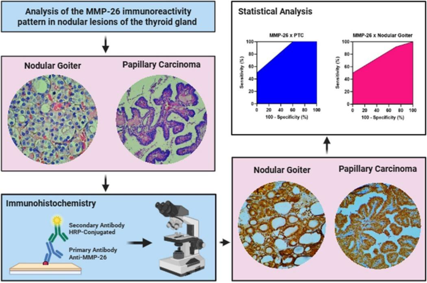
Thyroid cancer (TC) is the seventh most common cancer and the twenty-fourth most deadly, according to GLOBOCAN estimates for 2022. The diagnosis of TC is confirmed by histopathological analysis after surgical biopsy, in addition to previous ultrasound and cytopathological evaluations. Despite this, TC still lacks a definitive molecular classification, capable of not only identifying tumors in terms of their cellular origin, but also distinguishing them in terms of aggressiveness, benign or malignant nature and prognosis. This study investigated the potential of matrix metalloproteinase 26 (MMP-26) as a complementary biomarker for the diagnosis of nodular thyroid lesions. Fifty histological blocks, including healthy thyroid tissue, nodular goiter and papillary thyroid carcinoma (PTC), were analyzed using immunohistochemistry. The results showed significant differences in MMP-26 immunoreactivity between healthy thyroid tissue, nodular goiter and PTC, as shown by Spearman's analysis (p<0,001), Chi-square (p<0,001) and logistic regression (p<0,001). However, there were no significant differences between MMP-26 immunoreactivity and clinical-pathological data. MMP-26 showed good sensitivity (93,3% and 100%) and negative predictive values (90,91% and 100%) but low specificity (28,57% and 40%) for identifying nodular goiter and PTC, respectively. It is concluded that MMP-26 is a promising complementary biomarker for the diagnosis of thyroid tumors, highlighting its participation in the pathogenesis of lesions. However, more research is needed to determine its real impact as a diagnostic and prognostic biomarker.
Keywords:
Matrix Metalloproteinase; Tumor Biomarkers; Thyroid Gland Neoplasms; Immunohistochemistry.
HIGHLIGHTS
There was a significant expression of MMP-26 in nodular goiter and papillary thyroid carcinoma.
MMP-26 showed good sensitivity for identifying nodular goiter and papillary thyroid carcinoma.
The results suggest the involvement of MMP-26 in the pathogenesis of thyroid tumors.
MMP-26 is a promising complementary biomarker for the diagnosis of nodular thyroid lesions.
INTRODUCTION
The thyroid, a fundamental endocrine gland for body homeostasis, regulates various physiological activities through the synthesis of the hormones triiodothyronine (T3) and thyroxine (T4) [1]. Imbalances in this process, induced by genetic mutations and behavioral risk factors, can lead to the development of nodular lesions, which prevalence varies between 50% and 67% when detected on ultrasound [2, 3]. Although the majority of thyroid nodules are benign, approximately 5% are carcinomas, with a predominance of well-differentiated lesions [2, 4-6]. According to GLOBOCAN estimates for 2022, thyroid cancer is the seventh most incident and the twenty-fourth most deadly, with a low mortality rate due to the good prognosis of well-differentiated lesions [7].
The diagnosis of TC is confirmed by histopathological analysis after surgical biopsy, in addition to previous ultrasound and cytopathological evaluations [8]. However, TC still lacks a definitive molecular classification capable of identifying tumors in terms of their cellular origin, aggressiveness, benign or malignant nature and prognosis [9]. The investigation of the genetic etiology of tumors through the analysis of oncogenic mutations in BRAF and RAS genes, as well as the search for biomarkers that aid in the diagnosis and prognosis of TC, has been an area of intense research, with the aim of improving the precision and personalization of treatment [10, 11].
Matrix metalloproteinases (MMPs) are a family of zinc-dependent endopeptidases involved in the degradation and remodeling of the extracellular matrix, processes that are essential for tumor progression [12]. MMP-26, a matrilysin devoid of a hemopexin (Hpx) domain and peptide ligand, acts by cleaving type IV collagen, fibrinogen and other constituents of the tissue matrix, as well as activating pro-MMP-9 [13]. Studies have shown that MMP-26 expression is altered in various types of cancer, including breast, prostate and endometrial cancers [14, 15].
However, the role of MMP-26 in the context of thyroid cancer remains poorly understood. Although some studies have suggested that MMP-26 may be involved in the tumor progression of TC, its mechanisms of action and its relationship with the clinical-pathological characteristics of patients remains unclear [12]. Given this gap in knowledge, this study set out to investigate the potential of MMP-26 as a complementary biomarker for the diagnosis of nodular thyroid lesions, seeking to answer the following research question: is the expression of MMP-26 associated with the clinical-pathological characteristics and tumor staging of patients with papillary thyroid carcinoma?
To answer this question, we analyzed the immunoreactive profile of MMP-26 in healthy thyroid tissues, nodular goiter and papillary thyroid carcinoma, seeking to identify significant differences in MMP-26 expression between these conditions and correlate them with the clinical-pathological characteristics of the patients and the tumor staging of the PTCs. The results of this study may contribute to a better understanding of the pathophysiology and microenvironment of thyroid lesions, as well as to the development of new diagnostic and treatment strategies for thyroid cancer.
MATERIAL AND METHODS
We selected 50 histological samples from the Pathology Department of the Hospital das Clínicas da Universidade Federal de Pernambuco (HC/UFPE). All were from 2015 to 2017 and presented thyroidectomy material compatible only with nodular thyroid lesions, including 15 cases of nodular goiter and 25 of papillary carcinoma, in addition to 10 cases of healthy adjacent thyroid tissue. Immunohistochemistry (IHC) was carried out at the Cytological and Molecular Research Laboratory (LPCM), and to complement the IHC and statistical analyses, information was collected on the sex of the patients and the staging of the malignant tumors. Histopathological diagnosis and tumor staging were carried out by HC/UFPE pathologists. This study was approved by the UFPE Research Ethics Committee (CAAE number: 47863515.0.0000.5208).
Immunohistochemistry
For immunohistochemistry, the slides made from the histological samples were deparaffinized and rehydrated, followed by antigen recovery in sodium citrate buffer (10 mM, pH 6.0). Endogenous peroxidase and non-specific proteins were blocked following the instructions in the Envision Flex kit (Dako Denmark A/S) and the slides were then incubated overnight at 4°C with polyclonal IgG anti-MMP-26 primary antibody (FNab05242, 100 μg, Fine Test®, 1:100). After incubation with secondary antibody conjugated to HRP (horseradish peroxidase), immunostaining with 3,3-diaminobenzidine (DAB) and counterstaining with Harris hematoxylin, the slides were observed under an optical microscope. The negative control was carried out on thyroid histological samples in the absence of the primary anti-MMP-26 antibody, while the positive control was carried out on endometrial tissue samples.
Image Capture and Analysis
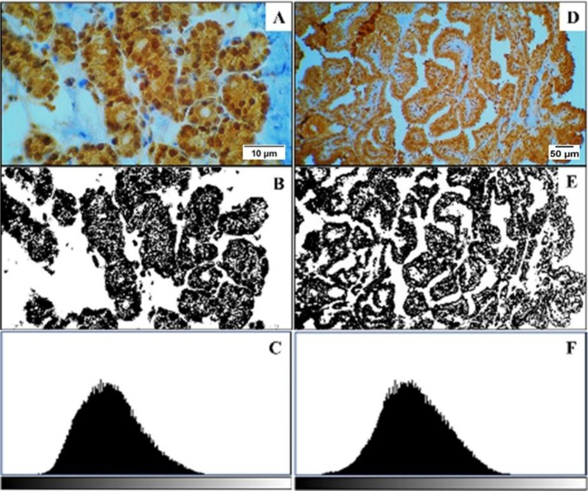
To analyze the immunoreactivity, a Kasvi® optical microscope (K55-OIT) with a CMOS sensor camera (½.33” 16MP HDMI FullHD 100x) was used to capture and record the images. The analysis method was semi-quantitative, by area and intensity of immunoreaction, as shown in Figure 1, using ImageJ software and the IHC TOOL BOX plugin. The average nuclear and/or cytoplasmic reaction intensity pattern was estimated by measuring the number of pixels corresponding to the DAB-positive areas. The values were analyzed and graphically represented in histograms according to a gray scale from 0 to 255, where 0 corresponded to black and 255 to white.
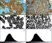
Analysis of the MMP-26 immunoreactivity pattern in thyroid gland lesions (a) MMP-26 immunoreactivity in nodular goiter (400✕); (b) Marking area (%) for MMP-26 in nodular goiter; (c) Histogram for MMP-26 reaction in nodular goiter, ranging from 0 to 255 on the gray scale; (d) MMP-26 immunoreactivity in PTC (100✕); (e) Marking area (%) for MMP-26 in PTC; (f) Histogram for MMP-26 reaction in PTC, ranging from 0 to 255 on the gray scale.
Finally, the values calculated for intensity and reaction area were adapted to different scoring scales, distributed over several levels. Intensity was interpreted based on four gradations, where: 0 is absent (0), between 1 and 85 weak (1), between 86 and 170 moderate (2) and above 170 strong (3). The percentage area of reaction was estimated using the following scale for the marked cells: 1 (up to 25%), 2 (26% to 50%), 3 (51% to 75%) and 4 (>75%). Finally, the final marking positivity score was obtained by multiplying the values assigned for the area and intensity of immunoreaction, ranging from 0 to 12, in the following intervals: 0 (absent), 1 to 4 (mild), 5 to 8 (intermediate) and 9 to 12 (marked).
Statistical Analysis
Statistical analysis was carried out using softwares Jamovi, v. 2.3.28 and GraphPad Prism®, v. 8.0.2. Initially, the Shapiro-Wilk test was realized to assess the normality of data distribution. In the absence of normality, non-parametric tests were applied. The Chi-Square test was used to compare MMP-26 expression between the groups (healthy thyroid tissue, nodular goiter and papillary thyroid carcinoma) and with the clinical-pathological characteristics of the patients. The strength of association between the variables was determined by Spearman's correlation coefficient, while logistic regression was applied to estimate the probability of influence of the marking pattern on tumor staging. Analyses of sensitivity, specificity and positive (PPV) and negative (NPV) predictive values were carried out, with the preparation of a Receiver Operating Characteristic (ROC) curve. The significance level adopted was 5% (p < 0.05).
RESULTS
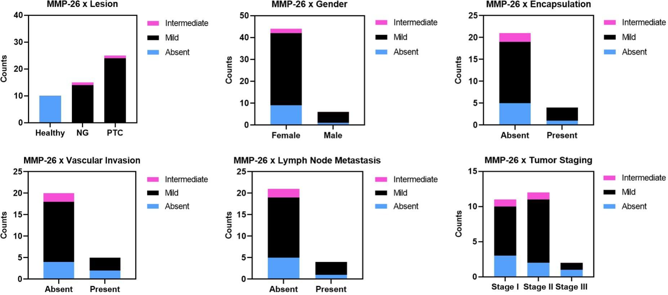
Initially, a descriptive analysis of the clinical-pathological information was carried out, including gender, tumor staging and the presence or absence of encapsulation, vascular infiltration and lymph node metastasis. The observed (OF) and expected frequencies (EF) of cases with absent, mild, or intermediate MMP-26 immunoreactivity and their distribution by gender, tumor stage, and presence or absence of encapsulation, vascular invasion and lymph node metastasis were analyzed using the Chi-Square test (Table 1). Significant differences were observed when comparing OF and EF frequencies between MMP-26 immunoreactivity and thyroid lesions, whether nodular goiter or PTC, as well as healthy adjacent thyroid tissue. There were no statistically significant relationships between the other variables.
The distribution of clinical-pathological data, separated by MMP-26 immunoreactivity, was represented graphically by stacked bars (Figure 2).
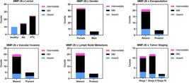
Distribution of clinical-pathological data by MMP-26 immunoreactivity. The graphs show the distribution of thyroid lesions and healthy adjacent thyroid tissue, gender, tumor staging and presence or absence of tumor encapsulation, vascular invasion and lymph node metastasis by levels of Absent, Mild and Intermediate immunoreactivity of MMP-26. NG: Nodular Goiter. PTC: Papillary Thyroid Carcinoma.
The analysis of the MMP-26 immunoreactivity pattern only considered nuclear and/or cytoplasmic reactions in thyroid follicular cells, and there were notable variations in the levels of intensity of expression and percentage area of MMP-26 marking between the different types of sample, whether healthy tissue, benign lesions with cases of nodular goiter, or malignant lesions with PTCs (Table 2). The final score, calculated by multiplying the intensity and area of labeling values, was between absent (0) and intermediate (5 to 8). No immunoreactivity for MMP-26 was observed in all the healthy tissue samples, while in nodular goiters and PTCs the predominant expression profile was mild (1 to 4), with no notable differences in frequency between the two lesions.
Spearman's correlation was applied to investigate the strength of the association between MMP-26 immunoreactivity, thyroid lesions and the clinical-pathological characteristics of the patients, as shown in Table 3. Spearman's Rho measures ranged from -1 to 1, with negative values indicating inversely proportional relationships and positive values directly proportional relationships. Statistically significant differences were observed when comparing MMP-26 expression and the different conditions of the thyroid gland, whether healthy or affected by nodular goiter or PTC. There were also significant associations between the presence or absence of lymph node metastasis and the presence or absence of vascular invasion. Correlations between tumor staging and the presence or absence of encapsulation, vascular invasion and lymph node metastasis also showed significant differences.
Correlation between MMP-26 immunoreactivity, thyroid lesions and clinical-pathological data.
Logistic regression assessed the probability of MMP-26 immunoreactivity influencing the thyroid lesion profile, whether benign or malignant, and statistical significance was observed (Table 4). When the probabilities associated MMP-26 expression with clinical-pathological variables, there were no significant differences. Odds ratio (OR) values analyzed the possibilities of MMP-26 expression being related to the presence or absence of encapsulation, vascular invasion and/or lymph node metastasis, as well as to different levels of tumor staging.
Regression analysis for MMP-26 immunoreactivity, thyroid lesions and clinico-pathological characteristics of the patients.
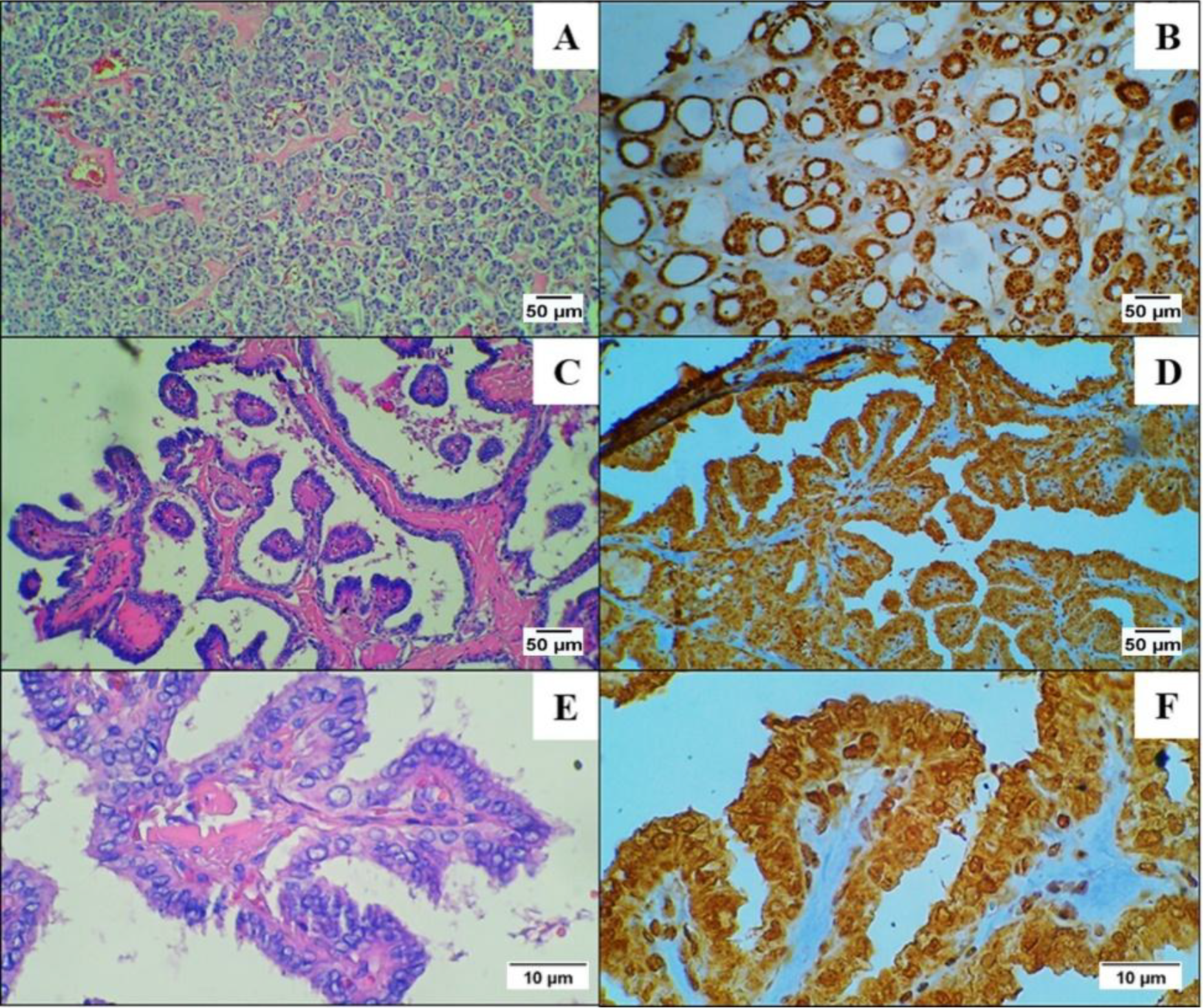
The histopathological pattern of the thyroid lesions, stained with Hematoxylin and Eosin (HE), was compared to the immunoreactivity for MMP-26 (Figure 3). There were no significant differences in expression intensity and marking area (%) between nodular goiter and PTC, with the final score (predominantly Mild, with only one Intermediate) being very similar for both lesions.
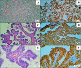
MMP-26 immunoreactivity in thyroid lesions. (a) Nodular goiter (HE, 100✕); (b) MMP-26 immunoreactivity in nodular goiter (100✕); (c) PTC (HE, 100✕); (d) MMP-26 immunoreactivity in PTC (100✕); (e) PTC (HE, 400✕); (f) MMP-26 immunoreactivity in PTC (400✕).
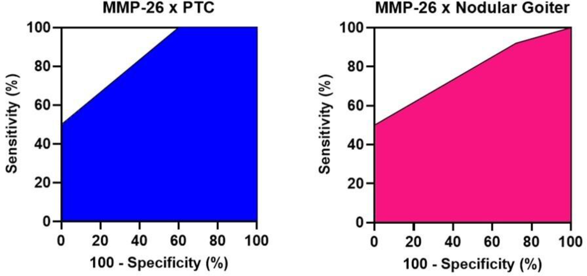
Analyses of sensitivity, specificity and positive (PPV) and negative predictive values (NPV) estimated the potential of MMP-26 for diagnosing nodular goiter and papillary thyroid carcinoma (Table 5). Reasonable performance of MMP-26 was observed, with good sensitivity and NPV measures for both nodular goiter and PTC diagnosis, but without significant differences in their performance profiles. The Youden’s Index (YI) estimated the optimum cut-off points for the ROC curves produced, while the Area Under the Curve (AUC) values made it possible to analyze the performance pattern of MMP-26 for each thyroid lesion.
ROC curves were drawn up to illustrate the performance of MMP-26 in both thyroid lesions, as shown in Figure 4. The ROC curves for both nodular goiter and PTC were very similar, with no significant differences between the two.
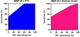
ROC curves for diagnostic performance of MMP-26 in thyroid lesions. PTC: Papillary Thyroid Carcinoma.
DISCUSSION
This study investigated the potential of MMP-26 as a complementary biomarker for the diagnosis of nodular thyroid lesions, comparing its expression in healthy thyroid tissues, nodular goiter and papillary thyroid carcinoma (PTC). The results revealed a significant increase in MMP-26 expression in nodular goiter and PTC tissues compared to healthy thyroid tissues, indicating a possible involvement of MMP-26 in the pathogenesis of nodular thyroid lesions.
As shown in the results, there was a predominance of nodules among female patients (Table 1), which may be associated with the effects of female sex hormones such as prolactin and estrogen [16, 17]. According to Zhang and coauthors (2022), the action of estrogen can favor the development of nodular lesions and goiter in women, stimulating the growth and proliferation of tumor cells [17]. Based on this, Piskór and coauthors (2021) suggest that MMP-26 may be involved in the development of estrogen-dependent cancers, including breast cancer, whose neoplastic cells expressing MMP-26 tend to have a more prominent malignant phenotype [13].
In addition, Nishi, Kuroda and Isaka (2013) suggest that MMP-26 is under-expressed in endometrial carcinomas. According to the authors, estrogen may not be directly involved in endometrial carcinogenesis, which would attenuate the expression of MMP-26, whose action depends on the transactivation of its gene promoters by estrogen receptors [18]. In contrast, Cheng and coauthors (2017) identified positive immunoreactivity for MMP-26 in prostate cancer and absent in hyperplasia, which demonstrates the versatility of expression of this biomarker in various lesions, whether benign or malignant and whether or not associated with estrogenic action [19].
When MMP-26 immunoreactivity was compared to other clinical-pathological characteristics, such as tumor staging and the presence or absence of encapsulation, vascular invasion and lymph node metastasis, no statistically significant differences were observed. This corroborates our hypothesis that positive MMP-26 expression alone is not enough to recognize specific thyroid lesions and should be associated with other diagnostic, aggressive and prognostic tumor biomarkers. Table 2 shows the absence of MMP-26 expression in healthy thyroid tissue and positive immunoreactivity in nodular goiter and PTC, although there were no significant differences between both.
Table 3 shows significant correlations between MMP-26 immunoreactivity and thyroid nodular lesions, with a strong association. The directly proportional profile of this correlation suggests that MMP-26 tends to be expressed more intensely in areas of nodular lesions, whether nodular goiter or PTC, than in healthy adjacent thyroid tissue. The regression analyses, in Table 4, show that the pattern of MMP-26 expression can directly influence the profile of thyroid lesions, without, however, distinguishing them between benign and malignant lesions.
When analyzing the potential correlation and influence of MMP-26 on clinical-pathological variables, no statistically significant differences were identified either. This reinforces our argument that, although it is efficient at detecting lesions, the low specificity of MMP-26 makes it unlikely to be associated with capsular and angiolymphatic invasion, lymph node metastasis and more advanced tumor stages, compatible with more clinically aggressive lesions.
Although the specificity of MMP-26 to distinguish between nodular goiter and PTC was not demonstrated in this study, as presented in Table 5, the identification of a significant increase in MMP-26 expression in nodular thyroid lesions compared to healthy tissues suggests a potential diagnostic value. The growing evidence of the involvement of MMPs in extracellular matrix remodeling and tumor progression in various types of cancer suggests that MMP-26 could be used as a complementary biomarker in conjunction with other diagnostic methods, such as cytopathology and ultrasound, to help identify nodular lesions with a higher risk of malignancy [19]. This is corroborated by the excellent sensitivity of MMP-26 for diagnosing nodular goiter and PTC, as demonstrated by the ROC curves in Figure 4.
MMP-26, in particular, has demonstrated proteolytic activity on components of the extracellular matrix, such as type IV collagen and fibrinogen, which can facilitate the invasion and metastasis of tumor cells [20]. Thus, MMP-26 could act by intensifying thyroid tumorigenesis, either through its association with different genetic mutations and molecular pathways, or by creating a favorable microenvironment for lesions to advance.
Based on this, it is important to highlight the immunoreactive profile of MMP-26, which is useful for detecting thyroid lesions, but has a low capacity to distinguish between benign and malignant tumors, limiting the diagnostic potential of this enzyme as a solitary biomarker. This characteristic can be explained by the non-specific nature of MMPs, whose expression tends to rise in response to inflammatory and interstitial alterations, with synthesis by fibroblasts and macrophages [21-23]. Thus, the creation of diagnostic panels, with joint analysis of the IHQ expression of various biomarkers, could improve the specificity and PPV of MMP-26, facilitating the identification of thyroid lesions, whether benign or malignant.
In this sense, it is important to emphasize the need to understand the progression of these lesions in a multifactorial way, from the morphological to the molecular. Analysis of the expression of other biomarkers, including estrogen and its receptors, could contribute to the molecular classification of thyroid tumors, with a view to ensuring greater accuracy in prognosis and clinical intervention. Patients with more advanced tumors and higher MMP-26 expression could thus be identified as a higher risk group, which could benefit from more aggressive therapies and more rigorous monitoring.
Finally, it should be noted that this study has some limitations, such as its small sample size, which may have influenced statistical significance, and its retrospective nature. Future studies, with a larger sample size and a wide range of thyroid lesions, whether benign or malignant, and in addition to nodular goiter and PTC, such as follicular and oncocytic neoplasms, may provide more specific and concrete results. The association of malignant thyroid tumors with analyses of gene mutations, cell signaling pathways and molecular effectors will also contribute to the staging of lesions, with a view to elucidating their microenvironment and pathogenic characteristics. Prospective studies that combine the analysis of MMP-26 with that of other biomarkers, both in the pre-surgical and post-surgical diagnosis, can expand our understanding of MMP-26 and its relationship to tumor progression and prognosis, validating our findings and ensuring safer clinical decision-making. Thus, our findings may contribute to building a solid basis for future research investigating the role of MMP-26 in thyroid cancer.
CONCLUSION
The results of this study show that MMP-26 has potential as a complementary biomarker in the diagnosis of nodular thyroid lesions. The significantly elevated expression of MMP-26 in nodular goiter and papillary thyroid carcinoma tissues, compared to healthy thyroid tissues, suggests an active role for this metalloproteinase in the pathogenesis of nodular lesions, with possible involvement in disease progression. Although the limitations inherent to the sample size and retrospective design must be considered, these findings provide consistent evidence for future investigations into the role of MMP-26 in thyroid cancer. The validation of these results in prospective studies with a more robust sample size could contribute to the development of more accurate diagnostic and prognostic strategies, impacting the clinical management of patients with nodular thyroid lesions.
-
Use of Generative Artificial Intelligence: The authors declare that large language models and other generative artificial intelligence (AI) or AI-assisted technologies cannot be credited as authors and have not been listed as authors of this paper. The authors declare that no generative artificial intelligence (AI) or AI-assisted technologies were used to generate or modify the scientific content of this manuscript, including the conception of the study, data collection, data analysis, interpretation of results, or creation of original text, figures, tables or graphical abstracts, apart from routine tools for spelling, grammar checking and reference management that do not create original scholarly content.
-
Funding:
This research was funded by the Coordenação de Aperfeiçoamento de Pessoal de Nível Superior (CAPES)-Finance Code 001, and Conselho Nacional de Desenvolvimento Científico e Tecnológico (CNPq), grant numbers 382804/2025-2, 308687/2025-6.
-
Institutional Review Board Statement: The study was conducted in accordance with the Declaration of Helsinki, and approved by the Research Ethics Committee of Federal University of Pernambuco (Certificate of Submission for Ethical Appraisal - CAAE: 47863515.0.0000.5208 and Approval Number: 1214998, 04/09/2015).
-
Informed Consent Statement: Patient consent was waived because the samples and clinical-pathological information used in this study were obtained from archived material and medical records stored in the Pathological Anatomy Department of HC/UFPE and the Clinical Documentation Center (NDC) of HC/UFPE, respectively.
Acknowledgments:
The authors would like to thank the Hospital das Clínicas da Universidade Federal de Pernambuco (HC/UFPE) and the Conselho Nacional de Desenvolvimento Científico e Tecnológico (CNPq).
Data Availability Statement:
Research data are available in the body of the manuscript.
REFERENCES
- 1 Nilsson M, Fagman H. Development of the thyroid gland. Development. 2017 Jun;144(12):2133-40.
- 2 Kobaly K, Kim CS, Mandel SJ. Contemporary Management of Thyroid Nodules. Annu Rev Med. 2022 Jan;73:517-28.
- 3 Juhlin CC, Baloch ZW. Pitfalls in thyroid fine-needle aspiration cytopathology: an approach to atypical findings. Acta Cytol. 2024;68(3):179-93.
- 4 Nachiappan AC, Metwalli ZA, Hailey BS, Patel RA, Ostrowski ML, Wynne DM. The thyroid: review of imaging features and biopsy techniques with radiologic pathologic correlation. Radiographics. 2014 Mar-Apr;34(2):276-93.
- 5 Detweiler K, Elfenbein DM, Mayers D. Evaluation of Thyroid Nodules. Surg Clin North. 2019 Aug;99(4):571-86.
- 6 Wong R, Farrell SG, Grossmann M. Thyroid nodules: diagnosis and management. Med J Aust. 2018 Jul;209(2):92-8.
- 7 Bray F, Laversanne M, Sung H, Ferlay J, Siegel RL, Soerjomataram, et al. Global cancer statistics 2022: GLOBOCAN estimates of incidence and mortality worldwide for 36 cancers in 185 countries. CA Cancer J Clin. 2024 May-Jun;74(3):229-63.
- 8 Alexander EK, Cibas ES. Diagnosis of thyroid nodules. Lancet Diabetes Endocrinol. 2022 Jul;10(7):533-9.
- 9 Wylie D, Beaudenon-Huibregtse S, Haynes BC, Giordano TJ, Labourier E. Molecular classification of thyroid lesions by combined testing for miRNA gene expression and somatic gene alterations. J Pathol Clin Res. 2016 Feb;2(2):93-103.
- 10 Patel J, Klopper J, Cottrill EE. Molecular diagnostics in the evaluation of thyroid nodules: Current use and prospective opportunities. Front Endocrinol (Lausanne). 2023 Feb;14:1101410.
- 11 Crescenzi A, Baloch Z. Immunohistochemistry in the pathologic diagnosis and management of thyroid neoplasms. Front Endocrinol. 2023 May;14:1198099.
- 12 Ivković I, Limani Z, Jakovčević A, Huić D, Prgomet D. Role of Matrix Metalloproteinases and Their Inhibitors in Locally Invasive Papillary Thyroid Cancer. Biomedicines. 2022 Dec;10(12):31-78.
- 13 Piskór BM, Przylipiak A, Dąbrowska E, Sidorkiewicz I, Niczyporuk M, Szmitkowski M, et al. Plasma Concentrations of Matrilysins MMP-7 and MMP-26 as Diagnostic Biomarkers in Breast Cancer. J Clin Med. 2021 Apr;10(7):1436.
- 14 Yang H, Wang Y, Li Y, Zhang L, Deng Y, Qi D, et al. Roles of matrix metalloproteinase 26 in the growth, invasion and angiogenesis of breast cancer. Oncol Lett. 2012 Oct;4:832-6.
- 15 Silva RLD, Paes FLA, Silva SMSD, Santos F, Santana ESD, Silva Neto JDC. Specific immunohistochemical expression of Mmp-26 in prostatic adenocarcinoma. An Acad Bras Ciênc [Internet]. 2025;97(1):e20231380.
- 16 Liu J, Xu T, Ma L, Chang W. Signal Pathway of Estrogen and Estrogen Receptor in the Development of Thyroid Cancer. Front Oncol. 2021 Apr;11:593479.
- 17 Zhang F, Teng D, Tong N, Wang G, Li Y, Yu X, et al. Gender-Specific Associations Between Metabolic Disorders and Thyroid Nodules: A Cross-Sectional Population-Based Study from China. Thyroid. 2022 May;32(5):571-80.
- 18 Nishi H, Kuroda M, Isaka K. Estrogen and estrogen receptor induce matrix metalloproteinase-26 expression in endometrial carcinoma cells. Oncol Rep. 2013 Aug;30(2):751-6.
- 19 Cheng T, Li F, Wei R, Lv MQ, Zhou Y, Dai Y, et al. MMP26: A potential biomarker for prostate cancer. J Huazhong Univ Sci Technolog Med Sci. 2017 Dec;37:891-4.
- 20 Zhang Y, Zhao H, Wang Y, Lin Y, Tan Y, Fang X, et al. Non-small cell lung cancer invasion and metastasis promoted by MMP-26. Mol Med Rep. 2011 Jul;4:1201-9.
- 21 Wen J, Qin X, Zhang J, Wu X, Yan X, Lu K, et al. Clinical significance of matrix metalloproteinase-9 expression in papillary thyroid carcinoma: a meta-analysis. World J Surg Oncol. 2023 Jul;21(1):225.
- 22 Cabral-Pacheco GA, Garza-Veloz I, Castruita-De la Rosa C, Ramirez-Acuña JM, Perez-Romero BA, Guerrero-Rodriguez JF, et al. The Roles of Matrix Metalloproteinases and Their Inhibitors in Human Diseases. Int J Mol Sci. 2020 Dec;21(24):9739.
- 23 Lee HS, Kim WJ. The Role of Matrix Metalloproteinase in Inflammation with a Focus on Infectious Diseases. Int. J. Mol. Sci. 2022 Sep;23(18):10546.
-
Editor-in-Chief:
Paulo Vitor Farago
-
Associate Editor:
Paulo Vitor Farago