 Impact of Somatic Cell Count on Lipogenic and Antioxidant Gene Expression in Holstein Cattle
Abstract
Somatic cells result from desquamation of mammary epithelium and contribute to the synthesis and secretion of numerous milk components. Transcriptome analyses have demonstrated a high similarity between genes expressed in the mammary gland and those present in milk somatic cells, enabling the use of this sample in gene expression assays. In this study, we standardized methods for obtaining somatic cells from milk of Holstein cattle and for subsequent total RNA extraction. Then, we evaluated the relative gene expression of milk somatic cells from cows with low and high somatic cell counts (SCC), focusing on lipogenic (GPAM and FASN) and antioxidant (SOD1, SOD2, SOD3, GPx1, GPx3, and CAT) genes. qRT-PCR assays revealed lower gene expression of the GPAM gene in cows with high SCC, while no statistically significant differences were observed for the other genes analyzed. The obtained data reinforce the effect of SCC on altering milk characteristics, resulting from changes in gene expression in milk somatic cells. Thus, it is essential to consider evaluating numerous intrinsic/environmental factors to which the sample groups are subjected when analyzing differentially expressed genes. Additionally, improving non-invasive evaluation techniques, combined with animal welfare considerations, enables the advancement of scientific studies using gene expression methodologies.
Keywords:
fat metabolism; mastitis; milk somatic cells; qRT-PCR.
HIGHLIGHTS
Standardization of methods for obtaining somatic cells from Holstein cattle milk.
SCC was used as a criterion for analyzing differentially expressed genes in milk somatic cells.
mRNA quantification of lipogenic and antioxidant genes.
SCC in Holstein cow milk can significantly affect the expression of fat metabolism genes.
INTRODUCTION
Somatic cells correspond to a heterogeneous population of cells composed of epithelial cells, resulting from the desquamation of the mammary epithelium of the alveoli and ducts of the udder, and defense cells, such as lymphocytes, neutrophils, and macrophages [1]. Somatic cells represent the second line of defense against mammary infections [1,2]. They are also responsible for repairing damage to epithelial tissue, providing a consistent model for studies of lactogenesis, environmental interactions, immunity, and viral transmission [1,2]. The presence of these cells in milk is a normal physiological phenomenon, and they are responsible for the synthesis and secretion of numerous components, such as proteins, lipids, and oligosaccharides [1,3]. The number and composition of milk somatic cell count (SCC) are influenced by several factors, such as lactation stage, breed, parity, milking management, environmental factors, and mammary gland infection status [2,4].
In dairy cows, subclinical mastitis, characterized by increased SCC in milk, is a common disease that can reduce milk production and quality. Correlation analysis showed that higher SCC were associated with an increased number of mastitis pathogens per milliliter of milk, indicating that SCC was a good indicator of bacterial shedding from the mammary gland [5]. Some environmental bacteria can penetrate the cow's udder [6] and attack the mammary gland cells, triggering an immune response and generating white blood cells, key components of SCC [1]. The increase in the number of these cells in the alveolus is an indicator of an infectious response (mastitis) [7], which can affect the production, composition, and shelf life of milk [2].
Most farmers treat high SCC with systemic or intramammary antibiotics, but antibiotic resistance and residues pose challenges for farm management, cow welfare, and global health [8]. To achieve profitable udders, better dairy cow health and welfare, and effective treatment outcomes, udder infections should be detected as early as possible [9]. SCC, which is globally recognized as an indicator of udder health status [10,11], is widely employed for monitoring milk quality and determining the value of raw milk in dairy production [12]. According to the International Dairy Federation [13], a SCC threshold of 200,000 cells mL-1 indicates a possible intramammary bacterial infection.
Milk somatic cells are regarded as a non-invasive and potential diagnostic tool for monitoring mammary gland health, identifying subclinical mastitis, and conducting gene expression studies [14-17]. Transcriptome analyses have already demonstrated the high similarity of genes expressed in the mammary gland compared to somatic milk cells, showing that most genes expressed in the mammary gland were also present in somatic cells, enabling less invasive procedures for analyzing the gene expression profile of the tissue [14,15,18]. Among the main sets of genes expressed in milk somatic cells are those that encode caseins, whey proteins, enzymes involved in the lactose synthesis pathway, endogenous proteases, antioxidants, and fat metabolism, among others [18].
The expression of genes with an antioxidant profile and those related to fat metabolism in milk can be influenced by different situations [14,18]. Free radicals are produced because of pathogen phagocytosis when mastitis occurs, which may result in lesion of mammary epithelial cell and decreased milk secretion [19]. Antioxidant enzymatic machinery is important in the cellular environment [20,21], whose function is to prevent possible oxidative damage against generating reactive oxygen species [22]. The significant line of defense against reactive oxygen species consists of enzymes such as glutathione peroxidase (GPx), catalase (CAT), and superoxide dismutase (SOD) [23]. Thus, milk antioxidant systems can inhibit superoxide radicals, hydroxyl radicals, and peroxide radicals [24].
Milk fat content and fatty acid composition are important indicators of milk quality [25]. The biosynthesis of triacylglycerols (TAG) in milk occurs in mammary epithelial cells by gradually adding activated acyl-fatty groups to glycerol-3-phosphate by different acyltransferases [26]. The Glycerol-3-phosphate acyltransferase (GPAM) gene plays a role in the fatty acid esterification pathway [18]. The fatty acid synthase (FASN) gene is involved in the bovine mammary glands' de novo synthesis of milk fatty acids [27].
In this study, the influence of SCC in milk samples from Holstein cattle (groups with low and high SCC) was evaluated regarding the quantification of the expression of lipogenic (GPAM and FASN) and antioxidant (SOD1, SOD2, SOD3, GPx1, GPx3, and CAT) genes, highlighting their potential as biomarkers for mammary gland health and dairy cow welfare.
MATERIAL AND METHODS
Obtaining milk somatic cells
The protocols used in animal management and sample collection were in accordance with the Animal Ethics Committee of the State University of Ponta Grossa (UEPG) (process CEUA - SEI 23.000017438-0). Eight Holstein cows from the Fazenda Escola Capão da Onça (FESCON) - UEPG, Ponta Grossa, PR, Brazil, were used in the study. The experimental facility consisted of a semi-covered masonry barn with floors lined with rubber mats and sawdust. The facility was also equipped with individual stalls and feeders, as well as two waterers with a capacity of 250 L water each. The basal diet was formulated to meet the nutritional requirements of lactating cows, following the guidelines of NASEM [28]. The animals were fed twice daily: after the morning milking and before the afternoon milking.
During morning milking, individual milk samples were obtained by hand-milking. Milk samples were collected in 2023, and the cows were categorized as follows: i) animals with low SCC (n = 4) showed values of 113,33 ± 31,20 × 10³ cells mL-1, and days in milk (DIM) of 316 ± 39,06; ii) animals with high SCC (n = 4) presented 1284 ± 671,38 × 10³ cells mL-1 and DIM of 271,25 ± 48,47. The SCC analyzes were performed by flow cytometry (Somacount 500, Bentley Instruments), according to Paula and coauthors [29].
Somatic cells were obtained according to the protocol of Mura and coauthors [30], with modifications. An aliquot of 300 mL of milk was collected from each cow in sterile bottles and kept refrigerated until the start of the centrifugation processes for cell separation. Somatic cell precipitation was performed by centrifugation at 2,700 g at 4 °C for 15 min. The fat layer was removed with a spatula and the supernatant by overturning. The pellet was washed three times in 5 mL of 1X Phosphate-Buffered Saline (PBS) and 0.5 M EDTA at 2,700 g at 4 °C for 10 min. Then, 250 µL of 1X PBS was added, and the cell precipitate was obtained by centrifugation at 6,000 g at 4°C for 15 min.
qRT-PCR assays
Total RNA from milk somatic cells was extracted using the Trizol reagent (Invitrogen, Waltham, MA, USA), according to the manufacturer's specifications. The quantification and purity of the isolated RNA were determined using a NanoDrop Ultra (Thermo Fisher Scientific, Waltham, MA, USA), measuring absorbance ratios at 260/280 nm and 260/230 nm. Only RNA samples with an A260/A280 ratio between 1.8 and 2.0 were used. The GoScript™ Reverse Transcription System kit (Promega, Madinson, WI, USA) was used for cDNA synthesis, following the manufacturer's instructions. Gene expression analysis was performed using qRT-PCR assays with the GoTaq® qPCR Master Mix kit (Promega, Madinson, WI, USA), according to the manufacturer’s instructions, on the QuantStudio™ 5 Real-Time PCR System (Applied Biosystem, Foster City, CA, USA). cDNA samples were diluted to the same concentration (20 ng µL-1), and all assays were performed in duplicate. qRT-PCR assays were performed under the following conditions: denaturation at 95 °C for 2 min; 40 cycles of denaturation at 95 °C for 15 s and annealing/extension at 60 °C for 30 s; followed by a melting curve (0.01 °C/s) to confirm the presence of unique amplicons.
Based on available Bos taurus transcriptome data in the National Center for Biotechnology Information (NCBI) database (http://www.ncbi.nlm.nih.gov), qRT-PCR primers were designed using Primer Blast (https://www.ncbi.nlm.nih.gov/tools/primer-blast/) for the following genes: Superoxide Dismutase 1 (SOD1), Superoxide dismutase 2 (SOD2), Superoxide dismutase 3 (SOD3), Catalase (CAT), Glutathione Peroxidase 1 (GPx1), Glutathione Peroxidase 3 (GPx3), Glycerol-3-Phosphate Acyltransferase (GPAM), and Fatty acid synthase (FASN) (Table 1). β-actin (ACTB) or Glyceraldehyde-3-phosphate dehydrogenase (GAPDH) genes were used as endogenous reference (Table 1). The stability of reference genes was evaluated using NormFinder [31]. The expression of the target genes SOD1, SOD2, SOD3, CAT, GPx1, GPx3, and FASN was normalized using GAPDH (stability value 0,130) as the reference gene, whereas GPAM expression was normalized to ACTB (stability value 0,075), since both presented similar values of stability among the samples. Before the real-time quantification, PCRs were optimized for all genes, and the primer efficiencies were determined according to Bustin and coauthors [32], by constructing a standard curve plotting serial cDNA dilutions against their respective cycle thresholds (Ct).
Primer sequences (orientation 5´→3´), amplicon sizes (in base pairs - bp), and amplification efficiency values (Eff, in %) of SOD1, SOD2, SOD3, CAT, GPx1, GPx3, GPAM, FASN, ACTB, and GAPDH genes.
Statistical analysis
The Ct mean of each sample was obtained for each gene, and the relative gene expression values were calculated by the 2-∆∆Ct method of Livak and Schmittgen [33]. The significance of the values obtained for the low SCC samples was determined as 1, and the relative expression of the high SCC group was presented as a fold-change relative to the low SCC group. Values were presented as mean ± standard error of the mean (SEM). Data normality was determined by the Shapiro-Wilk test (p < 0.05), and the difference between the groups was tested by the Student's t-test (p < 0.05). Statistical analyses were performed using GraphPad Prism® 8 software (GraphPad Software, San Diego, CA, USA).
RESULTS
The primers ranged from 18 to 23 bp and were designed to amplify amplicons between 75 and 153 bp in length (Table 1). Primers designed in the present study showed similar amplification efficiency, ranging from 95.18 to 107.01% (Table 1), and the dissociation curves showed no peaks corresponding to primer dimers or nonspecific products.
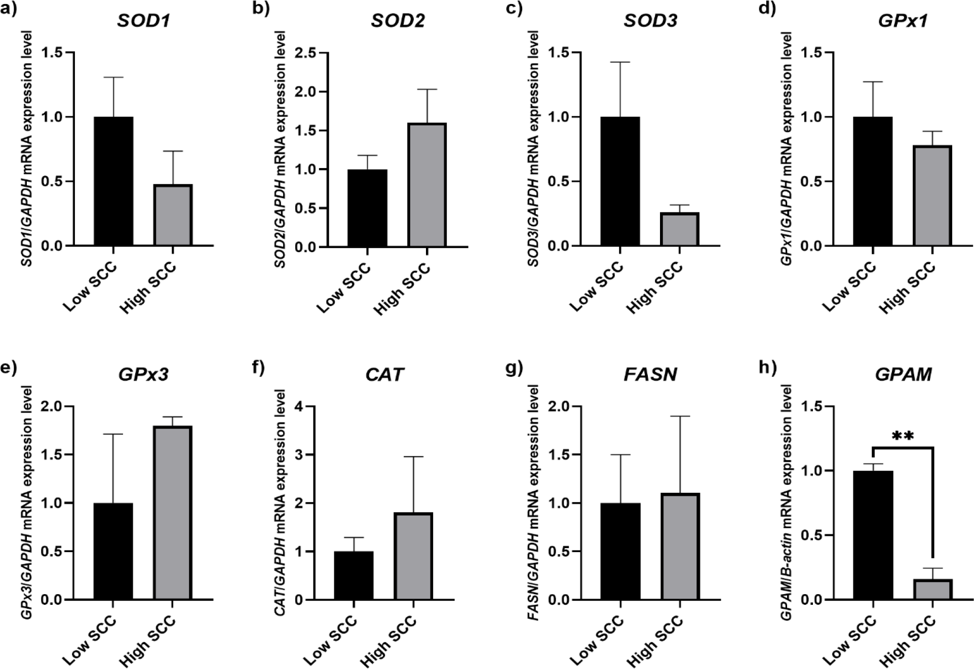
Analysis of the relative gene expression of SOD1, SOD2, SOD3, GPx1, GPx3, CAT, and FASN genes from mRNA of milk samples obtained from cows with low versus high SCC did not indicate a statistically significant differential expression between the samples analyzed (p > 0.05) (Figure 1a-g). However, analysis of the GPAM gene showed a significantly lower gene expression level in cows with high SCC (p < 0.01) (Figure 1h).
Analysis of relative gene expression of milk somatic cell samples obtained from Holstein cattle presenting low (black bars) and high (gray bars) SCC. Values were presented as means ± SEM. (a) SOD1: p = 0.2617; (b) SOD2: p = 0.2627; (c) SOD3: p = 0.1594; (d) GPx1: p = 0.4993; (e) GPx3: p = 0.3296; (f) CAT: p = 0.5352 (g) FASN: p = 0.9172; (h) GPAM: p = 0.0011. (** p < 0.01).
DISCUSSION
The Holstein cows with high and low SCC used in this study were matched for DIM and were in the late lactation stage, minimizing potential biases in the analyses. The role of SCC in relation to DIM is controversial, as some studies report that SCC gradually increases toward the end of lactation [34], while others indicate that lactation stage has no significant effect on SCC [35].
According to Wickramasinghe and coauthors [18], compared with the mammary gland, milk somatic cells expressed a higher number of genes, including sets uniquely related to immunity, organ development, and behavior. Thus, identifying and characterizing genes expressed in milk somatic cells is an important step toward understanding the complex biological properties and species-specific variations of milk [18]. The comparison of gene expression in cows with low and high SCC can help identify potential biomarkers for the diagnosis of mastitis, especially subclinical mastitis, which is considered a major challenge in herd health monitoring.
Our study consisted of the initial evaluation and standardization of methods for obtaining somatic cells from the milk of Holstein cattle, and subsequent extraction of total RNA. In our study, the protocol for RNA extraction from somatic cells in sheep milk, described by Mura and coauthors [30], was used with modifications, applying an additional step of washing the cells with PBS/EDTA solutions, and using Trizol reagent. The standardization of extraction and the improvement of non-invasive evaluation techniques, combined with animal welfare considerations, enable the advancement of scientific studies using gene expression evaluation methodologies. The possibility of obtaining somatic cells to evaluate gene expression, without the need for a biopsy of mammary tissue, allows the evaluation of expression levels using the same animals throughout experiments, such as those involving dietary supplementation, lactation time, among others [14,18]. Thus, the analysis of milk samples from cows with different nutritional treatments and/or different stages of lactation allows a better understanding of production, udder health, welfare, and nutrigenomics.
The evaluation of differentially expressed genes under certain conditions requires recognition of the numerous intrinsic or environmental variables to which sample groups are subjected. An important factor to evaluate is the SCC for the analysis of differentially expressed genes in dairy cattle based on somatic cells [2]. SCC in milk can be influenced by various factors, including cow productivity, health status, parity order, lactation stage, and breed [1,36,37]. Any changes in environmental conditions, management practices, and stressful situations significantly increase SCC in milk [1,37].
Changes in SCC values in cow’s milk significantly affect milk production, protein, and lactose, but not milk fat composition [1,11,38]. However, the decrease in milk fat concentration in cows with mastitis may be due to the reduction in the synthetic and secretory capacity of the mammary gland, with an increase in free fatty acids being observed [1]. For the genes analyzed in this study, only GPAM showed downregulated expression levels in milk samples from cows with high SCC. The enzyme encoded by the GPAM gene catalyzes the first committed step in the biosynthesis of triglycerides and phospholipids, using free fatty acids in this process [39,40]. The increase in free fatty acids in cows with mastitis may be an effect of the low number of glycerol-3-phosphate acyltransferase protein in milk somatic cells. Higher GPAM expression in cows with low SCC suggests greater lipid synthesis activity and better mammary gland health.
The relative gene expression of the antioxidant and the FASN genes demonstrated different patterns of increase or decrease in mRNA in milk samples from animals with low and high SCC values, although the values did not show statistical significance. The SOD1, SOD3, and GPx1 genes showed a downregulated profile in relation to the animals with low SCC, while the SOD2, GPx3, CAT, and FASN genes showed an upregulated profile in the high SCC condition. Some studies related that SOD enzyme activity was not correlated with the SCC in milk and was not influenced by an elevated number of cells [41,42], consistent with our findings. Hamed and coauthors [42] found high catalase activity in bulk milk with high SCC. Additionally, the increased activity of GPx in bulk milk indicated the presence of free radicals (H₂O₂) in milk with a high cellular charge, indicating that catalase and GPx are the main enzymatic defenses responsible for coping with free radicals induced by neutrophils. Wickramasinghe and coauthors [18] related significantly higher expression of genes with antioxidant activity during peak lactation, indicating that, compared to peak lactation, somatic cells in late-lactation milk were more involved in immune activity and the involution process. In addition, a significant reduction in FASN expression was also observed throughout lactation [18]. Our gene expression data did not reveal any significant association between SOD, GPx, CAT, and FASN gene expression levels and SCC, indicating that assessing the mRNA expression of multiple genes and their networks within a pathway is essential for drawing meaningful conclusions about a metabolic process. According to Mu and coauthors [43], milk fat synthesis is a highly complex regulatory process that involves not only the expression of a relatively small number of genes encoding essential proteins, but also intricate interactions among multiple organs and their integration within metabolic networks. Therefore, further analysis of additional milk fat-related genes, along with quantitative assessments of triglyceride and fatty acid levels, is required to support our findings. Gene expression studies involving milk samples with low and high SCC are still rare and focused on analyzing genes related to the immune system response. Fonseca and coauthors [17] quantified the relative expression of the IL-2, IL-4, IL-6, IL-8, IL-10, IFN-γ, and TNF-α genes in milk cells from healthy cows and cows with clinical mastitis, and no significant differences were observed for most of the analyzed genes. Alhussein and coauthors [44] analyzed the expression of CXCR1, CXCR2, IL-8, CD62L, and CD11b genes in milk samples from healthy cows and cows with subclinical and clinical mastitis, all genes were involved in neutrophil recruitment in cases of infections, and were upregulated in animals with mastitis, except for the CD-62L gene.
In this study, animal selection for gene expression evaluation was based exclusively on SCC values. However, analysis of milk control demonstrated that animals exhibited high or low SCC, regardless of the number of days of lactation and parity order. Thus, the high SCC observed in some animals is likely due to problems related to infections, as also proposed by Damm and coauthors [36], or related to milking management and herd stress [2]. In this context, the present study validates the importance of determining SCC as a factor that can interfere with gene expression analysis when using somatic cell-based methodologies from milk samples. Additionally, using somatic cells from milk to quantify mRNA levels in cattle is an effective and non-invasive alternative source of mammary tissue cells. This approach significantly improves the use of biopsy or post-mortem sampling to assess gene expression. By comparing the expression of genes encoding proteins in cow’s milk between somatic cells and post-mortem tissues collected from the same animal, Hayashi and coauthors [45] found that somatic cells in milk accurately reflect the gene expression observed in lactating mammary tissue. Furthermore, using somatic cells from milk allows for the reproducibility of the study throughout the lactation period, using the same animal [14,15,18].
CONCLUSION
The data obtained demonstrated that SCC in the milk of Holstein cows is a factor capable of causing significant changes in the expression of fat metabolism genes. In contrast, besides upor down-regulated changes observed in the animals, these were not significant for antioxidant genes. This finding reinforces the effect of SCC on altering milk characteristics by altering gene expression in milk somatic cells.
-
Funding:
This research was funded by Projeto Universal/CNPq - Process n. 403.555/2021-3, and Programa de Pesquisa Básica e Aplicada/FUNDAÇÃO ARAUCÁRIA - CP 09/2021.
-
Institutional Review Board Statement:
The animal study protocol was approved by the Animal Ethics Committee of the State University of Ponta Grossa (process CEUA - SEI 23.000017438-0).
-
Informed Consent Statement:
Not applicable
Acknowledgments:
We are grateful to Fazenda Escola Capão da Onça (FESCON) - UEPG, Ponta Grossa, PR, Brazil, and Laboratório de Análises de Leite of the Associação Paranaense de Criadores de Bovinos da Raça Holandesa (APCBRH) for their technical support.
Use of Generative Artificial Intelligence
The authors declare that large language models and other generative artificial intelligence (AI) or AI-assisted technologies cannot be credited as authors and have not been listed as authors of this paper.
The author declare that did not use the artificial intelligence.
Data Availability Statement:
Research data are available in the body of the manuscript.
REFERENCES
- 1 Alhussien MN, Dang AK. Milk somatic cells, factors influencing their release, future prospects, and practical utility in dairy animals: An overview. Vet World. 2018;11:562-77.
- 2 Boutinaud M, Jammes H. Potential uses of milk epithelial cells: a review. Reprod Nutr Dev. 2002;42:133-47.
- 3 Lindmark-Månsson H, Bränning C, Aldén G, Paulsson M. Relationship between somatic cell count, individual leukocyte populations and milk components in bovine udder quarter milk. Int Dairy J. 2006;16:717-27.
- 4 Li N, Boutinaud M, Martin P, Gagnaire V. Role of somatic cells on dairy processes and products: a review. Dairy Sci Technol. 2014;94:517-38.
- 5 Lopes Júnior JEF, Lange CC, Brito MAVP, Santos FR, Silva MAS, Moraes LCD, et al. Relationship between total bacteria counts and somatic cell counts from mammary quarters infected by mastitis pathogens. Cienc Rural. 2012;42:691-6.
- 6 Cedeño CB, Domínguez RL, Ordoñez VV, Carranza BV, Vázquez AG. [Importance of somatic cell count in determining udder health and rinde milk quality]. Braz J Anim Environ Res. 2022;5:104-23.
- 7 Santos MV, Ma Y, Barbano DM. Effect of somatic cell count on proteolysis and lipolysis in pasteurized fluid milk during shelf-life storage. J Dairy Sci. 2003;86:2491-503.
- 8 Haxhiaj K, Wishart DS, Ametaj BN. Mastitis: what it is, current diagnostics, and the potential of metabolomics to identify new predictive biomarkers. Dairy. 2022;3:722-46.
- 9 Ruegg PL. A 100-Year Review: Mastitis Detection, Management, and Prevention. J Dairy Sci. 2017;100:10381-97.
- 10 Guo JZ, Liu XL, Xu AJ, Xia Z. Relationship of somatic cell count with milk yield and composition in Chinese Holstein population. Agric Sci China. 2010;9:1492-6.
- 11 Cinar M, Serbester U, Ceyhan A, Gorgulu M. Effect of somatic cell Count on milk yield and composition of first and second lactation dairy cows. Ital J Anim Sci. 2015;14:105-8.
- 12 Sert D, Mercan E, Aydemir S, Civelek M. Effects of milk somatic cell counts on some physicochemical and functional characteristics of skim and whole milk powders. J Dairy Sci. 2016;99:5254-64.
- 13 IDF (International Dairy Federation). Guidelines for the use and interpretation of bovine milk somatic cell count. Bull IDF. 2013;466.
- 14 Medrano JF, Rincon G, Islas-Trejo A. Comparative analysis of bovine milk and mammary gland transcriptome using RNA-Seq. In: 9th World congress on genetics applied to livestock production, Leipzig, Germany. 2010;0852.
- 15 Cánovas A, Rincón G, Bevilacqua C, Islas-Trejo A, Brenaut P, Hovey RC, et al. Comparison of five different RNA sources to examine the lactating bovine mammary gland transcriptome using RNA-Sequencing. Sci Rep. 2014;4:5297.
- 16 Rainard P, Riollet C. Innate immunity of the bovine mammary gland. Vet Res. 2006;37:369-400.
- 17 Fonseca I, Silva PV, Lange CC, Guimarães MFM, Weller MMDCA, Sousa KRS, et al. Expression profile of genes associated with mastitis in dairy cattle. Genet Mol Biol. 2009;32:776-81.
- 18 Wickramasinghe S, Rincon G, Islas-Trejo A, Medrano JF. Transcriptional profiling of bovine milk using RNA sequencing. BMC Genomics. 2012;13:45.
- 19 Barbano DM, Ma Y, Santos MV. Influence of raw milk quality on fluid milk shelf life. J Dairy Sci. 2006;89:E15-9.
- 20 Wang X, Falcone T, Attaran M, Goldberg JM, Agarwal A, Sharma RK. Vitamin C and vitamin E supplementation reduce oxidative stress-induced embryo toxicity and improve the blastocyst development rate. Fertil Steril. 2002;78:1272-7.
- 21 Choi WJ, Banerjee J, Falcone T, Bena J, Agarwal A, Sharma RK. Oxidative stress and tumor necrosis factor-alpha-induced alterations in metaphase II mouse oocyte spindle structure. Fertil Steril. 2007;88:1220-31.
- 22 Nasiri N, Moini A, Eftekhari-Yazdi P, Karimian L, Salman-Yazdi R, Arabipoor A. Oxidative stress statues in serum and follicular fluid of women with endometriosis. Cell J. 2017;18:582-7.
- 23 Karakaya S, El SN, Tas AA. Antioxidant activity of some foods containing phenolic compounds. Int J Food Sci Nutr. 2001;52:501-8.
- 24 Khan IT, Nadeem M, Imran M, Ullah R, Ajmal M, Jaspal MH. Antioxidant properties of Milk and dairy products: a comprehensive review of the current knowledge. Lipids Health Dis. 2019;18:41.
- 25 Bernard L, Leroux C, Chilliard Y. Expression and nutritional regulation of lipogenic genes in the ruminant lactating mammary gland. Adv Exp Med Biol. 2008;606:67-108.
- 26 Coleman RA, Mashek DG. Mammalian triacylglycerol metabolism: synthesis, lipolysis, and signaling. Chem Rev. 2011;111:6359-86.
- 27 Bionaz M, Loor JJ. Gene networks driving bovine milk fat synthesis during the lactation cycle. BMC Genomics. 2008;9:366.
- 28 NASEM. National Academies of Sciences, Engineering, and Medicine. Nutrient Requirements of Dairy Cattle. 8th rev. ed. Washington DC: The National Academies Press. 2021.
- 29 Paula MC, Ribas NP, Monardes HG, Arce JE, Andrade UVC. [Somatic Cell Count in Milk Samples]. Rev Bras Zootec. 2004;33:1303-8.
- 30 Mura MC, Daga C, Bodano S, Paludo M, Luridiana S, Pazzola M, et al. Development of a RNA extraction method from milk for gene expression study in the mammary gland of sheep. Mol Biol Rep. 2013;40:2169-73.
- 31 Andersen CL, Jensen JL, Ørntoft TF. Normalization of Real-Time Quantitative Reverse Transcription-PCR Data: A Model-Based Variance Estimation Approach to Identify Genes Suited for Normalization, Applied to Bladder and Colon Cancer Data Sets. Cancer Res. 2004;64:5245-50.
- 32 Bustin SA, Benes V, Garson JA, Hellemans J, Huggett J, Kubista M, et al. The MIQE guidelines: minimum information for publication of quantitative real-time PCR experiments. Clin Chem. 2009;55:611-22.
- 33 Livak KJ, Schmittgen TD. Analysis of relative gene expression data using real-time quantitative PCR and the 2-∆∆CT method. Methods. 2001;25:402-8.
- 34 Kul E, Şahİn A, Atasever S, Uğurlutepe E, Soydaner M. The effects of somatic cell count on milk yield and milk composition in Holstein cows. Vet Arhiv. 2019;89:143-54.
- 35 Atasever S, Stádník L. Factors affecting daily milk yield, fat and protein percentage, and somatic cell count in primiparous Holstein cows. Indian J Anim Res. 2015;49:313-6.
- 36 Damm M, Holm C, Blaabjerg M, Bro MN, Schwarz D. Differential somatic cell count: A novel method for routine mastitis screening in the frame of Dairy Herd Improvement testing programs. J Dairy Sci. 2017;100:4926-40.
- 37 Gonçalves JL, Cue RI, Botaro BG, Horst JA, Valloto AA, Santos MV. Milk losses associated with somatic cell counts by parity and stage of lactation. J Dairy Sci. 2018;101:4357-66.
- 38 Danieli B, Schogor ALB, Zucchi J, Neto AT. Cows with High SCC Exhibit Poorer Performance and Milk Quality, Regardless of the Season. Dairy. 2025;6:46.
- 39 Lewin TM, Wang S, Nagle CA, Van Horn CG, Coleman RA. Mitochondrial glycerol-3-phosphate acyltransferase-1 directs the metabolic fate of exogenous fatty acids in hepatocytes. Am J Physiol Endocrinol Metab. 2005;288:E835-44.
- 40 Brockmöller SF, Bucher E, Müller BM, Budczies J, Hilvo M, Griffin JL, et al. Integration of metabolomics and expression of glycerol-3-phosphate acyltransferase (GPAM) in breast cancer-link to patient survival, hormone receptor status, and metabolic profiling. J Proteome Res. 2012;11:850-60.
- 41 Hicks CL. Occurrence and consequence of superoxide dismutase in milk products: a review. J Dairy Sci. 1980;63:1199-204.
- 42 Hamed H, Feki AE, Gargouri A. Total and differential bulk cow milk somatic cell counts and their relation with antioxidant factors. C R Biol. 2008;331:144-51.
- 43 Mu T, Hu H, Ma Y, Feng X, Zhang J, Gu Y. Regulation of Key Genes for Milk Fat Synthesis in Ruminants. Front Nutr. 2021;8:765147.
- 44 Alhussien M, Manjari P, Mohammed S, Sheikh AA, Reddi S, Dixit S, et al. Incidence of mastitis and activity of milk neutrophils in Tharparkar cows reared under semi-arid conditions. Trop Anim Health Prod. 2016;48:1291-5.
- 45 Hayashi AA, McCoard SA, Roy NC, Barnett MPG, Mackenzie DDS, McNabb WC. Gene expression in bovine mammary somatic cells isolated from milk. J Anim Feed Sci. 2004;13:401-4.
-
Editor-in-Chief:
Paulo Vitor Farago
-
Associate Editor:
Luiz Gustavo Lacerda