Physicochemical and antibacterial properties of an experimental paste associated with ozonized oil
ABSTRACT
Background Apexification is a key treatment for immature teeth with pulp necrosis.
Objective This study’s objective was to characterize, analyze, and conduct an initial evaluation of a novel filling material composed of calcium hydroxide, zinc oxide, zirconium oxide, and ozonized sunflower oil.
Material and Methods Four experimental formulations were prepared: (1) calcium hydroxide, zinc oxide, zirconium oxide, ozonized oil; (2) calcium hydroxide, zinc oxide, ozonized oil; (3) calcium hydroxide, zinc oxide, zirconium oxide, non-ozonized oil; and (4) calcium hydroxide, zinc oxide, non-ozonized oil. Physicochemical properties were assessed through pH dynamic over time (1–720 hours), material flow (ISO 6876/2012), radiopacity (aluminum equivalence), and SEM/EDS analysis. Antimicrobial efficacy was tested using agar diffusion and direct contact methods against Enterococcus faecalis and Porphyromonas gingivalis.
Results All groups exhibited sustained alkaline pH levels; groups containing ozonized oil showed notably higher flow rates than those with non-ozonized oil (p < 0.0001). Radiopacity surpassed ISO requirements across all formulations (p = 0.0441). No inhibition halos were observed in agar diffusion tests; however, direct contact tests demonstrated extended antimicrobial activity for up to 48 hours in groups with ozonized oil compared to 24 hours for non-ozonized groups. SEM/EDS analysis revealed calcium as the predominant element in the material and at the dentin interface, with reduced calcium content observed after 30 days.
Conclusion These findings indicate that this novel paste may represent a viable alternative for apexification in traumatized immature teeth.
KEYWORDS:
Antimicrobial activity; Calcium hydroxide; Dental trauma; Paste; Physicochemical properties
RESUMO
Contexto A apicificação é um tratamento essencial para dentes imaturos com necrose pulpar.
Objetivo O objetivo deste estudo foi caracterizar, analisar e conduzir uma avaliação inicial de um novo material obturador composto de hidróxido de cálcio, óxido de zinco, óxido de zircônio e óleo de girassol ozonizado.
Material e Métodos Quatro formulações experimentais foram preparadas: (1) hidróxido de cálcio, óxido de zinco, óxido de zircônio e óleo ozonizado; (2) hidróxido de cálcio, óxido de zinco e óleo ozonizado; (3) hidróxido de cálcio, óxido de zinco e óxido de zircônio e óleo não ozonizado; e (4) hidróxido de cálcio, óxido de zinco e óleo não ozonizado. As propriedades físico-químicas foram avaliadas por meio da dinâmica do pH ao longo do tempo (1 a 720 horas), fluxo de material (ISO 6876/2012), radiopacidade (equivalência de alumínio) e análise SEM/EDS. A eficácia antimicrobiana foi testada usando métodos de difusão em ágar e contato direto contra Enterococcus faecalis e Porphyromonas gingivalis.
Resultados Todos os grupos exibiram níveis de pH alcalinos sustentados; os grupos contendo óleo ozonizado apresentaram taxas de fluxo notavelmente maiores do que aqueles com óleo não ozonizado (p < 0,0001). A radiopacidade superou os requisitos ISO em todas as formulações (p = 0,0441). Nenhum halo de inibição foi observado nos testes de difusão em ágar; no entanto, os testes de contato direto demonstraram atividade antimicrobiana prolongada por até 48 horas nos grupos com óleo ozonizado, em comparação com 24 horas nos grupos não ozonizados. A análise SEM/EDS revelou o cálcio como o elemento predominante no material e na interface da dentina, com redução do teor de cálcio observada após 30 dias.
Conclusão Esses achados indicam que esta nova pasta pode representar uma alternativa viável para apicificação em dentes imaturos traumatizados. Esta versão garante que os nomes bacterianos estejam em itálico para precisão científica, ao mesmo tempo em que apresenta os principais resultados de forma concisa.
PALAVRAS-CHAVE:
Atividade antimicrobiana; Hidróxido de cálcio; Traumatismo dentário; Pasta; Propriedades físico-químicas
INTRODUCTION
Traumatic dental injuries are a significant clinical concern, often leading to pulp necrosis in developing permanent teeth [1]. These teeth, characterized by incompletely formed roots with thin and fragile walls [2], pose unique challenges in endodontic management. Among the available therapeutic strategies, apexification remains a widely adopted approach to facilitate the formation of an apical barrier.
Although effective, traditional apexification techniques, which require frequent replacement of intracanal medications, present notable drawbacks [3,4]. These include increased risk of treatment interruptions due to patient noncompliance, potential microbial contamination between sessions, and heightened fragility of root structures over time. Such limitations underscore the need for innovative materials and protocols that can streamline the process while enhancing clinical outcomes [5].
Calcium hydroxide (CH) has been widely used as an intracanal medication due to its high pH (12.5–12.8). Its mechanism of action involves the dissociation of Ca2+ and OH− ions, which depends on the vehicle used to deliver CH. These vehicles can be aqueous, viscous, or oily. In vitro studies have shown that the vehicle directly influences ion release rates and antibacterial activity [6].
Soares et al. [7] proposed a filling paste consisting of calcium hydroxide, zinc oxide, and 2% chlorhexidine gel as an intracanal medicament for long-term apexification without the need for frequent replacements. This formulation demonstrated favorable clinical outcomes [8-12]; however, it also revealed limitations such as challenging handling and insertion, as well as reduced dimensional stability when exposed to moisture [13,14]. The incorporation of zinc oxide in these formulations aims to enhance physicochemical properties, including pH stability and dimensional integrity, given that prolonged use of calcium hydroxide is associated with inadequate sealing ability and high solubility in oral fluids [15,16].
Radiopacity is a critical feature that minimizes the need for periodic replacement since these materials must be easily visualized during radiographic examinations [17]. Zirconium oxide has been proposed as a radiopacifying agent in repair cements due to its excellent radiopacity without compromising physical properties [18,19].
Ozonized sunflower oil has demonstrated antimicrobial properties due to its composition rich in linoleic and oleic acids. The ozone reaction with unsaturated fatty acids produces oxygenated compounds [20] that provide wound-healing and antibacterial effects. In vitro, studies have associated ozonized oil with calcium hydroxide as an intracanal medication and reported slower ion release compared to non-ozonized oil [21]. This finding suggests its potential use in large chronic periapical lesions requiring long-term calcium hydroxide therapy to induce mineralization while maintaining an antimicrobial environment.
To propose a paste for filling traumatized immature teeth without requiring periodic changes, this study evaluated the physicochemical and antimicrobial properties of a novel material composed of calcium hydroxide, zinc oxide, zirconium oxide, and ozonized sunflower oil. The null hypothesis tested whether the presence of ozonized sunflower oil and zirconium oxide alters the physicochemical or antimicrobial characteristics of the paste.
MATERIAL AND METHODS
Experimental groups
The experimental groups were prepared with calcium hydroxide, zinc oxide, zirconium oxide (Sigma-Aldrich, St. Louis, MO, USA), and ozonized and non-ozonized sunflower oils (Philozon LTDA., Balneário Camboriú, Santa Catarina, Brazil) (Table I). The consistency of each experimental group was determined by using the minimum amount of vehicles necessary to achieve a homogeneous mixture of all components. This formulation aimed to provide a filling material for traumatized teeth that would allow apexification without requiring frequent intracanal medication changes. The vehicle, being denser than conventional materials, was specifically chosen to ensure that the paste would remain in the root canal for a prolonged period, thus reducing the need for multiple applications and enhancing the long-term stability of the treatment.
The evaluations encompassed physicochemical tests (pH, flow, and radiopacity) and antimicrobial assessments conducted through agar diffusion and direct contact methods.
Bovine teeth
The bovine teeth used in this study were kindly provided by Frigorífico Angelelli, located at Rua João Pedro Corrêa, 1111, Piracicaba, SP, Brazil, ZIP code: 134111-142. The teeth were obtained from cattle slaughtered for consumption, and their use was approved by the relevant ethics committee. The origin of the teeth was carefully documented to ensure compliance with ethical and quality standards required for the study.
Physicochemical analysis of the material
Scanning electron microscopy/energy dispersive x-ray spectroscopy (SEM/EDS) analysis. Selected roots for SEM/EDS analysis were sectioned longitudinally after the insertion of the material corresponding to the evaluated group into 3-mm-thick discs. The sections were immersed in Hank's balanced salt solution, and discs from each group were removed at intervals of 24 hours, 15 days, and 30 days. The sections were air-dried at room temperature, dehydrated in a silica gel desiccator for one week, and mounted on aluminum stubs. Subsequently, they were sputter-coated with carbon (Bal-Tec, Balzers, Liechtenstein). Six specimens from each group were analyzed using SEM/EDS (JSM-5600/LvJEOL, Tokyo, Japan). SEM allowed visualization of the material's insertion and its interface with dentin. Following this, EDS analysis was performed to determine the percentage peaks of chemical elements present in each sample. Four fields within the material and four at the material/dentin interface were evaluated. Images were acquired at a magnification of 1000x.
pH assessment
The pH evaluation was conducted following a previously established methodology [22]. A total of 40 acrylic teeth (n=10) were prepared using an R40 reciprocating file (VDW, Munich, Germany), with the apical foramen standardized to a diameter of 400 µm. The specimens were divided into groups based on the additives incorporated into the experimental material and filled using a Lentulo spiral (Dentsply Sirona, Maillefer, Ballaigues, Switzerland). Subsequently, each specimen was placed in a fresh test tube containing an equal volume of deionized water. The water was systematically replaced at predetermined intervals: 1, 3, 7, 12, 24, 168, 360, and 720 hours. The pH of the solutions was measured using a pH meter (371; Micronal, São Paulo, Brazil), which had been calibrated with buffer solutions at pH levels of 4, 7, and 9 to ensure accuracy. Throughout the experiment, the room temperature was maintained at a constant 25 °C. Deionized water served as the control medium across all analyzed periods, providing a baseline for comparison.
Radiopacity and flow assessments
Radiopacity was assessed using aluminum equivalence, while flow properties were evaluated following the guidelines specified by the International Organization for Standardization (ISO) 6876/2012 [23]. For the radiopacity assessment, the material was meticulously spatulated under standardized conditions in a controlled environment, maintaining constant temperature and humidity for two minutes to ensure the homogeneity of the paste. The homogenized material was then inserted into stainless steel rings with dimensions of 10 mm in diameter and 1 mm in thickness, placed on three acrylic plates (57 mm x 76 mm x 2.5 mm). Radiographic exposures were performed three times using phosphor plates (6 x 8 cm) positioned adjacent to an aluminum step wedge with thicknesses ranging from 2 mm to 16 mm in 2 mm increments. The x-ray unit was operated at 60 kVp, 8 mA, 0.3 pulses per second, with a focus-film distance set at 30 cm. The radiographs were subsequently processed using a standardized automatic processor (Dürr Dental, Bietigheim-Bissingen, Germany). The resulting images were imported into the Image J software, which was used to calculate radiopacity values expressed as equivalent thicknesses of aluminum. For the flow analysis, 0.05 ml of the material was carefully placed at the center of a 40 mm x 40 mm glass slab using a 1.5 ml disposable syringe. Three minutes after the mixing process began, a second glass slab weighing 20 g was positioned on top of the material, followed by an additional weight of 100 g, resulting in a total load of 120 g applied for a duration of ten minutes. The largest and smallest diameters of the material spread were measured with a digital caliper (Mitutoyo MTI Corporation, Tokyo, Japan). If the difference between the diameters was less than 1 mm, their average was recorded. The test was repeated in triplicate.
Antibacterial activity test
Agar diffusion
E. faecalis (ATCC 29212) and P. gingivalis (ATCC 49417) were utilized to evaluate inhibition zones. The microorganisms were subcultured in suitable culture media under optimal temperature and gaseous conditions to verify purity [24]. Each strain was individually inoculated into tubes containing 5 ml of sterile 0.85% saline solution. Subsequently, 500 µL of each bacterial suspension was added to glass bottles with 50 ml of BHI agar maintained at 46 °C. These mixtures were homogenized using a vortex mixer before being poured into plates containing brain heart infusion (BHI) agar supplemented with 5% defibrinated sheep blood (ANILAB, Paulínia, São Paulo, Brazil). Plates inoculated with E. faecalis were incubated aerobically at 37 °C for 18 hours, while those with P. gingivalis were transferred to fastidious anaerobe agar (FAA) supplemented with sheep blood and incubated anaerobically for 48 hours inside an anaerobic chamber (Don Whitley Scientific Ltd., Bradford, United Kingdom) under a controlled atmosphere (80% N2; 10% CO2; 10% H2). Eight sterile steel cylinders measuring 4 x 1 x 1 mm (inner diameter: 6 mm) were filled with freshly mixed pastes from each experimental group [24]. Sterile saline solution was used as the negative control, and a felt disc soaked in 2% chlorhexidine gel (Drogal LTDA., Piracicaba, São Paulo, Brazil) served as the positive antimicrobial control, alongside tests with ozonized and non-ozonized oils. The plates were incubated under appropriate conditions, and microbial growth inhibition zones were measured with digital calipers after 24 hours for E. faecalis or up to 48 hours for P. gingivalis.
Direct contact
The E. faecalis bacterial suspension in BHI broth was standardized using a spectrophotometer at 800 nm to achieve a transmittance of 90 T, corresponding to 0.5 McFarland scale (approximately 1.5 x 10⁸ CFU). The experimental design included tubes 1 to 4 containing freshly mixed pastes, tubes 5 and 6 with 90 mg of ozonized and non-ozonized oil, respectively, tube 7 with a felt disc saturated in 2% chlorhexidine gel (positive control), and tube 8 containing sterile BHI solution for spectrophotometer calibration (negative control). All tubes were vortexed for five seconds and then incubated under suitable conditions. Each evaluation involved six serial dilutions (10−1 to 10−6), from which 50 µL aliquots were plated onto agar plates containing BHI supplemented with 5% defibrinated sheep blood to monitor bacterial growth. Antimicrobial efficacy was determined by counting colony-forming units (CFUs) immediately after sample preparation and at 3, 24, and 48-hour time points.
Statistical analysis
GraphPad Prism software (version 8.01) [25] was used for statistical analysis. The Shapiro-Wilk test was applied to assess sample normality. One-way ANOVA followed by Tukey's post hoc test was used to analyze radiopacity and flow data. The Shapiro-Wilk and Bartlett tests were applied to test the null hypothesis for pH data. Two-way ANOVA followed by Tukey's post hoc test was used to analyze pH values over time. A significance level (α) of 5% was adopted for all analysis. The power of the study (β) was maintained at 80% to detect significant differences between groups.
RESULTS
pH Test
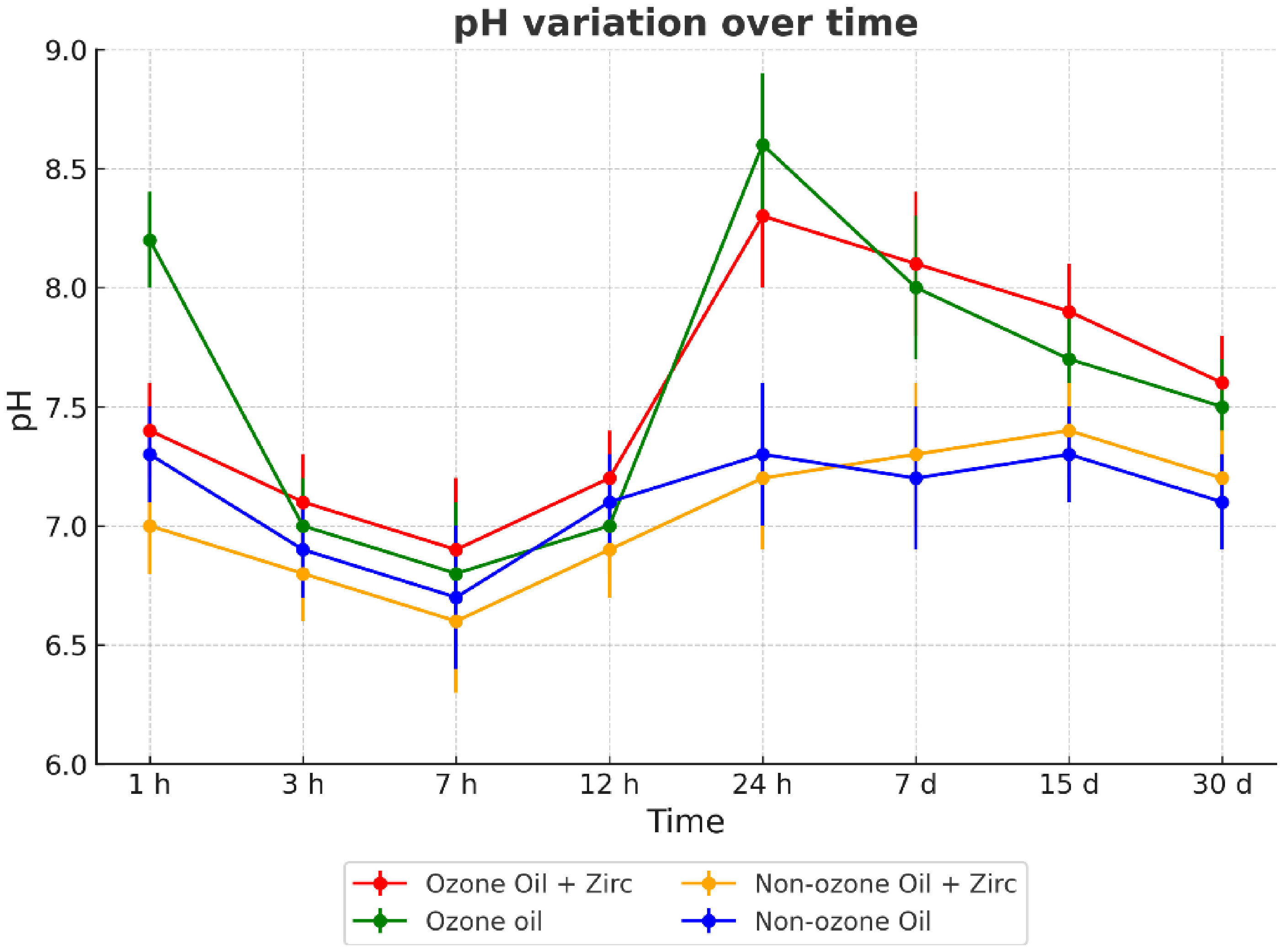
The pH data followed a time-dependent variation pattern, influenced by the composition of each experimental paste. All groups started with alkaline values close to or slightly above neutral (between 7 and 8), followed by a transient drop around 7 hours, and then a gradual recovery or stabilization over the evaluated period. Group 1 (ozonized sunflower oil + zirconium oxide) consistently exhibited higher pH values throughout the entire 30-day evaluation. After an initial decrease, this group showed a significant rise in pH, peaking around 7 days (approximately pH 8.7), followed by a slow decline, remaining above pH 8 by day 30.
Group 2 (ozonized oil) also reached a notable peak around 7 days but started and ended with lower pH values compared to Group 1. Group 3 (non-ozonized oil + zirconium oxide) had an initial pH above 8 at 1 hour but experienced a marked drop by 3–7 hours and maintained only mildly alkaline pH values (~7.2–7.5) over time. Group 4 (non-ozonized oil) showed the lowest and most stable pH, remaining close to neutral (~7.0–7.2) throughout the 30-day evaluation. Figure 1 presents the pH variations across groups over time.
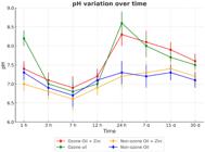
- Sample pH variations according to time. Circles = sample means; whiskers = standard deviation.
Flow test
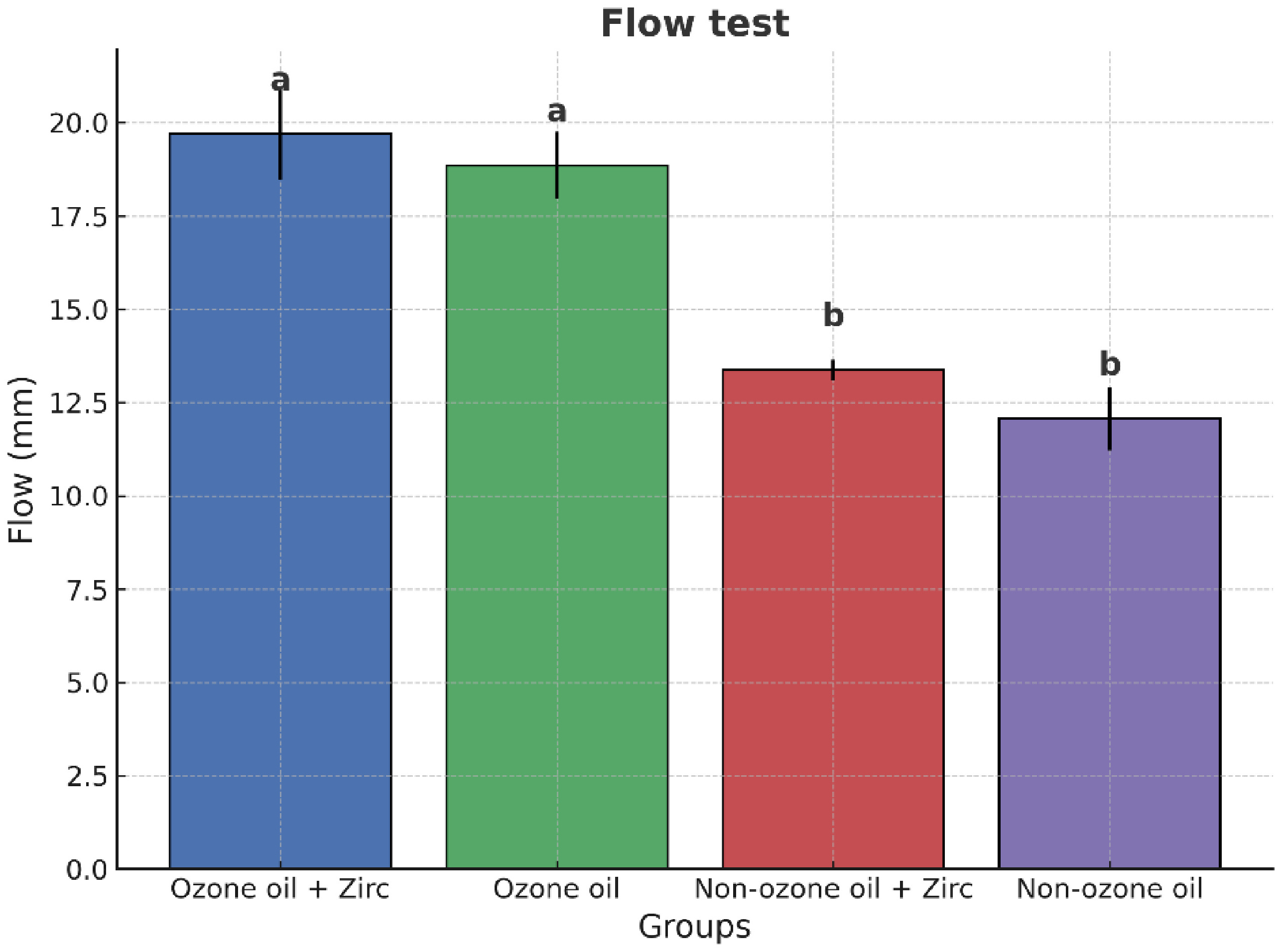
The data did not reject the null hypothesis of the Shapiro-Wilk and Bartlett tests (p > 0.05), indicating normality and homogeneity of variances. One-way ANOVA revealed significant differences between groups (p < 0.0001). Tukey's post hoc test showed that G1 and G2 had significantly higher flow values compared to G3 and G4 (p < 0.0001), while no significant differences were observed between G1 and G2 (p = 0.3727) or between G3 and G4 (p = 0.0765). A reference line at 17 mm was included in Figure 2 to indicate the minimum flow requirement according to ISO 6876/2012 for endodontic materials.
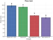
- Flow in millimeters. Different letters mean statistically significant differences between groups.
Increased viscosity of ozonized sunflower oil likely contributed to the higher flow values observed in the corresponding groups, as shown in Figure 2. This resulted in flow values above the 17 mm threshold set by ISO standards, while the non-ozonized oil groups showed lower flow values (12 and 13 mm), reflecting their lower viscosity. Increased viscosity likely led to decreased flowability, which aligns with the goal of enhancing the material's retention within the canal.
In this scenario, the increased viscosity of the ozonized sunflower oil likely caused a reduction in flowability, which, although decreasing the material's ability to flow easily, is advantageous in terms of ensuring prolonged retention within the root canal. This behavior is in line with the proposed benefits of using ozonized oil, as the material needs to remain in place for extended periods without frequent replacement.
Figure 2 illustrates the statistically significant differences in flow between the experimental groups, as well as the reference line indicating the ISO 6876/2012 minimum requirement.
Radiopacity
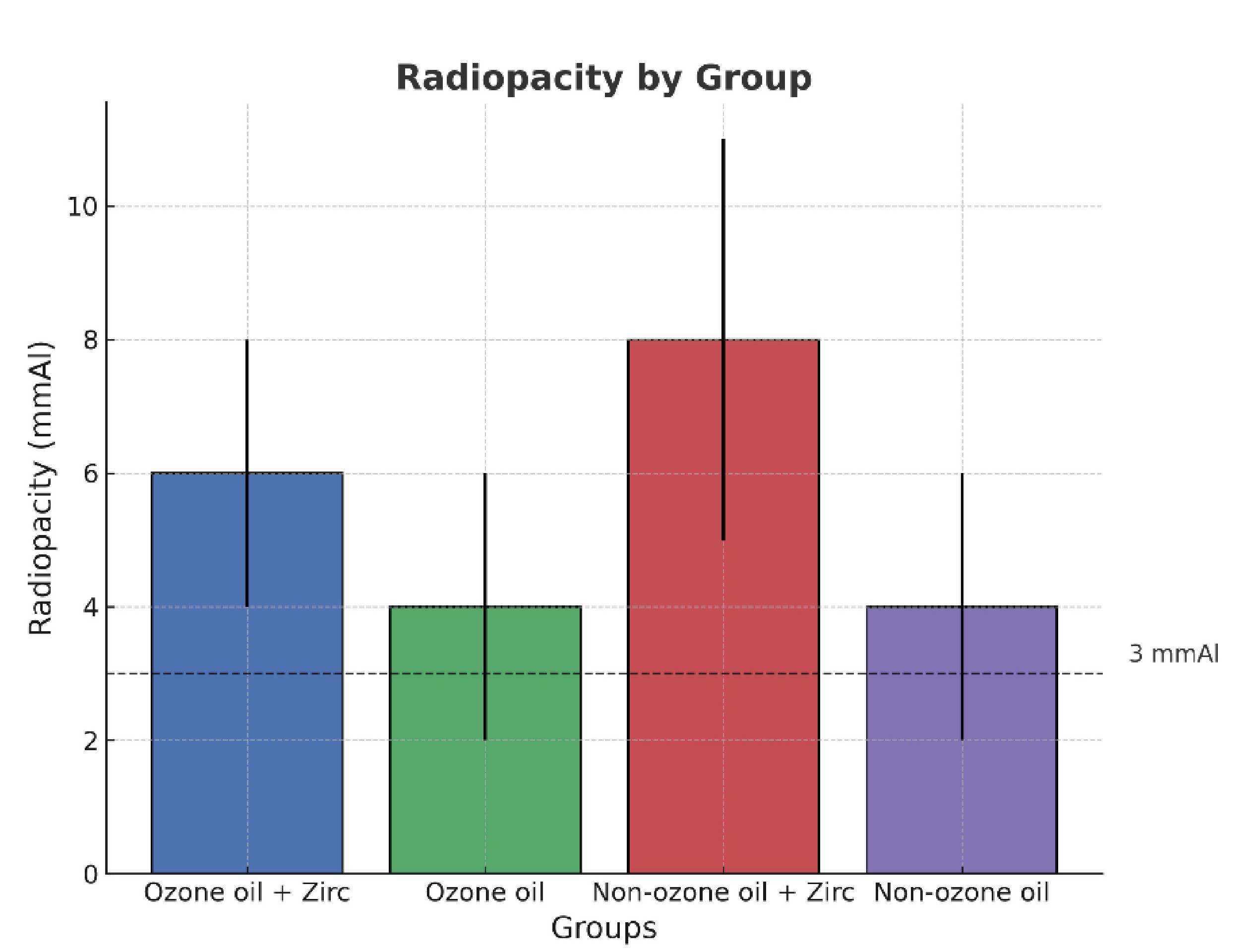
Radiopacity results are presented in Figure 3. Since the data did not follow a normal distribution (Shapiro–Wilk test, p < 0.05), comparisons among groups were performed using the Kruskal–Wallis test, which revealed statistically significant differences (H = 8.095; p = 0.0441).
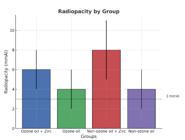
The experimental formulation containing non-ozonized oil and zirconium oxide (Group 3) exhibited the highest radiopacity, with a median value of 8 mmAl (first and third quartiles: 4–9). The formulation containing ozonized oil and zirconium oxide (Group 1) showed intermediate radiopacity values, with a median of 6 mmAl (4–6). In contrast, the formulations without zirconium oxide (Groups 2 and 4) presented lower radiopacity values, both with a median of 4 mmAl, although still above the minimum requirement of 3 mmAl established by ISO 6876/2012.
Overall, the presence of zirconium oxide was associated with increased radiopacity, and all experimental groups met the ISO standard for radiographic detectability.
Antimicrobial activity
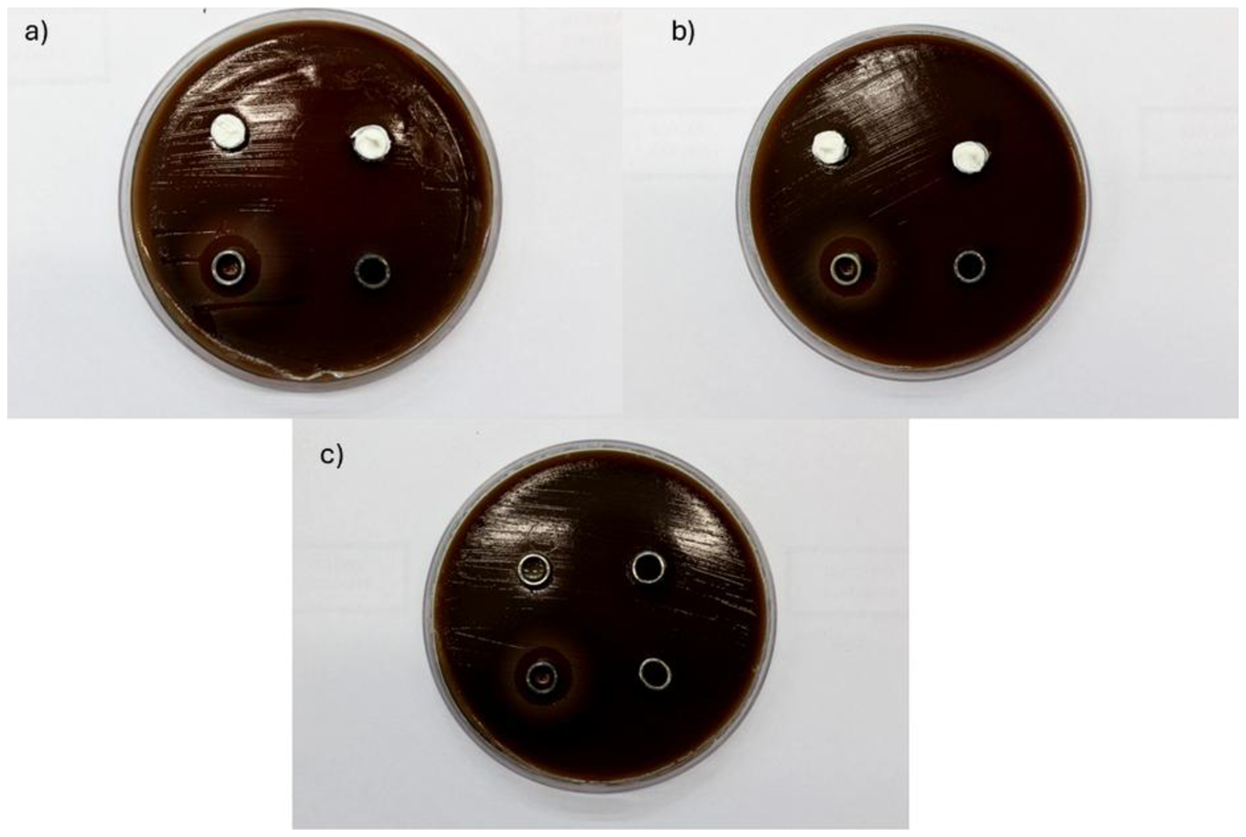
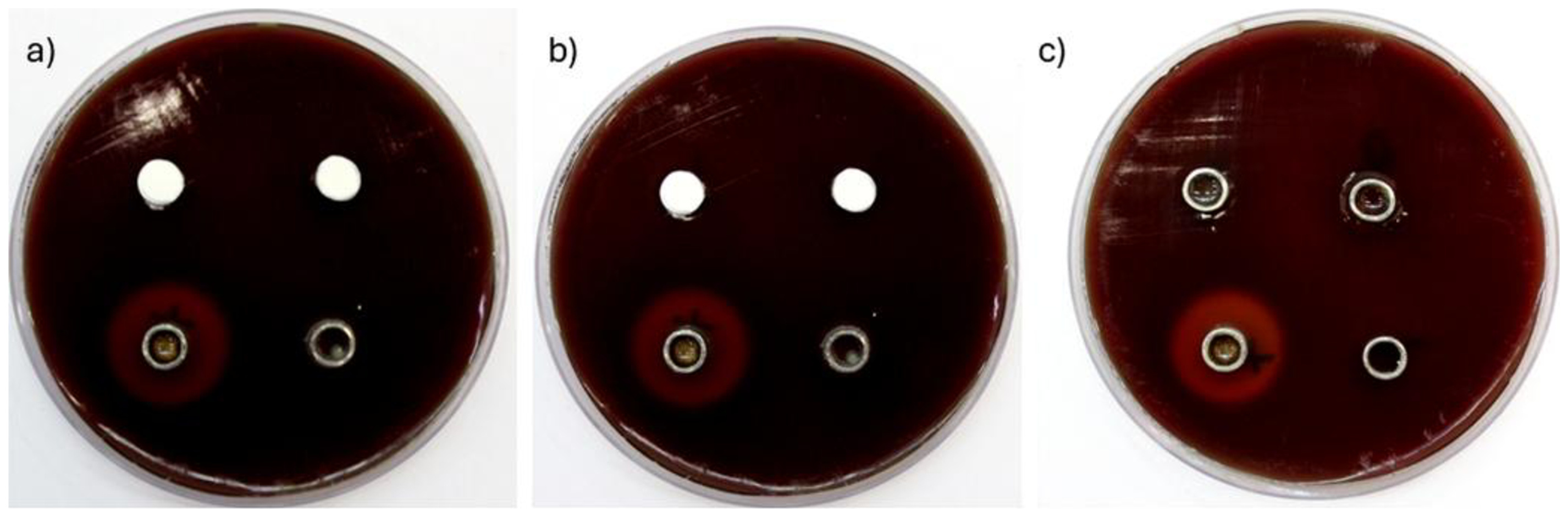
The antimicrobial potential of the experimental pastes was assessed using two complementary methods: agar diffusion and direct contact test. In the agar diffusion test, none of the experimental groups exhibited inhibition halos against E. faecalis or P. gingivalis, indicating limited diffusion of antimicrobial agents into the agar medium. This result may reflect the high viscosity or low solubility of the paste components, which restrict the leaching of active substances. In contrast, the positive control (2% chlorhexidine gel) produced clear inhibition halos, with mean diameters of 14.1 mm for E. faecalis and 17.45 mm for P. gingivalis, confirming the reliability of the method (Figures 4 and 5).
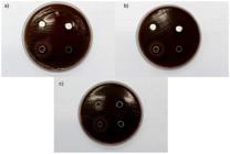
- Inhibition zones of the antibacterial test. (a) Representative image of a Petri dish with brain heart infusion (BHI) + 5% defibrinated sheep blood with 1.5 x 10⁸ CFU/mL of E. faecalis, representing the inhibition halo. (a) Groups Ozone oil + Zirc Ozone oil, 10 µL of 2% chlorhexidine gel (positive control), and negative control. (b) Groups non-ozonized oil + Zirc and Non-ozonized oil, 10 µL of 2% chlorhexidine gel (positive control), and negative control. (c) Ozonized and non-ozonized sunflower oils, 10 µL of 2% chlorhexidine gel, and negative control.

- Fastidious anaerobe agar (FAA) = 5% defibrinated sheep blood with 1.5 x 10⁸ CFU/mL of P. gingivalis. (a) Groups Ozone oil + Zirc and Ozone oil, 10 µL of 2% chlorhexidine gel, and negative control. (b) Groups non-ozonized oil + Zirc and Non-ozonized oil, 10 µL of 2% chlorhexidine gel (positive control), and negative control. (c) Ozonized and non-ozonized sunflower oils, 10 µL of 2% chlorhexidine gel, and negative control.
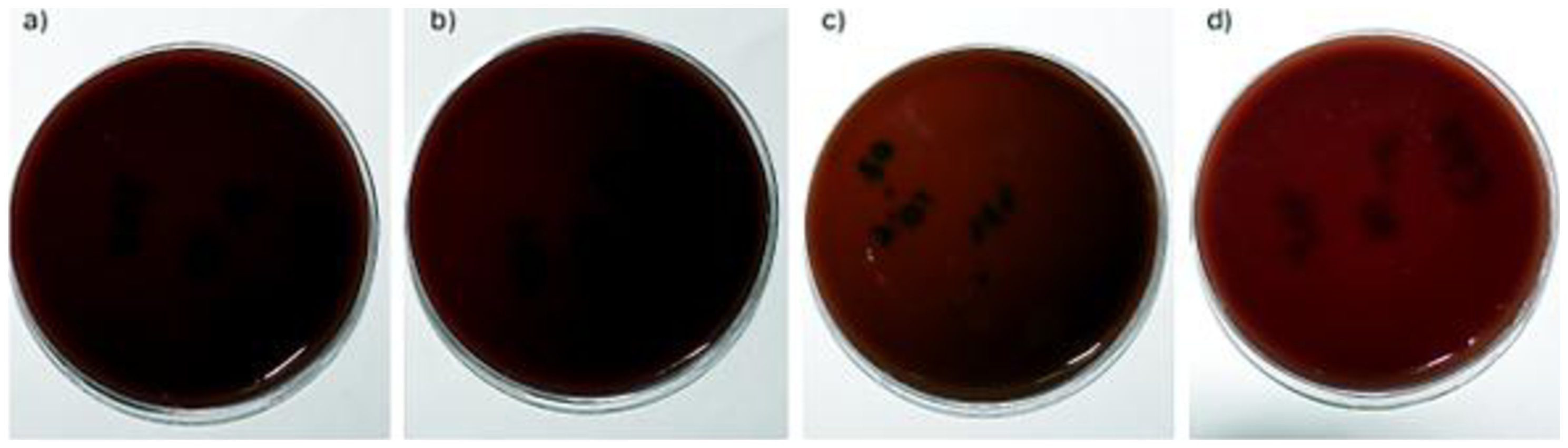
To evaluate direct antibacterial efficacy, the direct contact test (DCT) was performed. This method revealed progressive reductions in CFU counts over time for all experimental pastes. Formulations containing ozonized oil showed a slower reduction profile, with detectable viable bacteria up to 48 hours, while non-ozonized oil groups eliminated CFUs by 24 hours. Despite this difference in kinetics, all groups achieved 100% bacterial reduction by 48 hours. CFU reductions were quantified both in absolute values and log10 reductions (Table II). The positive control (chlorhexidine) again showed complete inhibition at all time points, while the negative control (saline) exhibited bacterial growth over time.
Complementing these findings, plate count assays confirmed the DCT results. Experimental groups showed a time-dependent decrease in bacterial colonies on agar plates following serial dilution. These results reinforce the direct contact findings and suggest that the materials' antimicrobial effect depends more on surface interaction than on diffusibility.
Overall, while the experimental pastes showed no antimicrobial halo formation, they demonstrated effective bactericidal activity upon direct exposure, particularly against E. faecalis. The inclusion of ozonized oil contributed to sustained activity, though with delayed bacterial elimination compared to non-ozonized formulations (Figures 6 and 7).
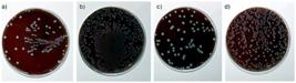
- Direct contact immediately, UFC count. (a) group Ozone oil + Zirc (b) group ozone oil. (c) group non-ozonized oil + Zirc. (d) group non-ozonized oil.
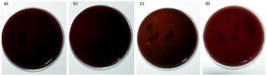
- Direct contact after 48 hours, UFC count. (a) group Ozone oil + Zirc (b) group ozone oil. (c) group non-ozonized oil + Zirc. (d) group non-ozonized oil.
Depict the log10 reductions in CFU, and Tables 2 summarize the quantitative microbiological data.
SEM/EDS analysis
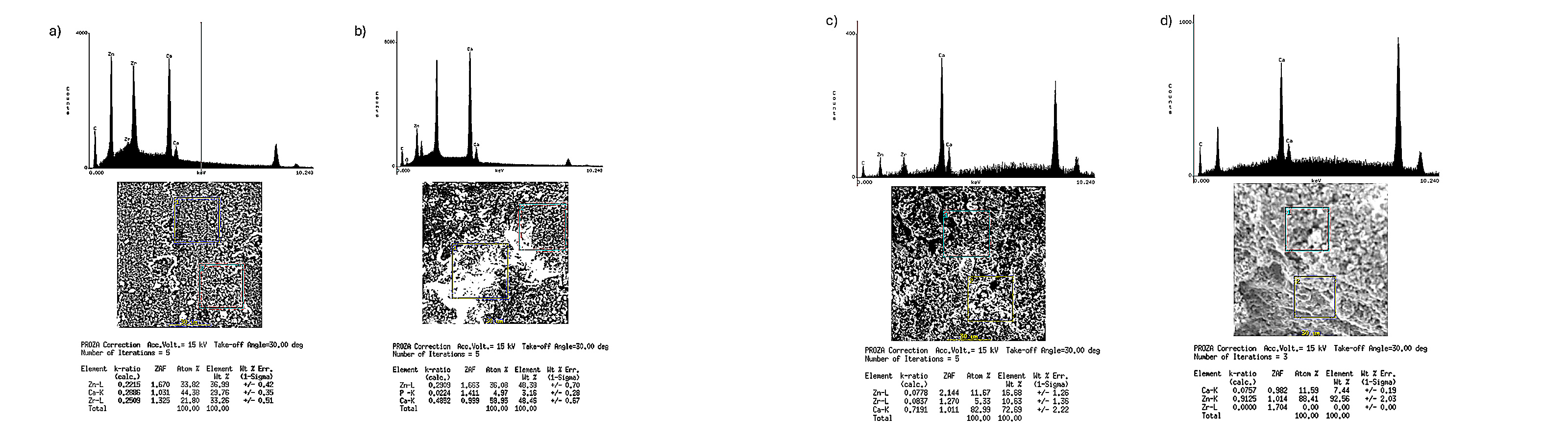
The chemical elements identified in both the material and its interface with dentin were calcium, zirconium, and zinc. Calcium was consistently the most abundant element across all samples, followed by zinc and zirconium when present in the group composition. At the material-dentin interface, calcium remained the predominant element. After a storage period of 30 days, a decrease in calcium content was observed across all groups (Figure 8).
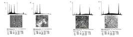
- Representative SEM/EDS image of the material surface; 1000µm scale. (a) group Ozone oil + Zirc. (b) group ozone oil. (c) group non-ozonized oil + Zirc. (d) group non-ozonized oil.
DISCUSSION
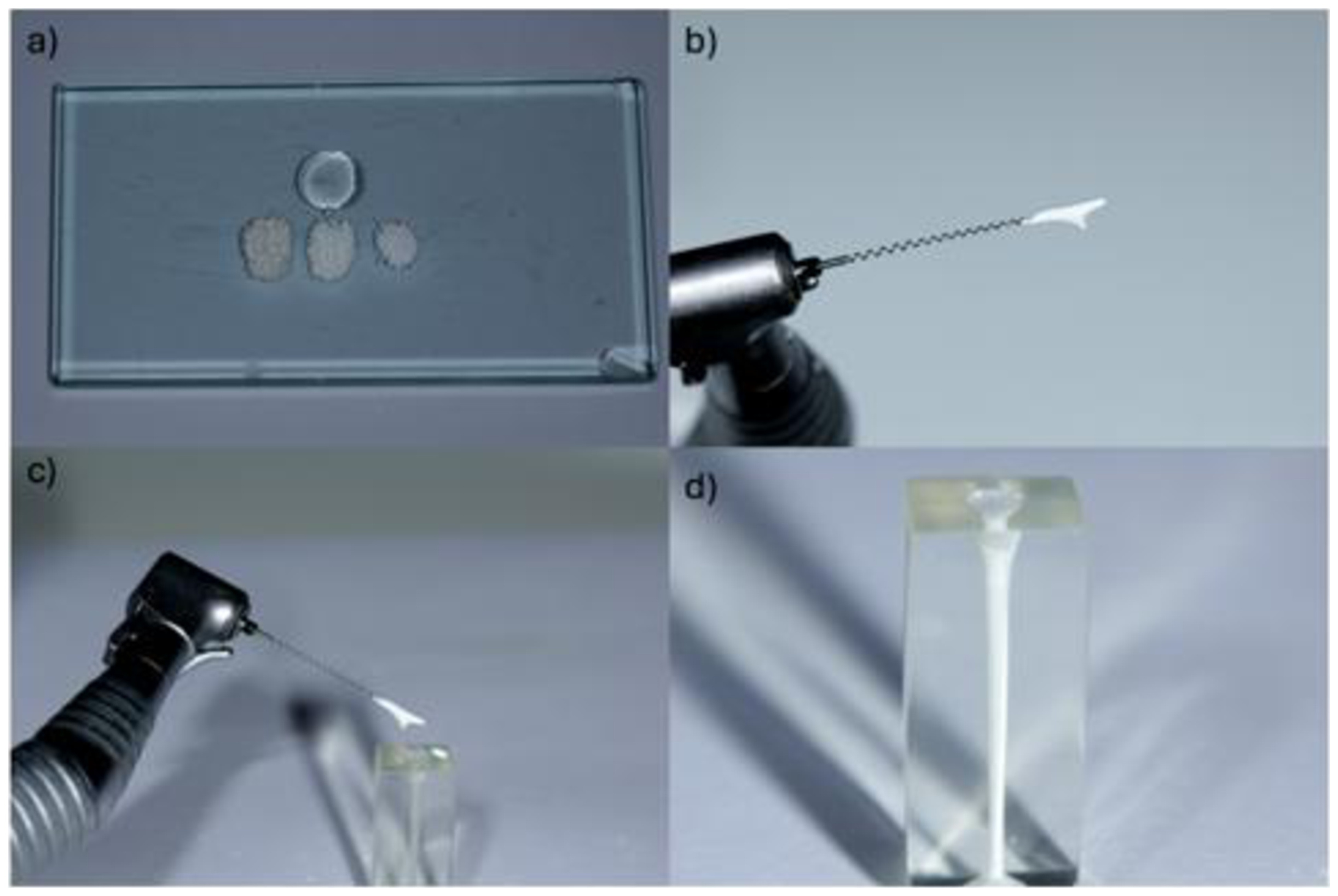
This research introduces an intracanal medication intended for apexification without the need for frequent replacements, achieved by combining calcium hydroxide, zinc oxide, zirconium oxide, and ozonized sunflower oil. To evaluate its potential, parameters such as pH variations, flow, radiopacity, and antibacterial activity were analyzed. The material developed in this investigation aims to balance clinical efficacy with ease of application. MTA has been widely recognized as a standard apical barrier in apexification treatments. Despite strong evidence supporting its effectivenes [26 –29], MTA is associated with challenges such as complex placement due to its low viscosity, grainy texture, and limited working time. These issues necessitate advanced skills and appropriate tools for proper handling [30]. Beyond ease of application, intracanal medications must also ensure effective contact with the canal walls [31]. This novel material proposes a more straightforward insertion method, utilizing either a lentulo spiral or manual endodontic instruments [Figure 9].

- Insertion of the experimental material into a simulated root canal using a Lentulo spiral, ensuring homogeneous distribution and minimizing voids. (a) Components of the experimental paste; (b) material captured with the Lentulo spiral; (c) insertion of the material into the simulated canal; and (d) simulated root canal completely filled with the experimental material.
The experimental material described in this study was specifically formulated for intracanal applications, with the added benefit of eliminating the need for periodic replacement. The primary focus of this study was to characterize the properties of the new material. Consequently, a positive control such as MTA was not used, as the goal was to evaluate the performance and potential of the material itself, without comparison to existing materials.
Soares et al. [11] proposed a filling paste composed of calcium hydroxide, zinc oxide, and 2% chlorhexidine gel that must be manipulated and used in a putty consistency [7,8]. Several studies have demonstrated its clinical effectiveness [8,12,32,33] and its ability to increase pH [34] and diffuse through dentinal tubules [35]. However, like MTA, its consistency complicates manipulation and insertion [14], requiring experienced clinicians and adequate materials. The ease of insertion of the proposed paste is related to its high flow degree. Using ozonized sunflower oil promotes increased viscosity, resulting in a flow value above 17 mm for the paste, meeting the ISO 6876/2012 standard recommendation [23]. Groups with non-ozonized sunflower oil showed flow values below the standard (12 and 13 mm). This difference may be attributed to ozonized sunflower oil's higher viscosity due to ozonation.
Ozonation forms polymeric peroxides by reacting ozone with carbon-carbon double bonds in fatty acids within the oil. These peroxides increase viscosity and create a highly viscous mass [36]. The ozonized oil used in this study serves as an alternative vehicle. Ozone has been widely used in dentistry in various forms—gas, ozonized water, or oil due to its ability to accelerate healing and provide antimicrobial activity [37-39]. While gaseous ozone has more potent antimicrobial properties than aqueous ozone, its toxic effects when inhaled make liquid forms more commonly used in dentistry [38]. Because aqueous vehicles lead to faster ion dissociation compared to oily ones, this study selected the oily form for better control of ion release. Previous studies have demonstrated the antimicrobial capacity of ozonized oil and its associations, particularly against species related to periradicular diseases [40].
This study assessed the antimicrobial activity of pastes using two methods: agar diffusion and direct contact tests. When testing ozonized and non-ozonized sunflower oils alone, both methods showed limited capacity to inhibit microbial growth, consistent with previous studies [41,42]. The antibacterial activity was evaluated against two pathogens: E. faecalis a Gram-positive facultative aerobic microorganism associated with persistent infections due to its resistance to alkaline environments (pH 9.6) and biofilm formation [43-45] — and P. gingivalis a Gram-negative anaerobic bacillus linked to periodontal disease and symptomatic primary endodontic infections [46,47]. None of the groups exhibited antibacterial activity in agar diffusion tests or inhibition halos after 24 hours for E. faecalis or 48 hours for P. gingivalis.
In direct contact tests, bacterial growth inhibition was observed after 24 hours of exposure to the medium. This outcome can be attributed to the slower ion release typically associated with pastes containing oily vehicles. By contrast, in agar diffusion tests, only the basal surface of the specimen interacts with the bacterial inoculum (area), whereas in direct contact tests, the entire specimen is in contact with the medium (volume) [48]. Previous studies have demonstrated notable differences in solubility among aqueous, viscous, and oily vehicles. Specifically, pastes prepared with aqueous vehicles exhibit higher solubility due to the more rapid dissociation of Ca2+ and OH− ions [34]. The release of OH− ions is directly related to increases in pH, a finding consistent with the results of this study: all groups showed elevated pH after 24 hours, with peak levels observed between one and seven days, followed by a gradual decline over time. Even after 30 days, all groups maintained pH levels above 7.0. Notably, groups containing ozonized sunflower oil exhibited higher pH values, with peaks exceeding 8.0 within 24 hours, a result potentially influenced by the by-products of ozonized oil. However, further research is required to confirm this hypothesis.
Additionally, SEM/EDS analysis provided complementary evidence of the material’s ionic activity by detecting a reduction in calcium content over time, particularly at the material with dentin interface. This finding is consistent with the hypothesis of gradual calcium ion release, which may contribute to the sustained alkalinization observed in the pH assessments. Similar ionic release behavior has been reported in bioceramic scaffolds, supporting the role of such physicochemical characteristics in the development of bioactive dental materials for endodontic use [30,49].
Radiopacity is a critical characteristic for endodontic materials as it allows visualization during radiographic or tomographic evaluations to identify lateral canals, accessory canals, resorption defects, fractures, apex overflow fillings, or material removal when necessary [50]. All tested compositions demonstrated radiopacity above 3 mmAl as recommended by ISO standards. Groups 1 and 3 showed higher radiopacity values (means of 6 mmAl and 8 mmAl), while groups 2 and 4 had lower values. This finding may be attributed to zirconium oxide's presence in groups 1 and 3 as a radiopacifier a result consistent with previous studies that highlight zirconium oxide's effectiveness as a radiopacifying agente [30,31,51].
Study limitations
Among this study's limitations is the need for further investigation into the interactions between ozonized sunflower oil and zirconium oxide on physicochemical properties. Future research should explore dimensional stability over time as well as cellular and tissue interactions with these components. Although agar diffusion and direct contact tests are widely used, these methods have limitations, particularly in quantitatively assessing antimicrobial activity. Chemical interactions between antimicrobial agents and the culture medium may lead to misinterpretation of results. Additionally, the viscosity and/or hydrophobicity of the material can affect the leachability of antimicrobial components, influencing diffusion and antimicrobial activity measurements. Furthermore, while the acquired salivary pellicle plays a crucial role in the oral environment and influences microorganism adhesion, most studies fail to incorporate pellicle formation in their samples, which may attenuate the assessment of antimicrobial activity under conditions. Lastly, most studies use mono-species inocula, which do not accurately represent the complex, polymicrobial biofilm found in the oral cavity. Incorporating polymicrobial biofilms in future studies would provide a more precise evaluation of antimicrobial efficacy, better reflecting the dynamic nature of the oral environment [52,53].
CONCLUSION
The experimental paste containing calcium hydroxide, zinc oxide, zirconium oxide, and ozonized sunflower oil showed adequate physicochemical properties—namely sustained alkalinity, satisfactory flow, and radiopacity within ISO standards. While no inhibition zones were detected in agar diffusion, the direct contact test revealed time-dependent antibacterial activity, particularly in formulations with ozonized oil. SEM/EDS findings suggested gradual ionic release. These preliminary results indicate potential for use in apexification without the need for replacement; however, further studies are necessary to assess long-term dimensional stability, biocompatibility, and efficacy against polymicrobial biofilms.
Acknowledgements
We would like to thank the support of the Coordenação de Aperfeiçoamento de Pessoal de Nível Superior (CAPES) (88887.885695/2023-00), Conselho Nacional de Desenvolvimento Científico e Tecnológico (CNPq) and Fundação de Amparo à Pesquisa do Estado de São Paulo (Fapesp).
-
How to cite:
Gabriel PH, Vieira WA, Santos VAB, Silva MAM, Gomes BPFA, Soares AJ. Physicochemical and antibacterial properties of an experimental paste associated with ozonized oil. Braz. Dent. Sci. 2026;29:e4967. https://doi.org/10.4322/bds.2026.e4967
-
Data availability
Institutional repositories.
-
Funding
We would like to thank the support of the Coordenação de Aperfeiçoamento de Pessoal de Nível Superior (CAPES) (88887.885695/2023-00).
-
Regulatory Statement
This study does not require ethics committee approval since it did not use live animals or studies with human beings.
References
-
1 Tewari N, Devi P, Sampath S, Mathur VP, Tsilingaridis G, Wikström A, et al. Comparative effectiveness of regenerative endodontic treatment versus apexification for necrotic immature permanent teeth with or without apical periodontitis: an umbrella review. Dent Traumatol. 2025;41(3):263-82. https://doi.org/10.1111/edt.13028 PMid:39692071.
» https://doi.org/10.1111/edt.13028 -
2 Trope M. Treatment of the immature tooth with a non-vital pulp and apical periodontitis. Dent Clin North Am. 2010;54(2):313-24. https://doi.org/10.1016/j.cden.2009.12.006 PMid:20433980.
» https://doi.org/10.1016/j.cden.2009.12.006 -
3 Lin J, Zeng Q, Wei X, Zhao W, Cui M, Gu J, et al. Regenerative endodontics versus apexification in immature permanent teeth with apical periodontitis: a prospective randomized controlled study. J Endod. 2017;43(11):1821-7. https://doi.org/10.1016/j.joen.2017.06.023 PMid:28864219.
» https://doi.org/10.1016/j.joen.2017.06.023 -
4 Murray PE. Review of guidance for the selection of regenerative endodontics, apexogenesis, apexification, pulpotomy, and other endodontic treatments for immature permanent teeth. Int Endod J. 2023;56(Suppl 2):188-99. https://doi.org/10.1111/iej.13809 PMid:35929348.
» https://doi.org/10.1111/iej.13809 -
5 Ali MRW, Mustafa M, Bardsen A, Bletsa A. Fracture resistance of simulated immature teeth treated with a regenerative endodontic protocol. Acta Biomater Odontol Scand. 2019;5(1):30-7. https://doi.org/10.1080/23337931.2019.1570822 PMid:30775411.
» https://doi.org/10.1080/23337931.2019.1570822 -
6 Mohammadi Z, Dummer PM. Properties and applications of calcium hydroxide in endodontics and dental traumatology. Int Endod J. 2011;44(8):697-730. https://doi.org/10.1111/j.1365-2591.2011.01886.x PMid:21535021.
» https://doi.org/10.1111/j.1365-2591.2011.01886.x -
7 Soares AJ, Lima TFR, Nagata JY, Gomes BPFA, Zaia AA, Souza-Filho FJ. Intracanal dressing paste composed by calcium hydroxide, chlorhexidine and zinc oxide for the treatment of immature and mature traumatized teeth. Braz J Oral Sci. 2014;13(1):6-11. https://doi.org/10.1590/1677-3225v13n1a02
» https://doi.org/10.1590/1677-3225v13n1a02 - 8 Soares AJ, Souza-Filho FJ. Traumatized teeth submitted to a new intracanal medication protocol. Braz J Dent Traumatol. 2011;2:1-5.
- 9 Marion JJC, Nagata JY, Senko RAG, Lima TFR, Soares A. Therapeutic proposal for avulsed teeth using calcium hydroxide associated to 2% chlorhexidine gel and zinc oxide. Dent Press Endod. 2012;2:48-53.
- 10 Soares AJ, Nagata JY, Casarin RCV, et al. Apexification with a New Intra-Canal Medicament: A Multidisciplinary Case Report. Iran Endod J. 2012;7(3):165-70. PMid:23056138.
- 11 Soares AJ, Nagata JY, Lima TFR, Sonoda T, Souza-Filho FJ. Apexification without periodic changes of intracanal medicament and MTA apical plug: 5 years follow-up. Dent Press Endod. 2013;3:84-9.
-
12 Herrera DR, Herrera CM, Lima AR, Nagata JY, Pereira AC, Silva EJ, et al. Repair of apical root resorption associated with periodontitis using a new intracanal medicament protocol. J Oral Sci. 2014;56(4):311-4. https://doi.org/10.2334/josnusd.56.311 PMid:25500930.
» https://doi.org/10.2334/josnusd.56.311 -
13 Zou X, Zheng X, Liang Y, Zhang C, Fan B, Liang J, et al. Expert consensus on irrigation and intracanal medication in root canal therapy. Int J Oral Sci. 2024;16(1):23. https://doi.org/10.1038/s41368-024-00280-5 PMid:38429299.
» https://doi.org/10.1038/s41368-024-00280-5 -
14 Soares AJ, Lima TFR, Nagata JY, Gomes BPFA, Zaia AA, Souza-Filho FJ. Intracanal dressing paste composed by calcium hydroxide, chlorhexidine and zinc oxide for the treatment of immature and mature traumatized teeth. Braz J Oral Sci. 2014;13(1):6-11. https://doi.org/10.1590/1677-3225v13n1a02
» https://doi.org/10.1590/1677-3225v13n1a02 -
15 Gandolfi MG, Siboni F, Botero T, Bossu M, Riccitiello F, Prati C. Calcium silicate and calcium hydroxide materials for pulp capping: biointeractivity, porosity, solubility and bioactivity of current formulations. J Appl Biomater Funct Mater. 2015;13(1):43-60. https://doi.org/10.5301/jabfm.5000201 PMid:25199071.
» https://doi.org/10.5301/jabfm.5000201 -
16 Lima TFR, Ascendino JF, Cavalcante IO, D Assunção FLC, Salazar-Silva JR, Silva EJNLD, et al. Influence of chlorhexidine and zinc oxide in calcium hydroxide pastes on pH changes in external root surface. Braz Oral Res. 2019;33:e005. https://doi.org/10.1590/1807-3107bor-2019.vol33.0005 PMid:30758405.
» https://doi.org/10.1590/1807-3107bor-2019.vol33.0005 -
17 Duggal M, Tong HJ, Al-Ansary M, Twati W, Day PF, Nazzal H. Interventions for the endodontic management of non-vital traumatised immature permanent anterior teeth in children and adolescents: a systematic review of the evidence and guidelines of the European Academy of Paediatric Dentistry. Eur Arch Paediatr Dent. 2017;18(3):139-51. https://doi.org/10.1007/s40368-017-0289-5 PMid:28508244.
» https://doi.org/10.1007/s40368-017-0289-5 -
18 Cutajar A, Mallia B, Abela S, Camilleri J. Replacement of radiopacifier in mineral trioxide aggregate; characterization and determination of physical properties. Dent Mater. 2011;27(9):879-91. https://doi.org/10.1016/j.dental.2011.04.012 PMid:21571361.
» https://doi.org/10.1016/j.dental.2011.04.012 -
19 Camilleri J, Cutajar A, Mallia B. Hydration characteristics of zirconium oxide replaced Portland cement for use as a root-end filling material. Dent Mater. 2011;27(8):845-54. https://doi.org/10.1016/j.dental.2011.04.011 PMid:21571360.
» https://doi.org/10.1016/j.dental.2011.04.011 -
20 Gao X, Leng C, Zeng G, Fu D, Zhang Y, Liu Y. Ozone initiated heterogeneous oxidation of unsaturated carboxylic acids by ATR-FTIR spectroscopy. Spectrochim Acta A Mol Biomol Spectrosc. 2019;214:177-83. https://doi.org/10.1016/j.saa.2019.02.025 PMid:30776719.
» https://doi.org/10.1016/j.saa.2019.02.025 -
21 Vasavada K, Kapoor S. Evaluation of ozonized calcium hydroxide as an effective intracanal medicament during root canal procedures: an in vitro observational study. Med Gas Res. 2020;10(3):122-4. https://doi.org/10.4103/2045-9912.296042 PMid:33004709.
» https://doi.org/10.4103/2045-9912.296042 -
22 Cavenago BC, Pereira TC, Duarte MA, Ordinola-Zapata R, Marciano MA, Bramante CM, et al. Influence of powder-to-water ratio on radiopacity, setting time, pH, calcium ion release and a micro-CT volumetric solubility of white mineral trioxide aggregate. Int Endod J. 2014;47(2):120-6. https://doi.org/10.1111/iej.12120 PMid:23647286.
» https://doi.org/10.1111/iej.12120 - 23 ISO: International Organization for Standardization. ISO 6876:2012. Dentistry — Root canal sealing materials. Geneva: ISO; 2012.
-
24 Gomes BP, Pedroso JA, Jacinto RC, Vianna ME, Ferraz CC, Zaia AA, et al. In vitro evaluation of the antimicrobial activity of five root canal sealers. Braz Dent J. 2004;15(1):30-5. https://doi.org/10.1590/S0103-64402004000100006 PMid:15322642.
» https://doi.org/10.1590/S0103-64402004000100006 - 25 GraphPad. GraphPad Prism, version 8.01. San Diego: GraphPad Software; 2019.
-
26 Marciano MA, Costa RM, Camilleri J, Mondelli RFL, Guimarães BM, Duarte MAH. Assessment of color stability of white MTA Angelus and bismuth oxide in contact with tooth structure. J Endod. 2014;40(8):1235-40. https://doi.org/10.1016/j.joen.2014.01.044 PMid:25069940.
» https://doi.org/10.1016/j.joen.2014.01.044 -
27 Aguiar AS, Guerreiro-Tanomaru JM, Faria G, Leonardo RT, Tanomaru-Filho M. Antimicrobial activity and pH of calcium hydroxide and zinc oxide nanoparticles intracanal medication and association with chlorhexidine. J Contemp Dent Pract. 2015;16(8):624-9. https://doi.org/10.5005/jp-journals-10024-1732 PMid:26423497.
» https://doi.org/10.5005/jp-journals-10024-1732 -
28 Orucoglu H, Cobankara FK. Effect of unintentionally extruded calcium hydroxide paste including barium sulfate as a radiopaquing agent in treatment of teeth with periapical lesions: report of a case. J Endod. 2008;34(7):888-91. https://doi.org/10.1016/j.joen.2008.04.012 PMid:18571001.
» https://doi.org/10.1016/j.joen.2008.04.012 -
29 Lu H, Lu J, Guo J, Zeng B, Zeng Q, Zhao W, et al. Radiographic outcomes and prognostic factors in nonvital immature permanent teeth after apexification with modified calcium hydroxide paste: a retrospective study. Clin Oral Investig. 2022;26(7):5079-88. https://doi.org/10.1007/s00784-022-04481-2 PMid:35399138.
» https://doi.org/10.1007/s00784-022-04481-2 -
30 Pelepenko LE, Saavedra F, Bombarda GF, Gomes BPFA, DE-Jesus-Soares A, Zaia AA, et al. Dental discoloration caused by Grey-MTAFlow cement: analysis of its physicochemical, biological and antimicrobial properties. J Appl Oral Sci. 2020;28:e20200269. https://doi.org/10.1590/1678-7757-2020-0269 PMid:32785521.
» https://doi.org/10.1590/1678-7757-2020-0269 -
31 Sarunket C, Banomyong D, Vibulcharoenkitja P, Sutimuntanakul S. Radiopacity of Portland Cement and Calcium Silicate-Based Cement with Different Mixed Ratios of Radiopacifiers. Eur Endod J. 2022;7(1):52-7. https://doi.org/10.14744/eej.2021.72691 PMid:35353064.
» https://doi.org/10.14744/eej.2021.72691 -
32 Marion JJC, Felippe LS, Vilela CB, Nagata J, Lima T, Soares A. Suggesting a new therapeutic protocol for permanent teeth with incomplete root apex and lateral luxation: a case report. Dent Press Endod. 2014;4(2):56-61. https://doi.org/10.14436/2178-3713.4.2.056-061.oar
» https://doi.org/10.14436/2178-3713.4.2.056-061.oar -
33 Marion JJC, Martelosso L, Nagata J, Lima T, Soares A. Suggesting a new therapeutic protocol for traumatized permanent teeth: case report. Dent Press Endod. 2014;4(1):71-7. https://doi.org/10.14436/2178-3713.4.1.071-077.oar
» https://doi.org/10.14436/2178-3713.4.1.071-077.oar -
34 Zancan RF, Vivan RR, Milanda Lopes MR, Weckwerth PH, de Andrade FB, Ponce JB, et al. Antimicrobial activity and physicochemical properties of calcium hydroxide pastes used as intracanal medication. J Endod. 2016;42(12):1822-8. https://doi.org/10.1016/j.joen.2016.08.017 PMid:27776884.
» https://doi.org/10.1016/j.joen.2016.08.017 -
35 França MCM, Silva TM, Queiroz RC, Bin CV, Cardoso FGR, Valera MC. Diffusion of hydroxyl ion on external root surface using different irrigating solutions: an In vitro study. Indian J Dent Res. 2019;30(3):414-9. https://doi.org/10.4103/ijdr.IJDR_253_17 PMid:31397418.
» https://doi.org/10.4103/ijdr.IJDR_253_17 -
36 Puxeddu S, Scano A, Scorciapino MA, Delogu I, Vascellari S, Ennas G, et al. Physico-chemical investigation and antimicrobial efficacy of ozonated oils: the case study of commercial ozonated olive and sunflower seed refined oils. Molecules. 2024;29(3):679. https://doi.org/10.3390/molecules29030679 PMid:38338423.
» https://doi.org/10.3390/molecules29030679 -
37 Saini R. Ozone therapy in dentistry: A strategic review. J Nat Sci Biol Med. 2011;2(2):151-3. https://doi.org/10.4103/0976-9668.92318 PMid:22346227.
» https://doi.org/10.4103/0976-9668.92318 -
38 Irie MS, Dietrich L, Souza GL, Soares PBF, Moura CCG, Silva GRD, et al. Ozone disinfection for viruses with applications in healthcare environments: a scoping review. Braz Oral Res. 2022;36:e006. https://doi.org/10.1590/1807-3107bor-2022.vol36.0006 PMid:35081224.
» https://doi.org/10.1590/1807-3107bor-2022.vol36.0006 -
39 Vasavada K, Kapoor S. Evaluation of ozonized calcium hydroxide as an effective intracanal medicament during root canal procedures: an in vitro observational study. Med Gas Res. 2020;10(3):122-4. https://doi.org/10.4103/2045-9912.296042 PMid:33004709.
» https://doi.org/10.4103/2045-9912.296042 -
40 Elshinawy MI, Al-Madboly LA, Ghoneim WM, El-Deeb NM. Synergistic effect of newly introduced root canal medicaments; ozonated olive oil and chitosan nanoparticles, against persistent endodontic pathogens. Front Microbiol. 2018;9:1371. https://doi.org/10.3389/fmicb.2018.01371 PMid:30018599.
» https://doi.org/10.3389/fmicb.2018.01371 -
41 Moureu S, Violleau F, Ali Haimoud-Lekhal D, Calmon A. Ozonation of sunflower oils: impact of experimental conditions on the composition and the antibacterial activity of ozonized oils. Chem Phys Lipids. 2015;186:79-85. https://doi.org/10.1016/j.chemphyslip.2015.01.004 PMid:25623845.
» https://doi.org/10.1016/j.chemphyslip.2015.01.004 -
42 Cho KH, Kang DJ, Nam HS, Kim JH, Kim SY, Lee JO, et al. Ozonated sunflower oil exerted protective effect for embryo and cell survival via potent reduction power and antioxidant activity in HDL with strong antimicrobial activity. Antioxidants. 2021;10(11):1651. https://doi.org/10.3390/antiox10111651 PMid:34829522.
» https://doi.org/10.3390/antiox10111651 - 43 Lima RA, Carvalho CB, Ribeiro TR, Fonteles CS. Antimicrobial efficacy of chlorhexidine and calcium hydroxide/camphorated paramonochlorophenol on infected primary molars: a split-mouth randomized clinical trial. Quintessence Int. 2013;44(2):113-22. PMid:23444177.
-
44 Prabhakar J, Senthilkumar M, Priya MS, Mahalakshmi K, Sehgal PK, Sukumaran VG. Evaluation of antimicrobial efficacy of herbal alternatives (Triphala And Green Tea Polyphenols), MTAD, and 5% sodium hypochlorite against Enterococcus faecalis biofilm formed on tooth substrate: an in vitro study. J Endod. 2010;36(1):83-6. https://doi.org/10.1016/j.joen.2009.09.040 PMid:20003940.
» https://doi.org/10.1016/j.joen.2009.09.040 -
45 Selvi MM, Selvabalaji A, Chonat A, Ananthan V, Mani E, Chinnaswamy AD. A Comparative evaluation of antimicrobial efficacy of triphala and calcium hydroxide as intracanal medicament: an in vitro study. J Pharm Bioallied Sci. 2022;14(Suppl 1):S774-80. https://doi.org/10.4103/jpbs.jpbs_168_22 PMid:36110780.
» https://doi.org/10.4103/jpbs.jpbs_168_22 -
46 Rôças IN, Siqueira JF Jr. Distribution of Porphyromonas gingivalis fimA genotypes in primary endodontic infections. Oral Surg Oral Med Oral Pathol Oral Radiol Endod. 2010;109(3):474-8. https://doi.org/10.1016/j.tripleo.2009.11.009 PMid:20219603.
» https://doi.org/10.1016/j.tripleo.2009.11.009 -
47 Wang Q, Zhou XD, Zheng QH, Wang Y, Tang L, Huang DM. Distribution of Porphyromonas gingivalis fimA genotypes in chronic apical periodontitis associated with symptoms. J Endod. 2010;36(11):1790-5. https://doi.org/10.1016/j.joen.2010.08.018 PMid:20951289.
» https://doi.org/10.1016/j.joen.2010.08.018 -
48 Aguiar AS, Guerreiro-Tanomaru JM, Faria G, Leonardo RT, Tanomaru-Filho M. Antimicrobial activity and ph of calcium hydroxide and zinc oxide nanoparticles intracanal medication and association with chlorhexidine. J Contemp Dent Pract. 2015;16(8):624-9. https://doi.org/10.5005/jp-journals-10024-1732 PMid:26423497.
» https://doi.org/10.5005/jp-journals-10024-1732 -
49 Bakopoulou A, Papachristou E, Bousnaki M, Hadjichristou C, Kontonasaki E, Theocharidou A, et al. Human treated dentin matrices combined with Zn-doped, Mg-based bioceramic scaffolds and human dental pulp stem cells towards targeted dentin regeneration. Dent Mater. 2016;32(8):e159-75. https://doi.org/10.1016/j.dental.2016.05.013 PMid:27298239.
» https://doi.org/10.1016/j.dental.2016.05.013 -
50 Lu H, Lu J, Guo J, Zeng B, Zeng Q, Zhao W, et al. Radiographic outcomes and prognostic factors in nonvital immature permanent teeth after apexification with modified calcium hydroxide paste: a retrospective study. Clin Oral Investig. 2022;26(7):5079-88. https://doi.org/10.1007/s00784-022-04481-2 PMid:35399138.
» https://doi.org/10.1007/s00784-022-04481-2 -
51 Ciołek L, Jaegermann Z, Zaczyńska E, Czarny A, Biernat M, Gąsiński A, et al. Cytotoxicity and antibacterial activity of mineral trioxide aggregate cement with radiopacity introduced by ZrO2. Bioinorg Chem Appl. 2022;2022(1):9574245. https://doi.org/10.1155/2022/9574245 PMid:36111206.
» https://doi.org/10.1155/2022/9574245 -
52 The Editorial Board of the Journal of Endodontics. Wanted: a base of evidence. J Endod. 2007;33(12):1401-2. https://doi.org/10.1016/j.joen.2007.09.004 PMid:18037045.
» https://doi.org/10.1016/j.joen.2007.09.004 -
53 Ibrahim MS, Garcia IM, Kensara A, Balhaddad AA, Collares FM, Williams MA, et al. How we are assessing the developing antibacterial resin-based dental materials? A scoping review. J Dent. 2020;99:103369. https://doi.org/10.1016/j.jdent.2020.103369 PMid:32387506.
» https://doi.org/10.1016/j.jdent.2020.103369
Edited by
-
Editor-in-chief:
Sergio Eduardo de Paiva Gonçalves
-
Editor:
Bruno Mello de Matos
Institutional repositories.