Effect of mechanical aging on the bond strength to dentin of CAD/CAM materials
ABSTRACT
Objective To evaluate the bond strength of different CAD/CAM materials to dentin with and without mechanical aging.
Material and Methods Sixty human dentin specimens (4 mm height) were used, onto which blocks (4 mm height) of the following materials were cemented: fiberglass (FC; FIBER CAD; Angelus), vitroceramic composite (BB; Brava Block; FGM), and polyetheretherketone (PK; PEEK; JUVORA). Half of the specimens for each material were subjected to mechanical aging (n=10). Subsequently, sticks (1×1 mm) were prepared, subjected to the microtensile test (MPa). Then, the fracture pattern was evaluated using a stereoscopic magnifying glass (50×). Additionally, a scanning electron microscope was employed. Data were analyzed using a generalized linear model and Fisher's exact test (α=0.05).
Results BB exhibited the highest bond strength values (28.39 ± 7.49 MPa without aging; 29.89 ± 7.28 MPa with aging), while FC showed the lowest (18.18 ± 5.26 MPa without aging; 18.46 ± 2.73 MPa with aging) (ρ<0.05), regardless of aging. PK showed intermediate values (24.73 ± 5.17 MPa without aging; 21.89 ± 7.41 MPa with aging). No significant differences were observed between conditions with and without mechanical cycling (ρ>0.05).
Conclusion Within the limitations of this in vitro study, the type of material influenced adhesive performance, while aging did not affect bond strength. FC exhibited the lowest bond strength, with an increase in adhesive failures after aging.
KEYWORDS:
Adhesion; Ceramics; Composite resins; Computer-aided design; Polymers
RESUMO
Objetivo Avaliar a resistência de união de diferentes materiais CAD/CAM à dentina com e sem o envelhecimento mecânico.
Material e Métodos Sessenta espécimes de dentina humana (4 mm de altura) foram utilizados, sobre os quais blocos (4 mm de altura) dos seguintes materiais foram cimentados: fibra de vidro (FC; FIBER CAD; Angelus), compósito vitrocerâmico (BB; Brava Block; FGM) e poliéter-éter-cetona (PK; PEEK; JUVORA). Metade dos espécimes de cada material foi submetida ao envelhecimento mecânico (n = 10). Em seguida, palitos (1 × 1 mm) foram preparados e submetidos ao teste de microtração (MPa). Depois, o padrão de fratura foi avaliado com lupa estereoscópica (50×). Além disso, utilizou-se um microscópio eletrônico de varredura. Os dados foram analisados por meio de um modelo linear generalizado e do teste exato de Fisher (α = 0,05).
Resultados BB apresentou os maiores valores de resistência de união (28,39 ± 7,49 MPa sem envelhecimento; 29,89 ± 7,28 MPa com envelhecimento), enquanto o FC apresentou os menores (18,18 ± 5,26 MPa sem envelhecimento; 18,46 ± 2,73 MPa com envelhecimento) (ρ < 0,05), independentemente do envelhecimento. O PK apresentou valores intermediários (24,73 ± 5,17 MPa sem envelhecimento; 21,89 ± 7,41 MPa com envelhecimento). Não foram observadas diferenças estatisticamente significativas entre as condições com e sem envelhecimento mecânico (ρ > 0,05).
Conclusão Dentro das limitações deste estudo in vitro, o tipo de material influenciou o desempenho adesivo, enquanto o envelhecimento não afetou a resistência de união. FC apresentou a menor resistência de união, com aumento de falhas adesivas após o envelhecimento.
PALAVRAS-CHAVE:
Adesão; Cerâmicas; Resinas compostas; Design assistido por computador; Polímeros
INTRODUCTION
Adhesive failure in indirect restorations remains a significant challenge in restorative dentistry [1-4]. Mainly in the rehabilitation of teeth with extensively destroyed coronal structures, which require intraradicular retention using fiber posts [4] or retention within the walls of the pulp chamber, fabricated from resin-based, ceramic, or hybrid materials [5,6]. Adhesive failures in such situations compromise the longevity of restorations [7], leading to retreatments, patient frustration, or even loss of the dental element. Overall, the prognosis of an extensively compromised tooth depends on the characteristics of the remaining tooth structure, such as the number and thickness of the walls, as well as on patient habits, the sensitivity of the restorative technique, and the type of restorative material used [2,4,8,9].
The introduction of CAD/CAM technology has revolutionized the planning and execution of dental rehabilitations [5,10]. CAD/CAM technology enables the scanning and milling of single-piece restorations with high precision, ensuring proper adaptation to the cavity preparation. Consequently, this reduces cementation lines and the sensitivity of the restorative technique, factors that significantly impact the long-term prognosis of the restoration [1,3,4]. Several materials have been developed for CAD/CAM systems [1,3]. However, despite advancements in adhesive technologies, the establishment of reliable cementation protocols for emerging CAD/CAM materials such as fiber-reinforced composites, hybrid ceramics and polyether ether ketone (PEEK) remains critical to ensuring improved clinical outcomes. Nevertheless, despite the growing number of studies evaluating CAD/CAM materials, the adhesion between dentin and polymer-based materials remains controversial and not fully elucidated [4,11,12]. Previous studies have reported inconsistent results, which may be attributed to differences in material composition, surface energy, and responsiveness to surface treatments, as well as variations in experimental protocols and testing methodologies. These factors may directly influence bonding performance and contribute to the lack of consensus in the literature [13-16].
Among the materials that have gained prominence in CAD/CAM technology are hybrid materials [17], which combine desirable ceramic characteristics, such as mechanical strength and color stability, with those of composite resins, such as greater resilience and an elastic modulus closer to dentin [14,18,19]. Vitroceramic composites are composed of a polymeric matrix reinforced with inorganic glass particles [18], offering a viable alternative for a range of clinical applications such as inlays, onlays, endocrowns and crowns due to their favorable physicomechanical properties [20].
More recently, a polymeric material known as PEEK has attracted growing interest in dentistry. It is considered a high-performance polymer that stands out for its biocompatibility, resistance to chemical degradation, and an elastic modulus lower than that of dentin [3,9]. Materials with an elastic modulus similar to or lower than that of dentin induce non-catastrophic failures without loss of retention [9]. PEEK has potential for various clinical applications, including personalized posts and cores [21] manufactured using CAD/CAM systems, being identified as a viable alternative for effective and functional indirect rehabilitation [17,22-24]. Nevertheless, despite the use of materials suitable for the luting of PEEK, such as adhesives containing functional phosphate monomers and organic solvents [25], its luting protocol remains under debate due to its high resistance to surface treatments, meaning that its surface exhibits low chemical reactivity and limits the creation of micromechanical retention [23,26].
The durability of the adhesive bond in indirect restorations is a critical factor for long-term clinical success [7,14,17,27], influenced by factors related to the dental substrate, the amount of remaining dentin available for bonding, the intrinsic properties of the adhesive system, resin cement, and restorative material, the cavity configuration factor, patient habits, thickness of material, and the mechanical and chemical degradation of restorative materials when exposed to the oral environment [27-29]. Even though there are discussions about the biomechanics of these CAD/CAM materials in their use as intraradicular retainers, endocrowns, and cores [19,20,24,28], controversial adhesive protocols and the lack of studies [4,11,12] that compare the bonding of these materials to dentin, mean that the influence of material type on the performance of indirect restorations, particularly in the long term, remains unclear.
Therefore, this study aimed to analyze the dentin bond strength using the microtensile test of three CAD/CAM materials - fiberglass, vitroceramic composite and PEEK, both before and after mechanical aging, and to evaluate the fracture patterns. Accordingly, this study presents the following null hypotheses: (1) the type of material does not affect the bond strength to dentin or the fracture pattern, and (2) mechanical aging does not affect the bond strength to dentin or the fracture pattern.
MATERIAL AND METHODS
The study was approved by the Local Research Ethics Committee (CAAE 78649824100005418). Sixty sound, permanent human molars, without prior endodontic treatment, extracted less than six months earlier and stored in saline solution, were selected. The following materials were cemented onto teeth: fiberglass (FC; Fiber CAD; Angelus), vitroceramic composite (BB; Brava Block; FGM), and polyetheretherketone (PK; PEEK; JUVORA). Half of the specimens from each group were subjected to mechanical aging (n = 10).
The sample size calculation was performed using G*Power 3.1 software (Heinrich-Heine-Universität Düsseldorf, Düsseldorf, Germany), considering the ANOVA: Fixed effects, special, main effects and interactions function, compatible with the study design, consisting of six groups. A significance level of α = 0.05, statistical power (1−β) = 0.80, and an effect size of f = 0.47 (medium) were adopted [30,31]. The calculation indicated a minimum sample size of 8 specimens per group. Considering the in vitro nature of the study, as well as possible specimen loss during experimental procedures, 10 specimens per group were used to ensure greater reliability and similarity to similar laboratory studies [9].
Specimens were then sectioned into 1 × 1 mm2 sticks and submitted to microtensile testing (MPa), followed by fracture pattern analysis using a stereomicroscope at 50×. All methodological steps will be described in detail below. The study design is presented in Figure 1, and the materials used are described in Table I.
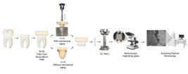
Specimen preparation
The teeth were sectioned parallel to the occlusal surface, 3 mm above the cemento-enamel junction, using a diamond abrasive disc (Diamond Abrasive Disc; Extec Corp; Connecticut; USA) mounted on a precision sectioning machine (IsoMet 1000 Precision Sectioning Machine; Buehler; Illinois; USA) under constant irrigation. The dentin surfaces were polished with 600-grit silicon carbide paper (Norton Abrasives; Saint-Gobain Abrasives Ltd.; Worcester; USA), under water cooling until the coronal remnants reached a height of approximately 2 mm, removing the occlusal enamel and exposing the mid-depth dentin surface [32]. Subsequently, the teeth were randomized by drawing lots and distributed into six groups (n=10) according to the type of material (FC, BB, and PK) and mechanical aging (with and without mechanical cycling) [9].
The blocks of restorative materials were sectioned into dimensions of 14 × 14 × 4 mm3 (length × width × height) in a square format for FC and BB and 18 × 4 mm (diameter × height) in a cylindrical format for PK. For cementation, the resin cementing agent was standardized. The blocks of each restorative material, as well as the dentin, underwent specific surface treatments as shown in Table II.
The dual-cure resin cement (RelyX Ultimate - A1; Solventum; St. Paul; USA) was prepared, applied to the surface of each material, positioned onto the dental structure, and light-cured according to the manufacturer's instructions. A corded LED light-curing unit (VALO Corded LED Curing Light – Ultradent; Ultradent, South Jordan, UT, USA) was used in standard mode at an irradiance of 1000 mW/cm2 to ensure stable irradiance during polymerization. The curing tip was positioned perpendicular to the specimen surface at a standardized distance of 1 mm. Each surface was light-cured for 20 s. All light-curing procedures were performed by a single trained operator.
The specimens were stored at 37°C for 24 hours before undergoing mechanical aging. For mechanical aging, polystyrene resin cylinders (Cristal; Redelease; Osasco; Brazil) with a polyether-based material (Impregum F; Solventum; St. Paul; USA) were fabricated to simulate the alveolar bone and periodontal ligament [21,33].
Mechanical cycling test
The samples were subjected to aging in a mechanical fatigue simulation machine (TD502 – Incon; Elquip; São Paulo; Brazil). A total of 300,000 cycles were performed using a rounded tip positioned on the occlusal surface of the block, aligned with the tooth's long axis, under a load of 50 N and a frequency of 1.2 Hz, while immersed in distilled water at 37 °C [3,9,11]. Which corresponds to approximately 1 year of clinical masticatory function [9].
Microtensile Bond Strength Test
The root portion of each tooth was sectioned 2 mm below the cemento-enamel junction [32,34]. Subsequently, the teeth were serially sectioned in both the buccolingual and mesiodistal directions using a low-speed diamond disc, yielding an average of nine dentin sticks per tooth, each approximately 1 × 1 mm in dimension [32,35]. The dentin sticks were mounted on a Geraldeli microtensile device using cyanoacrylate adhesive (Super Bonder Gel Loctite; Henkel Ltda.; Düsseldorf; Germany) at their ends, ensuring parallel alignment with the tensile direction. The specimens were tested using a universal testing machine (EZ Test L; Shimadzu; Kyoto; Japan) at a speed of 0.5 mm/min until failure occurred. The force required to fracture each stick was recorded in Newtons (N), and the dimensions of the adhesive interface were measured using a digital caliper. These values were then used to calculate the bond strength of the material to dentin in MPa (Bond Strength in MPa = Force in N / Area) [32,36,37]. Dentin sticks with pre-test failures were excluded from the bond strength analysis because measurable bond strength values could not be obtained.
Fracture pattern and scanning electron microscopy (SEM)
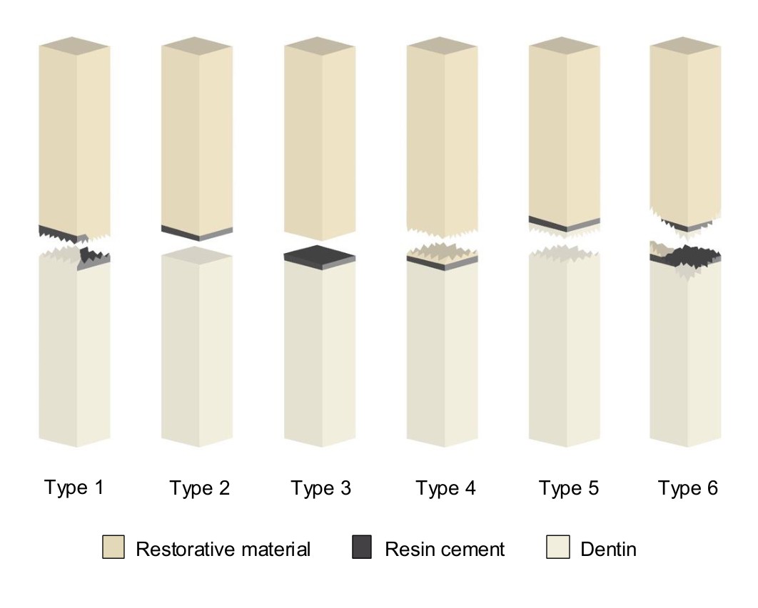
After the microtensile test, the fracture pattern was analyzed under a stereoscopic magnifying glass (MZ7.5; Leica Microsystems; Wetzlar; Germany) at 50× magnification [3]. The fracture types were documented and classified, as illustrated in Figure 2 [14,38].
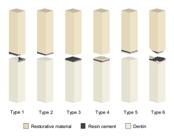
Illustration of the types of fracture patterns. Type 1 - Cohesive failure into resin cement; Type 2 - Adhesive failure between the dentin and the adhesive or resin cement; Type 3 - Adhesive failure between the adhesive or resin cement and the restorative material; Type 4 - Cohesive failure in the restorative material; Type 5 - Cohesive failure in the dentin; Type 6 - Mixed failure, when more than one type of failure occurs.
Subsequently, the fractured surfaces of representative dentin sticks were sputter-coated with gold using a sputter coater (SCD 050; BalTec; Balzers; Liechtenstein) to enhance conductivity. The coated specimens were then examined using a scanning electron microscope (SEM) (JSM 5600 Lv; JEOL; Akishima; Japan), which utilizes an electron beam to generate high-resolution images of the surface morphology, in 15kV.
Statistical analysis
To analyze bond strength, the mean value of all sticks from the same tooth was initially calculated, considering the tooth as the experimental unit. Descriptive and exploratory data analyses were conducted. Thus, a generalized linear model was employed to evaluate the effects of material type, aging conditions, and their interaction on bond strength values. For the analysis of fracture patterns using Fisher's exact test, all sticks within the same group were considered collectively, given the qualitative nature of the variable. Statistical analyses were performed using R software (R Foundation for Statistical Computing), with a significance level set at 5%.
RESULTS
Regardless of mechanical aging, bond strength was significantly higher in the BB group and lower in the FC group (ρ<0.0001). No statistically significant difference was observed between the groups with and without aging regarding bond strength (MPa), as presented in Table III (ρ=0.7623). Additionally, there was no interaction between material type and aging (ρ=0.5122). Pre-test failures were observed during specimen preparation and handling and were not included in the bond strength analysis. The number of pre-test failures recorded in each group was as follows: FC without mechanical aging (4 sticks), FC with mechanical aging (4 sticks), BB without mechanical aging (6 sticks), BB with mechanical aging (2 sticks), PK without mechanical aging (4 sticks), and PK with mechanical aging (8 sticks).
Mean (standard deviation) of bond strength (MPa) as a function of material and mechanical aging (without and with aging)
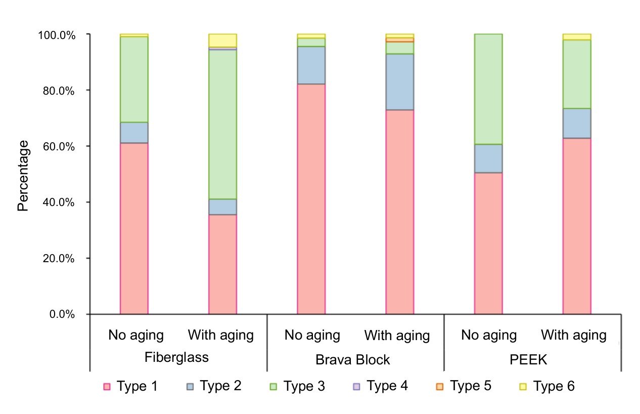
The fracture pattern distribution is presented in Figure 3, showing differences among materials and aging conditions. A significant association between aging and fracture patterns was observed for FC (ρ < 0.05). In the non-aged condition, most dentin sticks exhibited Type 1 failure (61.0%), whereas after aging this percentage decreased to 35.5%. Conversely, Type 3 failure increased from 30.5% in the non-aged group to 53.3% after aging. After aging, the frequency of Type 1 failure decreased for FC (35.5%) and BB (72.9%) but increased for PK (62.8%). In contrast, Type 3 failure increased for FC (53.3%) and BB (4.3%) and decreased for PK (24.5%).

- Fracture pattern as a function of the material and condition (with and without mechanical aging). Type 1 - Cohesive failure in the resin cement; Type 2 - Adhesive failure between the dentin and the adhesive or resin cement; Type 3 - Adhesive failure between the adhesive or resin cement and the restorative material; Type 4 - Cohesive failure in the restorative material; Type 5 - Cohesive failure in the dentin; Type 6 - Mixed failure.
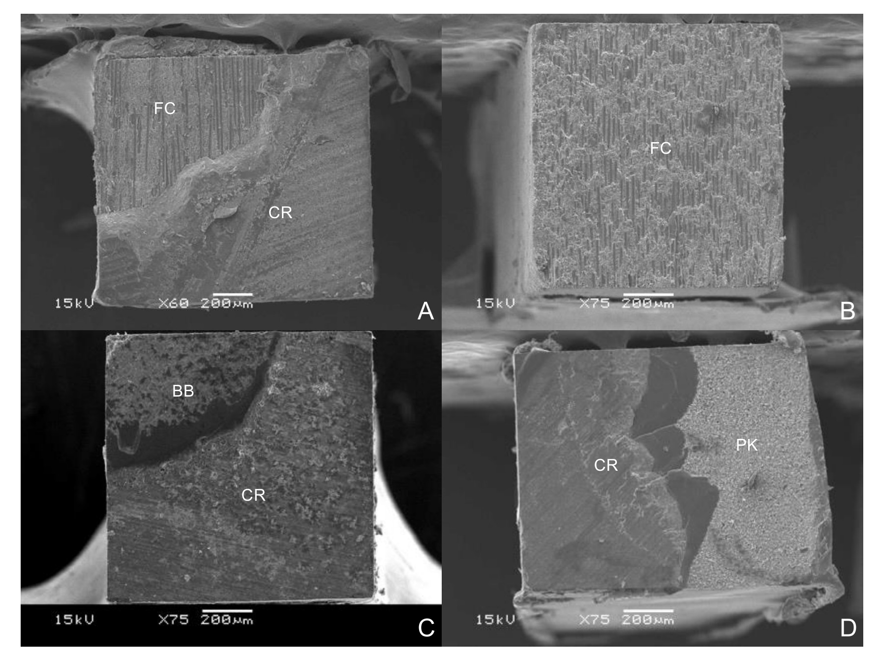
Figure 4 presents, through scanning electron microscopy (SEM) images, the fracture patterns observed for each tested material: (4A) FC without mechanical aging, exhibiting a type 1 fracture; (4B) FC subjected to mechanical aging, exhibiting a type 3 fracture; (4C) BB after mechanical aging, exhibiting a type 1 fracture; and (4D) PK after mechanical aging, also exhibiting a type 1 fracture. In images 4A, 4C, and 4D, the restorative material can be observed with resin cement partially adhered to its surface, corresponding to a type 1 fracture. In contrast, image 4B shows FC with complete detachment of the resin cement from its surface, corresponding to a type 3 fracture.
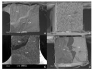
- Scanning electron microscope images of the fracture pattern. (A) FC without mechanical aging; (B) FC with mechanical aging; (C) BB with mechanical aging; (D) PK with mechanical aging. Resin Cement (CR); Fiberglass (FC); Brava Block (BB); and PEEK (PK).
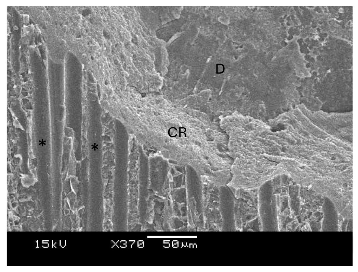
Figure 5, obtained via SEM at 370× magnification, illustrates the resin cement over dentin, revealing the imprint of glass fibers on its surface.
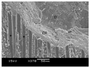
- Scanning electron microscope (SEM) images at 370× magnification, showing the imprint of glass fibers on the resin cement (*). Resin Cement (RC), Dentin (D).
DISCUSSION
The bond strength between the restorative material and dental tissue is fundamental to ensuring the stability of restoration [10,35]. Good bond strength indicates that the material is well-adhered to structure and less susceptible to adhesive failures, fractures, and infiltrations, factors that increase the likelihood of early failures [7]. This study was designed to evaluate the bonding performance of different CAD/CAM materials to dentin. Bond strength to microtensile stress and fracture patterns of CAD/CAM material blocks cemented to dentin were analyzed before and after mechanical aging. Regardless of aging, FC exhibited the lowest bond strength values, therefore, the first null hypothesis was rejected. Furthermore, bond strength values did not differ before and after aging, but the fracture pattern changed with aging, especially for FC, thus, the second null hypothesis was also rejected.
In the present study, the mean bond strength value was statistically higher for BB, followed by PK and FC. Materials used for rehabilitating teeth with extensive coronal destruction should possess physical characteristics and mechanical properties, such as an elastic modulus similar to that of dentin [1,2,4,21] to better absorb occlusal loads and distribute stresses more evenly, thereby preventing adhesive failures and fractures [4,9,11,21,24]. According to the manufacturers, the elastic moduli of the CAD/CAM blocks are 25 GPa for FC, 14 GPa for BB, and 4 GPa for PK [28]. Since the microtensile test induces stress at the adhesive interface and BB has an elastic modulus closest to of dentin, this could be related to its superior bond strength results and the higher incidence of cohesive failures within the cement, regardless of aging, considering that dentin has an elastic modulus of approximately 12.4 GPa [5]. Additionally, BB exhibits greater chemical compatibility with luting agents due to its resin matrix composition, which includes Bis-EMA, Bis-GMA, UDMA, and TEGDMA [14,19].
Beyond material composition, the increase in bond strength can also be attributed to surface treatment. BB demonstrated higher bond strength than PK, despite both materials exhibiting increased surface roughness after airborne-particle abrasion with aluminum oxide, which promotes micromechanical interlocking with the resin cement [22,26]. Although sandblasting improved PK surface roughness [22,24,26], it remains a low-surface-energy material [24] that is difficult to modify [3]. It has been reported that conditioning with 98% sulfuric acid could enhance its bond strength values [26], as sulfonation promotes benzene ring dissolution, improving surface hydrophilicity and facilitating bonding with the methacrylate groups in the resin cement [14,39], combined with the use of a specific hydrophobic adhesive system based on methyl methacrylate (MMA) [3]. However, this step presents risks and is not feasible for routine clinical use.
Unexpectedly, PK exhibited higher bond strength than FC, despite having a highly hydrophobic surface. PEEK demonstrates a more favorable stress distribution [3] than glass fiber retainers [9,22]. Additionally, the optimal surface treatment protocol for glass fiber blocks fabricated via CAD/CAM remains unclear [9,12]. Surface treatments using airborne abrasive particles compromise bond strength [9,12] by damaging the material’s fibers [9,12,37]. However, micromechanical interlocking occurs due to the partial removal of the epoxy resin, exposing the material's fibers [12] (Figure 5). Nonetheless, this interlocking is insufficient for fiber fixation within the cement, indicating inadequate and low adhesive retention between the fiber and the resin cement.
Conversely, the present findings contrast with those of Saisho et al. [9], who reported similar bond strength values for BB, FC, and PK. This difference could be attributed to the testing methodology employed by the authors, like the pull-out bond strength (POBS) test, which considers additional mediating factors, such as the cavity configuration factor, rather than focusing solely on the material type. Additionally, in their study, the resin cement and dentin treatment differed, and BB was not subjected to aluminum oxide sandblasting, which may explain its superior performance in the present study, regardless of mechanical aging.
Simulated mechanical aging did not affect the bond strength of any material, aligning with findings from previous studies [9,11]. However, it influenced the fracture pattern of FC. After aging, FC exhibited 53.3% adhesive failures at the resin cement–restorative material interface, a result similar to that reported by Garcia et al. [12], considering that this type of failure typically precedes structural damage [11]. Plastic deformations in the matrix can lead to microcracks, which are minimal in regions equidistant from the fibers and maximal in areas immediately adjacent to them [37., 2008]. This explains the altered fracture pattern following aging and the adhesive failure of the fibers (Figure 5). Furthermore, fiber orientation in this study may have played a significant role in these findings, as force propagation occurred perpendicular to the glass fiber structure rather than parallel to its longitudinal axis. Consequently, stress dissipation may have shifted the failure point to the adhesive interface between the resin cement and the restorative material, highlighting the need for further research exploring different methodologies and fiber configurations.
For the other groups, no significant changes in bond strength values or fracture patterns were observed after aging, which may be explained by the elastic modulus of the materials [9,11] and the absence of reinforcing fibers, allowing for more homogeneous force dissipation throughout the material.
As an in vitro study designed to evaluate the adhesion of different CAD/CAM materials to dentin, certain clinical variables were not addressed and should be considered. These include cavity preparation design, the effect of prior endodontic treatment of the restored tooth, dentin type (coronal or radicular), the distance between the light-curing unit and the dentin–restorative material interface, and fiber orientation. In addition, the different geometries of the CAD/CAM blocks (square and cylindrical) may have influenced stress distribution and should be considered when interpreting the results. In addition, although the experimental procedures were standardized, the surface treatment protocols followed the specific recommendations for each material, which may have influenced the bonding performance and should be considered when interpreting the results.
Furthermore, only mechanical aging was performed in the present study, without the inclusion of thermal or chemical aging protocols, which may differently affect the durability of the adhesive interface and contribute to long-term degradation. Therefore, the results should not be directly extrapolated to clinical conditions but should serve as a basis for future studies aimed at optimizing adhesive protocols for these materials. Further laboratory investigations incorporating combined aging methods, as well as subsequent randomized clinical trials, are necessary to better simulate the oral environment and to provide a more comprehensive understanding of the adhesive behavior and long-term performance of polymer-based CAD/CAM materials.
CONCLUSION
Within the limitations of this in vitro study, the type of CAD/CAM material influenced the bond strength to dentin in the presence of mechanical aging. The vitroceramic composite exhibited the highest bond strength values, while fiberglass showed the lowest. Simulated mechanical aging did not significantly affect the bond strength of the evaluated materials under the experimental conditions tested. However, changes in fracture pattern were observed for the fiberglass material, with an increased prevalence of adhesive failures between the restorative material and the resin cement.
List of abbreviations
CAD: Computer-Assisted Design
CAM: Computer-Assisted Manufacturing
CR: Resin Cement
FC: Fiber Cad
BB: Brava Block
PK: PEEK
SEM: Scanning Electron Microscope
MMA: Methyl Methacrylate
POBS: Pull-Out Bond Strength
Acknowledgements
The authors would like to thank Angelus for providing part of the materials used in this research.
Data availability
Data will be made available on request.
-
How to cite:
Branco TB, Vieira-Junior WF, Lima DANL, Marchi GM, Rischka K, Aguiar FHB. Effect of mechanical aging on the bond strength to dentin of CAD/CAM materials. Braz. Dent. Sci. 2026;29:e5173. https://doi.org/10.4322/bds.2026.e5173
-
Regulatory Statement
This study was conducted in accordance with all provisions of the local human subjects oversight committee guidelines and policies (CAAE: 78649824100005418). The study protocol was reviewed and approved by the CEP/FOP-UNICAMP.
-
Funding
This study was supported by Coordenação de Aperfeiçoamento de Pessoal de Nível Superior – Brazil (CAPES, code 001).
REFERENCES
-
1 Falcão Spina DR, Costa RG, Correr GM, Rached RN. Scanning of root canal impression for the fabrication of a resin CAD-CAM-customized post-and-core. J Prosthet Dent. 2018;120(2):242-5. https://doi.org/10.1016/j.prosdent.2017.08.009 PMid:29475760.
» https://doi.org/10.1016/j.prosdent.2017.08.009 -
2 Taha D, Spintzyk S, Schille C, Sabet A, Wahsh M, Salah T, et al. Fracture resistance and failure modes of polymer infiltrated ceramic endocrown restorations with variations in margin design and occlusal thickness. J Prosthodont Res. 2018;62(3):293-7. https://doi.org/10.1016/j.jpor.2017.11.003 PMid:29241944.
» https://doi.org/10.1016/j.jpor.2017.11.003 -
3 Monteiro LC, Pecorari VGA, Gontijo IG, Marchi GM, Lima DANL, Aguiar FHB. PEEK and fiberglass intra-radicular posts: influence of resin cement and mechanical cycling on push-out bond strength. Clin Oral Investig. 2022;26(12):6907-16. https://doi.org/10.1007/s00784-022-04645-0 PMid:35881239.
» https://doi.org/10.1007/s00784-022-04645-0 -
4 Costa TS, Brandão RMR, Farias Vajgel BC, Souto Maior JR. CAD-CAM glass fiber compared with conventional prefabricated glass fiber posts: a systematic review. J Prosthet Dent. 2024;132(2):337-42. https://doi.org/10.1016/j.prosdent.2022.01.007 PMid:35933174.
» https://doi.org/10.1016/j.prosdent.2022.01.007 -
5 El-Damanhoury HM, Haj-Ali RN, Platt JA. Fracture resistance and microleakage of endocrowns utilizing three CAD-CAM blocks. Oper Dent. 2015;40(2):201-10. https://doi.org/10.2341/13-143-L PMid:25268039.
» https://doi.org/10.2341/13-143-L -
6 Dartora NR, Maurício Moris IC, Poole SF, Bacchi A, Sousa-Neto MD, Silva-Sousa YT, et al. Mechanical behavior of endocrowns fabricated with different CAD-CAM ceramic systems. J Prosthet Dent. 2021;125(1):117-25. https://doi.org/10.1016/j.prosdent.2019.11.008 PMid:32057485.
» https://doi.org/10.1016/j.prosdent.2019.11.008 -
7 Sano H, Chowdhury AFMA, Saikaew P, Matsumoto M, Hoshika S, Yamauti M. The microtensile bond strength test: its historical background and application to bond testing. Jpn Dent Sci Rev. 2020;56(1):24-31. https://doi.org/10.1016/j.jdsr.2019.10.001 PMid:31827652.
» https://doi.org/10.1016/j.jdsr.2019.10.001 -
8 Sedrez-Porto JA, Rosa WL, da Silva AF, Münchow EA, Pereira-Cenci T. Endocrown restorations: a systematic review and meta-analysis. J Dent. 2016;52:8-14. https://doi.org/10.1016/j.jdent.2016.07.005 PMid:27421989.
» https://doi.org/10.1016/j.jdent.2016.07.005 -
9 Saisho H, Marcolina G, Perucelli F, Goulart da Costa R, Machado de Souza E, Rached RN. Fracture strength, pull-out bond strength, and volume of luting agent of tooth-colored CAD-CAM post-and-cores. J Prosthet Dent. 2023;129(4):599-606. https://doi.org/10.1016/j.prosdent.2022.06.012 PMid:36127164.
» https://doi.org/10.1016/j.prosdent.2022.06.012 -
10 Lümkemann N, Schönhoff LM, Buser R, Stawarczyk B. Effect of cleaning protocol on bond strength between resin composite cement and three different CAD/CAM materials. Materials (Basel). 2020;13(18):4150. https://doi.org/10.3390/ma13184150 PMid:32961873.
» https://doi.org/10.3390/ma13184150 - 11 Falcão Spina DR, Goulart da Costa R, Farias IC, Cunha LG, Ritter AV, Gonzaga CC, et al. CAD/CAM post-and-core using different esthetic materials: fracture resistance and bond strengths. Am J Dent. 2017;30(6):299-304. PMid:29251451.
-
12 Garcia PP, Costa RG, Garcia AV, Gonzaga CC, da Cunha LF, Rezende CE, et al. Effect of surface treatments on the bond strength of CAD/CAM fiberglass posts. J Clin Exp Dent. 2018;10(6):e591-7. https://doi.org/10.4317/jced.54904 PMid:29930778.
» https://doi.org/10.4317/jced.54904 - 13 Elsaka SE. Bond strength of novel CAD/CAM restorative materials to self-adhesive resin cement: the effect of surface treatments. J Adhes Dent. 2014;16(6):531-40. PMid:25516881.
-
14 Bellan MC, Cunha PFJSD, Tavares JG, Spohr AM, Mota EG. Microtensile bond strength of CAD/CAM materials to dentin under different adhesive strategies. Braz Oral Res. 2017;31(0):e109. https://doi.org/10.1590/1807-3107bor-2017.vol31.0109 PMid:29267670.
» https://doi.org/10.1590/1807-3107bor-2017.vol31.0109 - 15 Soares Machado P, Cadore Rodrigues AC, Chaves ET, Susin AH, Valandro LF, Pereira GKR, et al. Surface treatments and adhesives used to increase the bond strength between polyetheretherketone and resin-based dental materials: a scoping review. J Adhes Dent. 2022;24:233-45. PMid:35575656.
-
16 Soliman TA, Robaian A, Al-Gerny Y, Hussein EMR. Influence of surface treatment on repair bond strength of CAD/CAM long-term provisional restorative materials: an in vitro study. BMC Oral Health. 2023;23(1):342. https://doi.org/10.1186/s12903-023-03021-y PMid:37254207.
» https://doi.org/10.1186/s12903-023-03021-y -
17 Alamoush RA, Salim NA, Silikas N, Satterthwaite JD. Long-term hydrolytic stability of CAD/CAM composite blocks. Eur J Oral Sci. 2022;130(1):e12834. https://doi.org/10.1111/eos.12834 PMid:34897842.
» https://doi.org/10.1111/eos.12834 -
18 Ruse ND, Sadoun MJ. Resin-composite blocks for dental CAD/CAM applications. J Dent Res. 2014;93(12):1232-4. https://doi.org/10.1177/0022034514553976 PMid:25344335.
» https://doi.org/10.1177/0022034514553976 -
19 Awada A, Nathanson D. Mechanical properties of resin-ceramic CAD/CAM restorative materials. J Prosthet Dent. 2015;114(4):587-93. https://doi.org/10.1016/j.prosdent.2015.04.016 PMid:26141648.
» https://doi.org/10.1016/j.prosdent.2015.04.016 -
20 Goujat A, Abouelleil H, Colon P, Jeannin C, Pradelle N, Seux D, et al. Mechanical properties and internal fit of 4 CAD-CAM block materials. J Prosthet Dent. 2018;119(3):384-9. https://doi.org/10.1016/j.prosdent.2017.03.001 PMid:28552287.
» https://doi.org/10.1016/j.prosdent.2017.03.001 -
21 Gontijo IG, Ferretti MA, Monteiro LC, Caldas RA, Pecorari VGA, França FMG, et al. Influence of alveolar bone height on the biomechanical behavior of roots restored with custom-made posts-and-cores. J Esthet Restor Dent. 2023;35(4):677-86. https://doi.org/10.1111/jerd.13010 PMid:36602235.
» https://doi.org/10.1111/jerd.13010 -
22 Attia MA, Shokry TE, Abdel-Aziz M. Effect of different surface treatments on the bond strength of milled polyetheretherketone posts. J Prosthet Dent. 2022;127(6):866-74. https://doi.org/10.1016/j.prosdent.2020.08.033 PMid:33495042.
» https://doi.org/10.1016/j.prosdent.2020.08.033 -
23 Lima MO, Ferretti MA, Caldas RA, Barão VAR, França FMG, Lima DANL, et al. Application of polyetheretherketone (PEEK) posts: evaluation of fracture resistance and stress distribution in the root: in vitro and finite element analyses. Braz Oral Res. 2023;37:e047. https://doi.org/10.1590/1807-3107bor-2023.vol37.0047 PMid:37255067.
» https://doi.org/10.1590/1807-3107bor-2023.vol37.0047 - 24 Parate KP, Naranje N, Vishnani R, Paul P. Polyetheretherketone material in dentistry. Cureus. 2023;15(10):e46485. PMid:37927628.
-
25 Uhrenbacher J, Schmidlin PR, Keul C, Eichberger M, Roos M, Gernet W, et al. The effect of surface modification on the retention strength of polyetheretherketone crowns adhesively bonded to dentin abutments. J Prosthet Dent. 2014;112(6):1489-97. https://doi.org/10.1016/j.prosdent.2014.05.010 PMid:24993380.
» https://doi.org/10.1016/j.prosdent.2014.05.010 -
26 Pidhatika B, Widyaya VT, Nalam PC, Swasono YA, Ardhani R. Surface modifications of high-performance Polymer Polyetheretherketone (PEEK) to improve its biological performance in dentistry. Polymers (Basel). 2022;14(24):5526. https://doi.org/10.3390/polym14245526 PMid:36559893.
» https://doi.org/10.3390/polym14245526 -
27 Abad-Coronel C, Naranjo B, Valdiviezo P. Adhesive systems used in indirect restorations cementation: review of the literature. Dent J. 2019;7(3):71. https://doi.org/10.3390/dj7030071 PMid:31266163.
» https://doi.org/10.3390/dj7030071 -
28 Fathey IT, Azer AS, Abdelraheem IM. Fracture resistance and failure mode of three esthetic CAD-CAM post and core restorations. BMC Oral Health. 2024;24(1):523. https://doi.org/10.1186/s12903-024-04273-y PMid:38702708.
» https://doi.org/10.1186/s12903-024-04273-y -
29 Souza LS, Osório SVS, Monnerat MF, Caneppele TMF, Bresciani E. Evaluation of light transmission and uniformity of hardening of resin cements as a function of simulated indirect restoration thickness. Braz Dent Sci. 2025;28(3):e4815. https://doi.org/10.4322/bds.2025.e4815
» https://doi.org/10.4322/bds.2025.e4815 -
30 Chowdhury D, Mukherjee S, Maity I, Mazumdar P. Surface roughness and microhardness evaluation of composite resin restorations subjected to three different polishing systems immediately and after 24 h: an in vitro study. J Conserv Dent Endod. 2023;26(6):639-45. https://doi.org/10.4103/JCDE.JCDE_106_23 PMid:38292743.
» https://doi.org/10.4103/JCDE.JCDE_106_23 -
31 Zieliński G, Więckiewicz M. New effect size and sample size guidelines in dentistry. Dent Med Probl. 2025;62(5):907-17. https://doi.org/10.17219/dmp/210478 PMid:41159861.
» https://doi.org/10.17219/dmp/210478 -
32 Santos DBD, Araújo-Neto VG, Willers AE, André CB, Giannini M. Effect of argon plasma and dentin rehydration on bond strength of dental adhesives to etched dentin. Braz Dent J. 2024;35:e246029. https://doi.org/10.1590/0103-644020246029 PMid:39699494.
» https://doi.org/10.1590/0103-644020246029 -
33 Soares CJ, Pizi EC, Fonseca RB, Martins LR. Influence of root embedment material and periodontal ligament simulation on fracture resistance tests. Braz Oral Res. 2005;19(1):11-6. https://doi.org/10.1590/S1806-83242005000100003 PMid:16229350.
» https://doi.org/10.1590/S1806-83242005000100003 -
34 Phrukkanon S, Burrow MF, Tyas MJ. Effect of cross-sectional surface area on bond strengths between resin and dentin. Dent Mater. 1998;14(2):120-8. https://doi.org/10.1016/S0109-5641(98)00018-9 PMid:10023201.
» https://doi.org/10.1016/S0109-5641(98)00018-9 -
35 Haddad BA, da Silva TM, Gonçalves LL, Silva MR, Moreira C Jr, de Santis LR, et al. Analysis of adhesive bond strength under simulated pulpal pressure during laser irradiation on different dentin thicknesses. Braz Dent Sci. 2020;23(1):1858. https://doi.org/10.14295/bds.2020.v23i1.1858
» https://doi.org/10.14295/bds.2020.v23i1.1858 -
36 Rifane TO, Hirata R, Araújo-Neto VG, Ottoboni T, Feitosa VP, Giannini M. Effect of phosphoric acid etching and blasting with aluminum oxide on the enamel topography and adhesion of resin composite to intact or abraded enamel. Dent Mater. 2024;40(11):e102-11. https://doi.org/10.1016/j.dental.2024.08.003 PMid:39155186.
» https://doi.org/10.1016/j.dental.2024.08.003 -
37 Soares CJ, Santana FR, Pereira JC, Araujo TS, Menezes MS. Influence of airborne-particle abrasion on mechanical properties and bond strength of carbon/epoxy and glass/bis-GMA fiber-reinforced resin posts. J Prosthet Dent. 2008;99(6):444-54. https://doi.org/10.1016/S0022-3913(08)60106-7 PMid:18514666.
» https://doi.org/10.1016/S0022-3913(08)60106-7 -
38 Morais A, Santos AR, Giannini M, Reis AF, Rodrigues JA, Arrais CA. Effect of pre-heated dual-cured resin cements on the bond strength of indirect restorations to dentin. Braz Oral Res. 2012;26(2):170-6. https://doi.org/10.1590/S1806-83242012000200014 PMid:22473354.
» https://doi.org/10.1590/S1806-83242012000200014 -
39 Stawarczyk B, Jordan P, Schmidlin PR, Roos M, Eichberger M, Gernet W, et al. PEEK surface treatment effects on tensile bond strength to veneering resins. J Prosthet Dent. 2014;112(5):1278-88. https://doi.org/10.1016/j.prosdent.2014.05.014 PMid:24969411.
» https://doi.org/10.1016/j.prosdent.2014.05.014
Edited by
-
Editor-in-chief:
Sergio Eduardo de Paiva Gonçalves
-
Editor:
Sergio Eduardo de Paiva Gonçalves