Effects of incorporating natural substances into sodium alginate in the cultivation of human dental pulp stem cells: a systematic review with meta-analysis
ABSTRACT
Objective This study aimed to evaluate, through a systematic review with meta-analysis, the effects of incorporating natural substances into sodium alginate scaffolds on human dental pulp stem cells (hDPSCs), considering outcomes such as adhesion, proliferation, and differentiation.
Material and Methods A comprehensive literature search was conducted in PubMed, LILACS, Scopus, Web of Science, and Embase using the MeSH descriptors “Stem Cells” and “Alginates,” combined with the Boolean operator “AND” and their respective entry terms. In vitro studies without language or date restrictions that incorporated natural substances into alginate and evaluated their effects on hDPSCs were included. Two independent reviewers performed study selection and data extraction. Risk of bias was assessed, and Cohen’s d with 95% confidence intervals (CIs) was calculated for proliferation outcomes.
Results Out of 14,290 records retrieved, five studies met the inclusion criteria, all with a low risk of bias. Three studies (n = 48 samples) were included in the meta-analysis, which revealed a significant positive effect of incorporating natural substances on hDPSC proliferation (Cohen’s d = 2.66; 95% CI = 1.27–4.05). Substances such as hydroxyapatite, gelatin, agarose, plasma rich in growth factors (PRGF), growth factors, nano-hydroxyapatite, and lactose-modified chitosan (QTL) were used to enhance alginate properties. Notably, hydroxyapatite and PRGF improved both proliferation and osteogenic differentiation.
Conclusion The incorporation of natural substances into sodium alginate scaffolds appears to enhance biological responses of hDPSCs, supporting their potential use in regenerative endodontics and tissue engineering.
KEYWORDS:
Dental pulp; Sodium alginate; Stem cell; Tissue engineering; Tissue Scaffold
RESUMO
Objetivo Avaliar, por revisão sistemática com meta-análise, os efeitos da incorporação de substâncias naturais em scaffolds de alginato de sódio sobre células-tronco da polpa dental humana (hDPSCs), considerando desfechos como adesão, proliferação e diferenciação.
Materiais e Métodos Foi realizada uma busca abrangente nas bases PubMed, LILACS, Scopus, Web of Science e Embase utilizando os descritoresi MeSH “Stem Cells” e “Alginates”, combinados pelo operador booleano “AND” e seus termos de entrada. Foram incluídos estudos in vitro, sem restrição de idioma ou data, que incorporaram substâncias naturais ao alginato e avaliaram seus efeitos sobre hDPSCs. Dois revisores independentes realizaram a seleção dos estudos e a extração dos dados. O risco de viés foi avaliado, e o d de Cohen com intervalos de confiança (ICs) de 95% foi calculado para proliferação.
Resultados Dos 14.290 registros identificados, cinco atenderam aos critérios de inclusão, todos com baixo risco de viés. Três estudos (n = 48 amostras) foram incluídos na meta-análise, que revelou um efeito positivo significativo da incorporação de substâncias naturais na proliferação de hDPSCs (d de Cohen = 2,66; IC 95% = 1,27–4,05). Substâncias como hidroxiapatita, gelatina, agarose, plasma rico em fatores de crescimento (PRGF), fatores de crescimento, nano-hidroxiapatita e quitosana modificada por lactose (QTL) foram utilizadas para aprimorar propriedades do alginato. Hidroxiapatita e PRGF melhoraram a proliferação e a diferenciação osteogênica.
Conclusões A incorporação de substâncias naturais aos scaffolds de alginato de sódio parece aprimorar as respostas biológicas das hDPSCs, apoiando seu uso potencial em endodontia regenerativa e engenharia de tecidos.
PALAVRAS-CHAVE:
Polpa dentária; Alginato de sódio; Células-tronco; Engenharia tecidual; Alicerces teciduais
INTRODUCTION
The use of stem cells (SCs) has made it possible to reconstruct the structure and function of various tissues and organs in vitro, due to their unique capabilities of self-renewal and differentiation into different types of specialized cells. These characteristics make SCs a versatile source of cell substitutes with countless applications [1-3].
SCs can be classified, according to their nature, as embryonic stem cells and adult stem cells [4]. Due to the technical difficulties and ethical issues involved, stem cell studies have focused on using adult mesenchymal stem cells [5]. Although adult stem cells have less differentiation capacity compared to embryonic stem cells, they can be used as a source of autologous grafts throughout life [6].
In the oral cavity, several sources of adult stem cells are found, such as dental pulp, periodontal ligament, dental follicle, gums, bone, and dental papilla [7]. Human dental pulp stem cells (hDPSCs) are the easiest to obtain, have a greater capacity for differentiation, and are most commonly used in dental research4. Furthermore, adult stem cells can remain undifferentiated in the absence of signaling molecules, have the capacity to self-replicate for long periods, and maintain their ability to differentiate into multiple cell types [8,9].
Biomaterials act as scaffolds, which are three-dimensional structures that serve as support for cell growth. The ideal biomaterial must have the following characteristics: biocompatibility, low cost, mechanical and structural integrity, availability, and tissue biomimetization [10]. Therefore, several studies use biomaterials of natural origin, such as those based on proteins (like collagen and fibrin) as well as those based on polysaccharide materials (such as chitosan, sodium alginate, glycosaminoglycans, and hyaluronic acid). Because they are of natural origin, these biomaterials generally have good compatibility and low immunogenicity, which are very important factors in this área [11].
Sodium alginate, in turn, has been extensively studied due to its characteristics, which make it a biomaterial with great potential to promote the regeneration of tissues damaged by disease or trauma. Sodium alginate is a polysaccharide extracted from seaweed, and due to its biodegradability properties and lack of toxicity, it has been used in the manufacture of scaffolds. Chemically, it is a high molecular weight natural anionic copolymer composed of different combinations of the monomers guluronic acid and mannuronic acid, arranged in blocks, and has the ability, under the correct conditions, to form a type of hydrogel [12].
However, pure sodium alginate hydrogels show high degradation in vitro and in vivo due to their greater water absorption, poor mechanical stability, and low biological activity [13]. These properties can be improved by combining sodium alginate with other biomaterials, such as gelatin and hydroxyapatite [14,15]. Therefore, it is necessary to carry out studies that evaluate the effects of natural substances incorporated into sodium alginate, in order to analyze the direct influence of these materials on cell cultivation.
The present study aimed to analyze the results of in vitro tests, which evaluated the effects of adding natural substances to sodium alginate used as a scaffold in the cultivation of human dental pulp stem cells.
MATERIAL AND METHODS
This study is a systematic literature review conducted in accordance with the Preferred Reporting Items for Systematic Reviews and Meta-Analyses (PRISMA) [16] guidelines for conducting a systematic review and registered in the Prospective International Registry of Systematic Reviews (PROSPERO) database with the registration number CRD42023454407.
The search was carried out based on the PICOS elements:
-
Population (P): Dental pulp stem cells;
-
Intervention (I): Cultivation with sodium alginate incorporated with different natural substances;
-
Control (C): Sodium alginate without the substance or cells without scaffold;
-
Outcome (O): Viability, proliferation, and differentiation;
-
Type of study (S): In vitro studies,
To carry out the search, the descriptors “Stem Cells” and “Alginates,” registered in the Medical Subject Headings (MeSH), were combined using the Boolean operator “AND,” together with their entry terms. The search was conducted in the PubMed, Lilacs, Scopus, Web of Science, and Embase databases (Table I).
Eligibility criteria
The inclusion criteria established in vitro tests without language limitations or publication period, in which natural substances incorporated into sodium alginate were used in the cultivation of stem cells from human dental pulp. Studies such as literature reviews, in vitro research with synthetic substances or using other types of cells, and articles not available were excluded (Table II).
Study selection
The selection of studies was carried out in two stages. In phase 1, two of the review authors (LSA and SJMM) independently evaluated the titles and abstracts of all studies obtained with the search strategy using the Rayyan program (Qatar Foundation). Prior to the selection process, the reviewers were calibrated through a pilot screening of a random sample of studies to ensure consistency in applying the eligibility criteria. Inter-rater reliability was assessed using Cohen’s Kappa coefficient, which yielded k=0.921, indicating excellent agreement. All articles that did not meet the inclusion criteria (as per Table II) were excluded. In phase 2, the same reviewers independently applied the inclusion criteria to the full texts of the articles. Any disagreement in the first or second phase regarding the eligibility of the included studies was resolved through discussion, consensus, or by a third reviewer (PGBS). Only articles that met all the eligibility criteria were included in the systematic review.
Data extraction
Data were extracted using a standardized spreadsheet in Microsoft Office Excel 2016, recording the following information about the selected studies: year of publication, country, type of study, incorporated substance, concentration, geometric form, type of scaffold preparation, control used, and outcomes of interest for the systematic review.
Risk of bias
The risk of bias was assessed by two independent reviewers (LSA and SJMM). For this, the Joanna Briggs Institute critical appraisal checklist for experimental studies [17] was used, which consists of the following questions: 1) Was the study's objective clearly stated? 2) Was the sample size justified? 3) Was allocation to treatment groups truly random? 4) Were the outcome assessors blinded to the treatment allocation? 5) Were the control and treatment groups comparable at baseline? 6) Were the groups treated identically, except for the named interventions? 7) Was the preparation protocol clearly described? 8) Was the experimental protocol clearly described? 9) Were outcomes measured in the same way for all groups? 10) Were outcomes measured reliably? 11) Was appropriate statistical analysis used? If the authors reported the assessed parameter, the article received a "yes" for each question; if the information could not be found, the article received a "no." Articles where one to four items received a "yes" were classified as high risk of bias, five to eight items as moderate risk of bias, and nive to eleven items as low risk of bias.
Meta-analysis
As the quantitative data were heterogeneous (different proliferation assays), a meta-analysis was performed using the standardized mean difference based on the respective mean differences to calculate Cohen's d coefficient. The inverse variance method with random effects was applied to pool the quantitative data, and the inter-study heterogeneity coefficient (I2) was used to assess heterogeneity. Additionally, a sensitivity analysis was conducted using a leave-one-out approach, individually removing the results of each study to verify their influence on the overall estimates. All analyses were performed using RevMan software, adopting a 95% confidence interval (CI).
Quality of evidence
The quality of evidence was assessed according to the GRADE (Grading of Recommendations, Assessment, Development and Evaluation) approach using GRADE pro-GDT software [18]. Depending on the severity of certain aspects (study design, risk of bias, consistency, indirect evidence, precision, publication bias, and other aspects reported by studies included in the systematic review), the quality of evidence can be downgraded by one or two levels for each aspect.
RESULTS
Selected studies
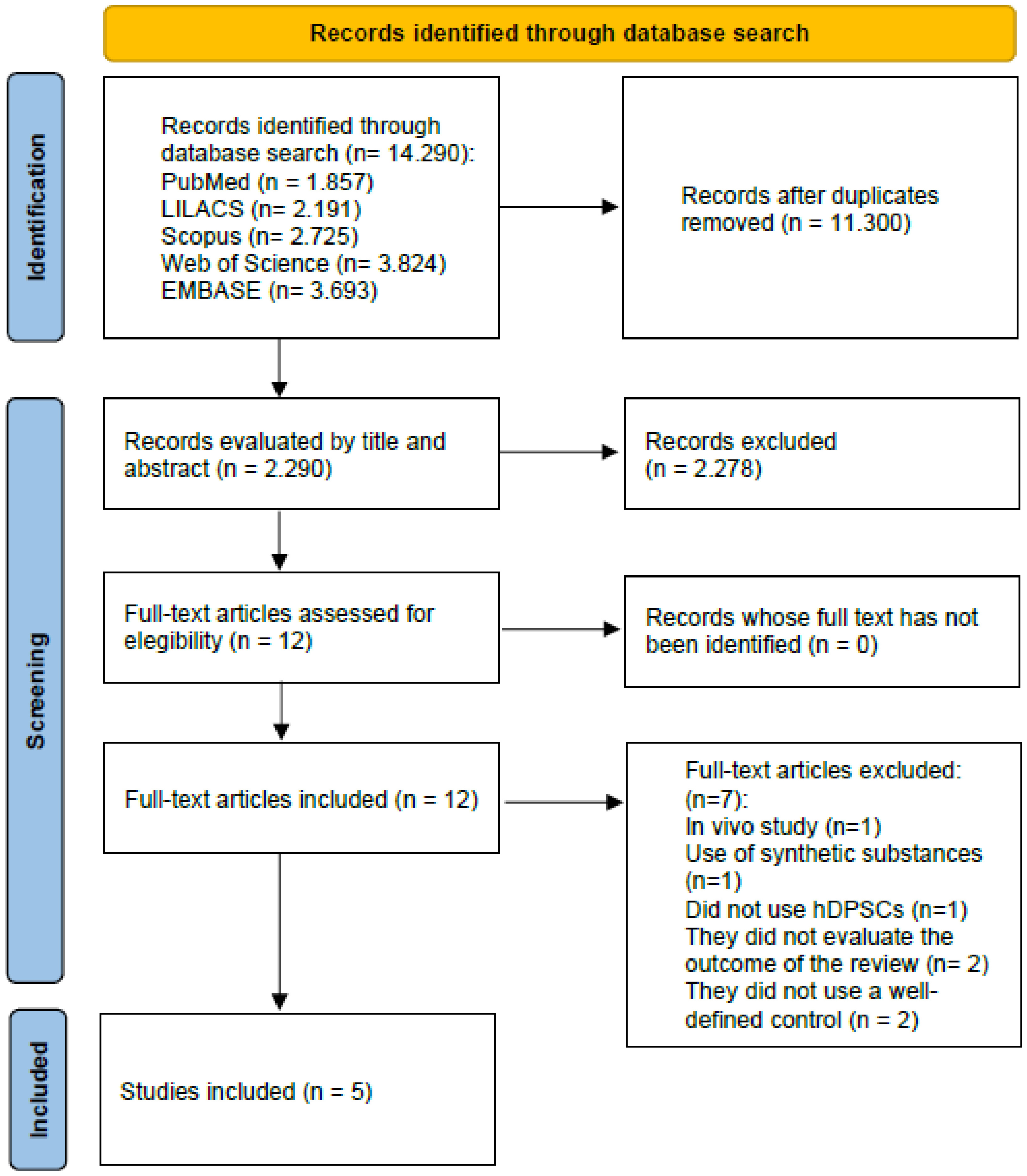
After applying the search strategies, a total of 14,290 articles were initially found in the six main databases. After removing duplicates and reading the titles and abstracts, 12 potentially relevant studies were selected for full-text reading. After applying the eligibility criteria, seven studies were excluded: one in vivo study, one study that used a synthetic substance, one study that did not use hDPSCs, two studies that did not evaluate the outcome of the systematic review, and two studies that did not use a well-defined control. In the end, five studies met the inclusion criteria and were considered for this systematic review (Figure 1).
Study characteristics
Seven types of natural substances were evaluated in this review, considering all the included studies, among which were: gelatin, hydroxyapatite, nano-hydroxyapatite, PRGF, agarose, lactose-modified chitosan, and growth factors. Additionally, different concentrations of sodium alginate, shapes, and types of scaffold preparation were reported in the selected studies. Among the control groups described in the studies, sodium alginate scaffolds without the addition of the evaluated substance and cells without the scaffold were used. Table III represents the general characteristics and outcomes of all the selected studies.
Risk of bias
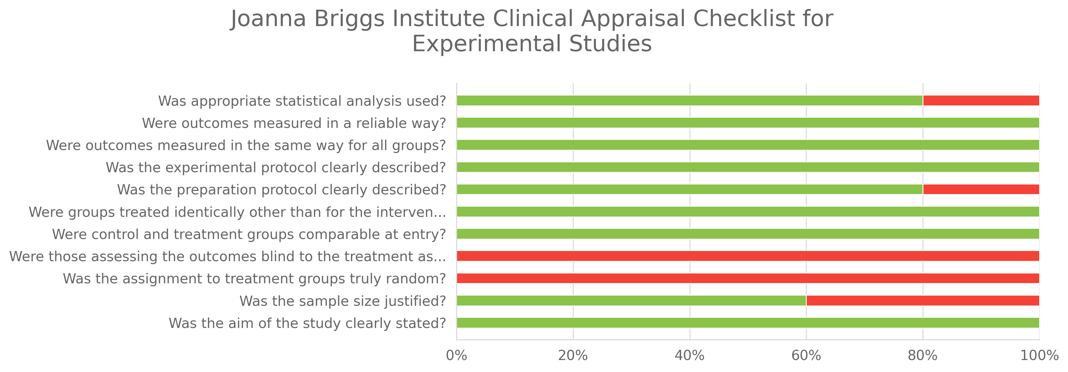
All articles reported a clear objective of the study, the control and treatment groups were comparable, the experimental protocols were clearly described, there was no randomization of groups or blinding of assessments, and the outcomes were measured reliably. However, 2 studies (40%) did not justify the sample size used, 1 study (20%) did not clearly describe the preparation protocol, and 1 study (20%) did not describe the statistical analysis. All articles presented a low risk of bias (Graphic 1).

Meta-analysis
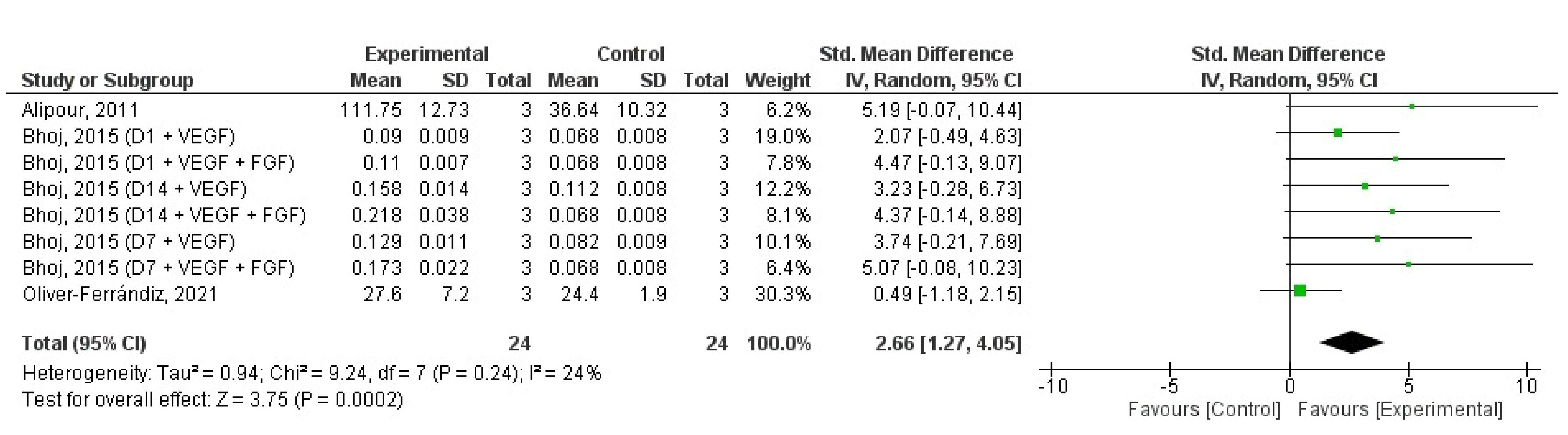
Out of the 5 studies included in the systematic review, only two were not included in the meta-analysis. The studies by Anitua et al. [19] and Porrelli et al. [23] were excluded for not specifying the number of samples used in each study group. The 3 included articles compared hDPSC proliferation between experimental and control groups after 1, 7, and 14 days [22], 3 weeks [21], and 4 weeks of cultivation [20].
In the meta-analysis, 48 specimens evenly distributed across the two study groups were evaluated. There was a significant benefit from incorporating natural substances into alginate with a Cohen's d of 2.66 [95% CI = 1.27 to 4.05]. There was no significant heterogeneity between studies (I2 = 24%, p = 0.240), and leave-one-out analysis showed that removing data from Bhoj et al. [22] significantly diluted the benefit found in the meta-analysis (Figure 2). Assessment of publication bias was not performed because the small number of included studies (n = 3) does not allow reliable application of funnel plots or regression-based tests.
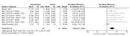
- Leave-one-out analysis of the effects of incorporating natural substances into sodium alginate on hDPSC proliferation.
Quality of evidence
According to the GRADE criteria assessment, the certainty of the evidence was considered moderate. Bias risk, imprecision, inconsistency, and indirect evidence were deemed "not serious"; however, publication bias was classified as highly suspicious. Thus, bias risk affected the overall quality of evidence. The evidence assessment can be seen in Table IV.
- Summary of findings from GRADE (Grading of Recommendations Assessment, Development and Evaluation criteria) table
DISCUSSION
This systematic review identified and analyzed the available scientific evidence on in vitro studies evaluating the effect of incorporating natural substances into sodium alginate used as a scaffold in the cultivation of human dental pulp stem cells (hDPSCs). According to the specific eligibility criteria, only five articles met the established inclusion criteria, highlighting the scarcity of studies using these materials.
For the meta-analysis, studies comparing the effects of substance incorporation into sodium alginate on cell proliferation were considered. As described in the results, 2 out of the 5 articles selected for the systematic review were excluded for not describing the number of samples used.
Dental pulp stem cells (DPSCs) are defined by their ability for self-renewal, proliferation, and differentiation into adult cells with various morphofunctional aspects. Stem cell populations are identified and characterized based on their tissue origins and differ in their differentiation capacities. Some stem cells have the potential to differentiate into various cell types composing human body tissues, while others differentiate into a limited number [24].
The dental pulp of permanent and deciduous teeth is a promising source of stem cells, namely hDPSCs, which can differentiate into various cell lineages such as osteoblasts, adipocytes, and chondrocytes [25]. Despite this versatile characteristic beneficial in tissue engineering, further research is needed to better understand their physiology, differentiation protocols, and thereby maximize their reparative potential [26].
Choosing the appropriate biomaterial for scaffold construction is crucial as it plays a fundamental role in providing cells with a suitable structure for adhesion, proliferation, and differentiation [27]. Natural origin biomaterials are interesting due to their microstructure, stability, biocompatibility, and ability to offer natural adhesion sites for cells. These can be biopolymers composed of amino acids or sugars, found both as components of the natural extracellular matrix (ECM) like collagen and fibrinogen, and in substances not naturally present in the ECM such as chitosan and sodium alginate. Moreover, when combined with other biopolymers, they allow for improvements in their properties [28-30].
Sodium alginate has been widely studied and used in tissue engineering due to its biocompatibility, biodegradability, ability to create three-dimensional structures tailored to specific applications, and use as scaffolds for cell culture and tissue regeneration matrices [31,32]. The ease of chemical modification of alginate also allows for the incorporation of bioactive compounds, growth factors, and cells to enhance its physical and biological properties [12].
Hydroxyapatite (Hap) is the main inorganic component of bones and teeth, comprising about 70% of the minerals present in bone structures. Chemically, hydroxyapatite belongs to the mineral group of apatites and is composed of calcium, phosphate, and hydroxyl ions in its crystalline matrix [33]. In tissue engineering, it has shown to be an important biomaterial due to its biocompatible and osteoconductive properties [34]. When incorporated into sodium alginate, Hap has been effective in promoting osteogenic proliferation and differentiation by providing biological signaling through the release of Ca and P ions [19,23].
Nanohydroxyapatite (nHap) is a nanocrystalline form of hydroxyapatite distinguished by its nanometric characteristics. Due to its small size and features like biocompatibility, osteoconductivity, lack of toxicity, and ability to be reabsorbed into bone tissue, nHap exhibits greater reactivity and interaction capability with cells and biological tissues at the molecular level, making it a valuable component in scaffolds for bone repair. nHap crystals enhance collagen fiber strength, facilitate and promote bone formation with increased cellular adhesion [21,35,36].
Gelatin, a natural biopolymer structurally similar to collagen, has been used over the years as a scaffold because it is biocompatible, mimics the ECM structure, and exhibits cell-responsive properties by allowing easy diffusion of growth factors and cytokines [37]. In seeking less invasive and more personalized therapies for tissue lesions, gelatin's ability to mimic the ECM and facilitate cell adhesion has been combined with sodium alginate's ability to support calcified matrix deposition, resulting in increased viability and proliferation of hDPSCs. This viability results from an appropriate pore size, providing a matrix with a permeable membrane that facilitates nutrient and oxygen transmission to cells [21].
Platelet-Rich Growth Factor (PRGF) is a platelet-derived product that involves separating and concentrating growth factors present in the patient's blood, which has been researched as an inducer of regenerative processes and healing. PRGF contains a variety of bioactive molecules, including growth factors and cytokines, which play crucial roles in modulating the cellular environment and regulating the inflammatory response [38,39]. Scaffolds with added PRGF have shown significant improvements in cell adhesion, chemotaxis, and proliferation. Additionally, this platelet derivative has been effective in promoting osteogenic differentiation due to the presence of growth factors and biological mediators involved in major tissue regeneration processes [19,40,41].
Agarose is a polysaccharide extracted from red seaweed that has been used in three-dimensional cell culture by allowing cellular growth and differentiation [42]. This compound has an adjustable capacity for water adsorption, and due to its rigidity and functional groups, it can also favor cell adhesion and proliferation [20]. Agarose combined with sodium alginate resulted in increased expression of collagen type II and aggrecan in hDPSC culture, consistent with its chondrogenic differentiation capacity described in the literature [43].
Chitosan, a polysaccharide derived from chitin, possesses interesting properties such as biocompatibility and biodegradability. When modified by introducing lactose groups, it can influence its interaction with proteins and tissues, facilitating controlled release of bioactive substances and regulation of inflammatory responses. Addition of lactose-modified chitosan (QTL) improved hDPSC adhesion on sodium alginate scaffold with hydroxyapatite [23]. It has been found to promote differentiation into chondrocytes [44] and neurons [45], but no effect was observed on cell proliferation. Furthermore, there was strong alkaline phosphatase expression, a marker of osteoblastic differentiation, when differentiation stimuli (dexamethasone, β-glycerophosphate, and potassium phosphate) were added. QTL acts as a multivalent ligand that, similar to ECM glycans, triggers a more responsive receptor clustering to differentiation stimuli [23].
Another approach being studied is the incorporation of growth factors into scaffolds due to their ability to direct cells to differentiate into specific cell types and stimulate proliferation. Studies have demonstrated the potential of adding growth factors to other types of biomaterials to induce cell homing, angiogenesis, and mineralized tissue formation due to their chemotactic effects [46-48].
One of the key elements in successful tissue engineering lies in maintaining vascularization of newly formed tissue to support its growth [49]. Some growth factors are more related to the onset of vasculogenesis, such as FGF-2 and VEGF, which are necessary for hemangioblast formation and allow differentiation and proliferation into angioblasts [50,51]. The effects of combining two types of growth factors (VEGF and FGF-2) in a sodium alginate scaffold model for use in pulp regeneration led to increased viable hDPSC numbers and cell proliferation [22].
Overall, the selection of a biomaterial used in scaffold creation represents a crucial step in ensuring an environment conducive to cell adhesion, proliferation, and differentiation, given that the materials used and their characteristics will have a direct influence on cell behavior [22]. Understanding the interaction between cells, biomaterials, and growth factors is essential to optimize the creation of more effective scaffolds [43,52,53].
This review has some limitations that should be considered. First, although the Joanna Briggs Institute checklist was used to assess methodological quality, some items such as randomization and blinding are not applicable to in vitro studies, which restricts the direct comparability of risk of bias across different study designs. Second, the number of eligible studies was limited, and only three could be included in the quantitative synthesis, which precluded a reliable assessment of publication bias through funnel plots or regression-based methods. These factors highlight the need for caution in interpreting the results and reinforce the importance of future investigations with larger and more standardized experimental designs.
CONCLUSION
Under the conditions of the present study, it can be concluded that the addition of different natural substances to sodium alginate, used as a scaffold, may contribute to creating a more favorable environment for the adhesion, proliferation, and differentiation of human dental pulp stem cells.
Data availability
The data that support the findings of this study are available from the corresponding author upon reasonable request.
-
How to cite:
Araújo LS, Maia SJM, Souza NO, Silva PGB, Saboia VPA. Effects of incorporating natural substances into sodium alginate in the cultivation of human dental pulp stem cells: a systematic review with meta-analysis. Braz. Dent. Sci. 2026;29:e4837. https://doi.org/10.4322/bds.2026.e4837
-
Funding
This research was supported by the Conselho Nacional de Desenvolvimento Científico e Tecnológico (CNPq), Brazil, through the provision of a scholarship to Lavina Sousa Araújo (Grant number: 130758/2021-3).
-
Regulatory Statement
This study did not involve human participants or animals and therefore did not require ethical approval. Regulatory compliance is not applicable.
REFERENCES
-
1 Avior Y, Sagi I, Benvenisty N. Pluripotent stem cells in disease modelling and drug discovery. Nat Rev Mol Cell Biol. 2016;17(3):170-82. https://doi.org/10.1038/nrm.2015.27 PMid:26818440.
» https://doi.org/10.1038/nrm.2015.27 -
2 McKee C, Chaudhry GR. Advances and challenges in stem cell culture. Colloids Surf B Biointerfaces. 2017;159:62-77. https://doi.org/10.1016/j.colsurfb.2017.07.051 PMid:28780462.
» https://doi.org/10.1016/j.colsurfb.2017.07.051 -
3 Rowe RG, Daley GQ. Induced pluripotent stem cells in disease modelling and drug discovery. Nat Rev Genet. 2019;20(7):377-88. https://doi.org/10.1038/s41576-019-0100-z PMid:30737492.
» https://doi.org/10.1038/s41576-019-0100-z -
4 Nuti N, Corallo C, Chan BMF, Ferrari M, Gerami-Naini B. Multipotent differentiation of human dental pulp stem cells: a literature review. Stem Cell Rev Rep. 2016;12(5):511-23. https://doi.org/10.1007/s12015-016-9661-9 PMid:27240827.
» https://doi.org/10.1007/s12015-016-9661-9 -
5 Devolder K. Complicity in stem cell research: the case of induced pluripotent stem cells. Hum Reprod. 2010;25(9):2175-80. https://doi.org/10.1093/humrep/deq176 PMid:20643694.
» https://doi.org/10.1093/humrep/deq176 -
6 Casagrande L, Cordeiro MM, Nör SA, Nör JE. Dental pulp stem cells in regenerative dentistry. Odontology. 2011;99(1):1-7. https://doi.org/10.1007/s10266-010-0154-z PMid:21271319.
» https://doi.org/10.1007/s10266-010-0154-z -
7 Egusa H, Sonoyama W, Nishimura M, Atsuta I, Akiyama K. Stem cells in dentistry – part II: clinical applications. J Prosthodont Res. 2012;56(4):229-48. https://doi.org/10.1016/j.jpor.2012.10.001 PMid:23137671.
» https://doi.org/10.1016/j.jpor.2012.10.001 -
8 Schmalz G, Smith AJ. Pulp development, repair, and regeneration: challenges of the transition from traditional dentistry to biologically based therapies. J Endod. 2014;40(4, Suppl):S2-5. https://doi.org/10.1016/j.joen.2014.01.018 PMid:24698689.
» https://doi.org/10.1016/j.joen.2014.01.018 -
9 Barry FP. Mesenchymal stem cell therapy in joint disease. In: Bock G, Goode J, editors. Novartis foundation symposia [Internet]. Chichester: John Wiley & Sons; 2008 [cited 2023 Aug 9]. p. 86-102. Available from: https://onlinelibrary.wiley.com/doi/10.1002/0470867973.ch7
» https://onlinelibrary.wiley.com/doi/10.1002/0470867973.ch7 -
10 Hospodiuk M, Dey M, Sosnoski D, Ozbolat IT. The bioink: A comprehensive review on bioprintable materials. Biotechnol Adv. 2017;35(2):217-39. https://doi.org/10.1016/j.biotechadv.2016.12.006 PMid:28057483.
» https://doi.org/10.1016/j.biotechadv.2016.12.006 -
11 Berthiaume F, Maguire TJ, Yarmush ML. Tissue engineering and regenerative medicine: history, progress, and challenges. Annu Rev Chem Biomol Eng. 2011;2(1):403-30. https://doi.org/10.1146/annurev-chembioeng-061010-114257 PMid:22432625.
» https://doi.org/10.1146/annurev-chembioeng-061010-114257 -
12 Lee KY, Mooney DJ. Alginate: properties and biomedical applications. Prog Polym Sci. 2012;37(1):106-26. https://doi.org/10.1016/j.progpolymsci.2011.06.003 PMid:22125349.
» https://doi.org/10.1016/j.progpolymsci.2011.06.003 -
13 Rastogi P, Kandasubramanian B. Review of alginate-based hydrogel bioprinting for application in tissue engineering. Biofabrication. 2019;11(4):042001. https://doi.org/10.1088/1758-5090/ab331e PMid:31315105.
» https://doi.org/10.1088/1758-5090/ab331e -
14 Giuseppe MD, Law N, Webb BA, Macrae R, Liew LJ, Sercombe TB, et al. Mechanical behaviour of alginate-gelatin hydrogels for 3D bioprinting. J Mech Behav Biomed Mater. 2018;79:150-7. https://doi.org/10.1016/j.jmbbm.2017.12.018 PMid:29304429.
» https://doi.org/10.1016/j.jmbbm.2017.12.018 -
15 Sancilio S, Gallorini M, Di Nisio C, Marsich E, Di Pietro R, Schweikl H, et al. Alginate/hydroxyapatite-based nanocomposite scaffolds for bone tissue engineering improve dental pulp biomineralization and differentiation. Stem Cells Int. 2018;2018:9643721. https://doi.org/10.1155/2018/9643721 PMid:30154869.
» https://doi.org/10.1155/2018/9643721 -
16 Page MJ, McKenzie JE, Bossuyt PM, Boutron I, Hoffmann TC, Mulrow CD, et al. The PRISMA 2020 statement: an updated guideline for reporting systematic reviews. BMJ. 2021;372:n71. https://doi.org/10.1136/bmj.n71 PMid:33782057.
» https://doi.org/10.1136/bmj.n71 -
17 Munn Z, Stone JC, Aromataris E, Klugar M, Sears K, Leonardi-Bee J, et al. Assessing the risk of bias of quantitative analytical studies: introducing the vision for critical appraisal within JBI systematic reviews. JBI Evid Synth. 2023;21(3):467-71. https://doi.org/10.11124/JBIES-22-00224 PMid:36476419.
» https://doi.org/10.11124/JBIES-22-00224 -
18 McMaster University. GRADE pro-GDT software [Internet]. Hamilton: McMaster University and Evidence Prime Inc.; 2023 [cited 2023 Aug 9]. Available from: http://gdt.guidelinedevelopment.org
» http://gdt.guidelinedevelopment.org -
19 Anitua E, Zalduendo M, Troya M, Erezuma I, Lukin I, Hernáez-Moya R, et al. Composite alginate-gelatin hydrogels incorporating PRGF enhance human dental pulp cell adhesion, chemotaxis and proliferation. Int J Pharm. 2022;617:121631. https://doi.org/10.1016/j.ijpharm.2022.121631 PMid:35247496.
» https://doi.org/10.1016/j.ijpharm.2022.121631 -
20 Oliver-Ferrándiz M, Milián L, Sancho-Tello M, Martín de Llano JJ, Gisbert Roca F, Martínez-Ramos C, et al. Alginate-agarose hydrogels improve the in vitro differentiation of human dental pulp stem cells in chondrocytes: a histological study. Biomedicines. 2021;9(7):834. https://doi.org/10.3390/biomedicines9070834 PMid:34356898.
» https://doi.org/10.3390/biomedicines9070834 -
21 Alipour M, Firouzi N, Aghazadeh Z, Samiei M, Montazersaheb S, Khoshfetrat AB, et al. The osteogenic differentiation of human dental pulp stem cells in alginate-gelatin/Nano-hydroxyapatite microcapsules. BMC Biotechnol. 2021;21(1):6. https://doi.org/10.1186/s12896-020-00666-3 PMid:33430842.
» https://doi.org/10.1186/s12896-020-00666-3 -
22 Bhoj M, Zhang C, Green DW. A first step in de novo synthesis of a living pulp tissue replacement using dental pulp mscs and tissue growth factors, encapsulated within a bioinspired alginate hydrogel. J Endod. 2015;41(7):1100-7. https://doi.org/10.1016/j.joen.2015.03.006 PMid:25958179.
» https://doi.org/10.1016/j.joen.2015.03.006 -
23 Porrelli D, Gruppuso M, Vecchies F, Marsich E, Turco G. Alginate bone scaffolds coated with a bioactive lactose modified chitosan for human dental pulp stem cells proliferation and differentiation. Carbohydr Polym. 2021;273:118610. https://doi.org/10.1016/j.carbpol.2021.118610 PMid:34561009.
» https://doi.org/10.1016/j.carbpol.2021.118610 -
24 Lemischka IR. Stem cell biology: a view toward the future. Ann N Y Acad Sci. 2005;1044(1):132-8. https://doi.org/10.1196/annals.1349.017 PMid:15958706.
» https://doi.org/10.1196/annals.1349.017 -
25 Gronthos S, Mankani M, Brahim J, Robey PG, Shi S. Postnatal human dental pulp stem cells (DPSCs) in vitro and in vivo. Proc Natl Acad Sci USA. 2000;97(25):13625-30. https://doi.org/10.1073/pnas.240309797 PMid:11087820.
» https://doi.org/10.1073/pnas.240309797 -
26 Volponi AA, Sharpe PT. The tooth: a treasure chest of stem cells. Br Dent J. 2013;215(7):353-8. https://doi.org/10.1038/sj.bdj.2013.959 PMid:24113958.
» https://doi.org/10.1038/sj.bdj.2013.959 -
27 Afewerki S, Sheikhi A, Kannan S, Ahadian S, Khademhosseini A. Gelatin-polysaccharide composite scaffolds for 3D cell culture and tissue engineering: towards natural therapeutics. Bioeng Transl Med. 2018;4(1):96-115. https://doi.org/10.1002/btm2.10124 PMid:30680322.
» https://doi.org/10.1002/btm2.10124 -
28 Guarino V, Cirillo V, Ambrosio L. Bicomponent electrospun scaffolds to design extracellular matrix tissue analogs. Expert Rev Med Devices. 2016;13(1):83-102. https://doi.org/10.1586/17434440.2016.1126505 PMid:26619260.
» https://doi.org/10.1586/17434440.2016.1126505 -
29 Ahadian S, Civitarese R, Bannerman D, Mohammadi MH, Lu R, Wang E, et al. Organ-on-a-chip platforms: a convergence of advanced materials, cells, and microscale technologies. Adv Healthc Mater. 2018;7(2):1700506. https://doi.org/10.1002/adhm.201700506 PMid:29034591.
» https://doi.org/10.1002/adhm.201700506 -
30 Ullah S, Chen X. Fabrication, applications and challenges of natural biomaterials in tissue engineering. Appl Mater Today. 2020;20:100656. https://doi.org/10.1016/j.apmt.2020.100656
» https://doi.org/10.1016/j.apmt.2020.100656 -
31 Draget KI, Smidsrød O, Skjåk‐Bræk G. Alginates from Algae. In: Steinbüchel A, editor. Biopolymers online. 1st ed. Weinheim: Wiley; 2002. https://doi.org/10.1002/3527600035.bpol6008
» https://doi.org/10.1002/3527600035.bpol6008 -
32 Draget KI, Taylor C. Chemical, physical and biological properties of alginates and their biomedical implications. Food Hydrocoll. 2011;25(2):251-6. https://doi.org/10.1016/j.foodhyd.2009.10.007
» https://doi.org/10.1016/j.foodhyd.2009.10.007 -
33 Deng Y, Wang H, Zhang L, Li Y, Wei S. In situ synthesis and in vitro biocompatibility of needle-like nano-hydroxyapatite in agar–gelatin co-hydrogel. Mater Lett. 2013;104:8-12. https://doi.org/10.1016/j.matlet.2013.03.145
» https://doi.org/10.1016/j.matlet.2013.03.145 - 34 Li Z, Sheng P, Li C, Yuan G, Deng Y. Osteoimmunology in bone regeneration. BioMed Res Int. 2020;2020:6297356. PMid:32766310.
-
35 Shadjou N, Hasanzadeh M, Khalilzadeh B. Graphene based scaffolds on bone tissue engineering. Bioengineered. 2018;9(1):38-47. https://doi.org/10.1080/21655979.2017.1373539 PMid:29095664.
» https://doi.org/10.1080/21655979.2017.1373539 -
36 Bordea IR, Candrea S, Alexescu GT, Bran S, Băciuț M, Băciuț G, et al. Nano-hydroxyapatite use in dentistry: a systematic review. Drug Metab Rev. 2020;52(2):319-32. https://doi.org/10.1080/03602532.2020.1758713 PMid:32393070.
» https://doi.org/10.1080/03602532.2020.1758713 - 37 Echave MC, Saenz del Burgo L, Pedraz JL, Orive G. Gelatin as biomaterial for tissue engineering. Curr Pharm Des. 2017;23(24):3567-84. PMid:28494717.
-
38 Luo Y, Lode A, Akkineni AR, Gelinsky M. Concentrated gelatin/alginate composites for fabrication of predesigned scaffolds with a favorable cell response by 3D plotting. RSC Adv. 2015;5(54):43480-8. https://doi.org/10.1039/C5RA04308E
» https://doi.org/10.1039/C5RA04308E -
39 Anitua E, De La Fuente M, Muruzabal F, Riestra A, Merayo-Lloves J, Orive G. Plasma rich in growth factors (PRGF) eye drops stimulates scarless regeneration compared to autologous serum in the ocular surface stromal fibroblasts. Exp Eye Res. 2015;135:118-26. https://doi.org/10.1016/j.exer.2015.02.016 PMid:25708868.
» https://doi.org/10.1016/j.exer.2015.02.016 -
40 Anitua E, Alkhraisat MH, Orive G. Perspectives and challenges in regenerative medicine using plasma rich in growth factors. J Control Release. 2012;157(1):29-38. https://doi.org/10.1016/j.jconrel.2011.07.004 PMid:21763737.
» https://doi.org/10.1016/j.jconrel.2011.07.004 -
41 Anitua E, Troya M, Zalduendo M, Orive G. Personalized plasma-based medicine to treat age-related diseases. Mater Sci Eng C. 2017;74:459-64. https://doi.org/10.1016/j.msec.2016.12.040 PMid:28254317.
» https://doi.org/10.1016/j.msec.2016.12.040 -
42 Salati MA, Khazai J, Tahmuri AM, Samadi A, Taghizadeh A, Taghizadeh M, et al. Agarose-based biomaterials: opportunities and challenges in cartilage tissue engineering. Polymers. 2020;12(5):1150. https://doi.org/10.3390/polym12051150 PMid:32443422.
» https://doi.org/10.3390/polym12051150 -
43 Mata M, Milian L, Oliver M, Zurriaga J, Sancho-Tello M, Martín de Llano JJ, et al. In vivo articular cartilage regeneration using human dental pulp stem cells cultured in an alginate scaffold: a preliminary study. stem cells Int. 2017;2017:8309256. https://doi.org/10.1155/2017/8309256 PMid:28951745.
» https://doi.org/10.1155/2017/8309256 -
44 Donati I, Stredanska S, Silvestrini G, Vetere A, Marcon P, Marsich E, et al. The aggregation of pig articular chondrocyte and synthesis of extracellular matrix by a lactose-modified chitosan. Biomaterials. 2005;26(9):987-98. https://doi.org/10.1016/j.biomaterials.2004.04.015 PMid:15369687.
» https://doi.org/10.1016/j.biomaterials.2004.04.015 -
45 Medelin M, Porrelli D, Aurand ER, Scaini D, Travan A, Borgogna MA, et al. Exploiting natural polysaccharides to enhance in vitro bio-constructs of primary neurons and progenitor cells. Acta Biomater. 2018;73:285-301. https://doi.org/10.1016/j.actbio.2018.03.041 PMid:29621637.
» https://doi.org/10.1016/j.actbio.2018.03.041 -
46 d’Aquino R, Graziano A, Sampaolesi M, Laino G, Pirozzi G, De Rosa A, et al. Human postnatal dental pulp cells co-differentiate into osteoblasts and endotheliocytes: a pivotal synergy leading to adult bone tissue formation. Cell Death Differ. 2007;14(6):1162-71. https://doi.org/10.1038/sj.cdd.4402121 PMid:17347663.
» https://doi.org/10.1038/sj.cdd.4402121 -
47 Mullane EM, Dong Z, Sedgley CM, Hu JCC, Botero TM, Holland GR, et al. Effects of VEGF and FGF2 on the revascularization of severed human dental pulps. J Dent Res. 2008;87(12):1144-8. https://doi.org/10.1177/154405910808701204 PMid:19029083.
» https://doi.org/10.1177/154405910808701204 -
48 Kim JY, Xin X, Moioli EK, Chung J, Lee CH, Chen M, et al. Regeneration of dental-pulp-like tissue by chemotaxis-induced cell homing. Tissue Eng Part A. 2010;16(10):3023-31. https://doi.org/10.1089/ten.tea.2010.0181 PMid:20486799.
» https://doi.org/10.1089/ten.tea.2010.0181 -
49 Jabbarzadeh E, Starnes T, Khan YM, Jiang T, Wirtel AJ, Deng M, et al. Induction of angiogenesis in tissue-engineered scaffolds designed for bone repair: a combined gene therapy–cell transplantation approach. Proc Natl Acad Sci USA. 2008;105(32):11099-104. https://doi.org/10.1073/pnas.0800069105 PMid:18678895.
» https://doi.org/10.1073/pnas.0800069105 -
50 Flamme I, Risau W. Induction of vasculogenesis and hematopoiesis in vitro. Development. 1992;116(2):435-9. https://doi.org/10.1242/dev.116.2.435 PMid:1286617.
» https://doi.org/10.1242/dev.116.2.435 - 51 Ferrara N. Vascular endothelial growth factor. ATVB. 2009;29(6):789-91.
-
52 Kumabe S, Nakatsuka M, Kim GS, Jue SS, Aikawa F, Shin JW, et al. Human dental pulp cell culture and cell transplantation with an alginate scaffold. Okajimas Folia Anat Jpn. 2006;82(4):147-55. https://doi.org/10.2535/ofaj.82.147 PMid:16526573.
» https://doi.org/10.2535/ofaj.82.147 -
53 Umemura E, Yamada Y, Nakamura S, Ito K, Hara K, Ueda M. Viable cryopreserving tissue-engineered cell-biomaterial for cell banking therapy in an effective cryoprotectant. Tissue Eng Part C Methods. 2011;17(8):799-807. https://doi.org/10.1089/ten.tec.2011.0003 PMid:21517691.
» https://doi.org/10.1089/ten.tec.2011.0003
Edited by
-
Editor-in-chief:
Sergio Eduardo de Paiva Gonçalves
-
Section Editor:
Renata Falchete do Prado