Patient-reported outcome measures between digital and conventional splints for bruxism: systematic review
ABSTRACT
Background The integration of digital workflows in dentistry has raised questions about the effectiveness of digital occlusal stabilizing splints (OSS) in bruxism management.
Objective This systematic review evaluates whether computer-aided design and computer-aided manufacturing (CAD-CAM) OSS provide superior Patient-Reported Outcome Measures (PROMs) compared to conventional splints.
Material and Methods This review was registered in PROSPERO (CRD42024536412). A comprehensive search was conducted in six databases (MEDLINE/PubMed, Scopus, Embase, Web of Science, CENTRAL, and BVS) up to March 2024. Eligible RCTs comparing digital and conventional OSS in PROMs were included. After screening 4,269 records, four studies met the inclusion criteria.
Results Primary outcomes such as comfort, appearance, and quality of life favored digital OSS. Secondary outcomes, including time spent, T-scan analysis, electromyography (EMG) activity, and stability assessed by the operator, also suggested advantages for digital splints. Although, the certainty of evidence of the included studies was assessed as low to very low for several outcomes.
Conclusion Digital OSS appears to improve PROMs in bruxism patients. However, variations in splint materials, thicknesses, and PROM assessments across studies complicate direct comparisons and result clustering, due to the limited and preliminary nature of the evidence. Further standardized research is needed to validate these findings.
KEYWORDS:
Bruxism; Computer-aided design and manufacturing; Digital workflows; Occlusal splints; Patient-reported outcome measures
RESUMO
Contexto A integração do fluxo digital na odontologia suscitou questões sobre a eficácia de placas oclusal estabilizadora (OSS) no manejo do bruxismo.
Objetivo Esta revisão sistemática avalia se as OSS confeccionadas por meio de design auxiliado por computador e manufatura auxiliada por computador (CAD-CAM) proporcionam melhores medidas de desfecho relatadas pelos pacientes (PROMs) em comparação com as placas convencionais.
Material e Métodos Essa revisão foi registrada no PROSPERO (CRD42024536412). A ferramenta de risco de viés de Cochrane (Rob-2) foi utilizada para avaliar o viés nos ensaios clínicos randomizados (RCTs), e a certeza das evidências foi determinada por meio da abordagem GRADE. Uma busca abrangente foi realizada em seis bases de dados (MEDLINE/PubMed, Scopus, Embase, Web of Science, CENTRAL e BVS) até março de 2024. Foram incluídos RCTs elegíveis que compararam OSS digitais e convencionais em termos de PROMs. Após a análise de 4,269 registros, quatro estudos atenderam os critérios de inclusão.
Resultados Os desfechos primários, como conforto, aparência e qualidade de vida foram mais favoráveis às OSS digitais. Os desfechos secundários, incluindo tempo gasto, análise cm T-scan, atividade eletromiográfica (EMG) relacionada ao sono e estabilidade avaliada pelo operador também sugeriram vantagens para as placas digitais. No entanto, a certeza da evidência dos estudos incluídos foi avaliada como baixa a muito baixa para vários desfechos.
Conclusão As OSS digitais demonstram potencial para melhorar PROMs em pacientes com bruxismo. Porém, variações em materiais das placas, espessura e métodos de avaliação de PROMs entre os estudos dificultam comparações diretas e agrupamento de resultados, devido à natureza limitada e preliminar das provas. São necessárias mais pesquisas padronizadas para validar estes resultados.
PALAVRAS-CHAVE:
Bruxismo; Desenho assistido por computador; Fluxo de trabalho digital; Placas oclusais; Medidas de resultados relatados pelo paciente
INTRODUCTION
The definition of bruxism has evolved from being considered a pathology or disorder to being seen as a motor activity and behavior. This activity can have potential physiological or protective significance and may even indicate underlying conditions acting as a risk factor for detrimental disorders [1-3]. In 2018, bruxism during sleep (sleep bruxism - SB) was defined as rhythmic (phasic) or non-rhythmic (tonic), while bruxism during wakefulness (awake bruxism - AB) was characterized by repetitive or sustained tooth contact and/or by bracing or thrusting of the mandible [2]. In both cases, it is not considered a movement or sleep disorder (in SB) in otherwise healthy individuals. As a masticatory muscle activity, bruxism can potentially lead to clinical consequences, such as mechanical tooth wear, musculoskeletal pain [4], muscle hypertrophy, tongue indentations, or lip and/or linea alba on the inner cheek.
The occlusal stabilizing splint (OSS) promotes posterior contacts, anterior guidance on anterior teeth, and lateral guidance on canines. The thickness must be the minimum possible to allow occlusal adjustments and achieve bilateral contacts [5-9]. The OSS prevents potential harmful effects caused by bruxism [5,7,9-11]. These splints can be produced on a conventional workflow using impression and acrylic resin-type materials. Some disadvantages of this method are time consumption, allergies, dimensional instability, and susceptibility to human errors [12,13]. Evolving digital technologies based on computer-aided design and computer aided manufacturing (CAD/CAM) allow a digital workflow with scanning and subtractive or additive manufacturing [13]. A digital workflow promises an optimized process, precision fit, and comfort, simplifying the number of materials and steps [12]. The choice of the splint material also plays a crucial role in determining splint longevity and performance, as the mechanical properties achieved with 3D-printed and milled materials are clinically relevant [12-24].
Recent literature on OSS [7,14] has suggested the importance of evaluating variables concerning patient satisfaction outcomes, known as Patient-Reported Outcome Measures (PROMs) when analyzing the efficacy of different treatment modalities and understanding the patient’s viewpoint within the care process [14]. Despite the growing adoption of digital procedures, there remains a literature gap regarding the comparative efficacy of digital OSS relative to traditional. Especially after the 2018 International consensus on the assessment of bruxism [2]. The authors are unaware of a previous systematic review that summarized the available evidence regarding the efficacy of digital OSS in a homogeneous population of patients with bruxism. A 2022 systematic review [25] reported the effectiveness of digital OSS in a heterogeneous population, which included arthrogenous and myogenous TMD mixed with bruxism patients. The inclusion of arthrogenous TMD in that 2022 systematic review [25], allows the potential consideration of other degenerative disorders and osteoarthritis, in which digital OSS wouldn't be the primary treatment, and the efficacy of this approach could not be adequately evaluated. The 2017 systematic review [26] included two studies that involved patients with disc displacement with and without reduction, muscle disorders, and arthralgia. Neither study reported the prevalence of bruxism in the population or the occurrence of osteoarthritis or joint degenerative disorders.
Therefore, this systematic review aims to assess the effectiveness of digital OSS in a population with bruxism. The research question is whether CAD/CAM OSS provides better PROMs than conventional splints.
MATERIAL AND METHODS
This systematic review was conducted by the Preferred Reporting Items for Systematic Reviews and Meta-Analyses (PRISMA) guidelines [27] (Supplementary Material 1) and is registered in the International Prospective Register of Systematic Reviews (PROSPERO) (CRD42024536412). The review utilized the Population, Intervention, Comparison, Outcome (PICO) framework. The PICO question was “In patients with bruxism, how do digital occlusal stabilization splints compare to conventional occlusal stabilization splints in terms of PROMs?”.
A comprehensive literature search was performed without language restrictions through March 2024 in six databases [(MEDLINE via PubMed, Scopus, Embase, Web of Science, CENTRAL, and Biblioteca Virtual em Saúde (BVS)] following specific inclusion and exclusion criteria. The inclusion criteria for this systematic review encompass randomized clinical trials (RCTs) involving patients with bruxism assessment, evaluating PROMs, that compared conventional and digital OSS.
Conversely, the exclusion criteria comprise animal studies, systematic reviews, retrospective studies, case reports, and case series. Furthermore, articles that were not available online, studies involving patients diagnosed with arthrogenous, myogenous, and mixed TMD symptoms were excluded. Research that does not evaluate OSS was excluded.
Medical subject heading terms and keywords were combined with Boolean operators to search the databases. The search strategy can be consulted in Table I.
In the first phase of study selection, the Rayyan web application was used to remove duplicates and to select articles by title and abstract. After removing duplicates, two reviewers (J.O. and M.K.) independently screened the titles and abstracts of the articles. Disagreements were resolved via discussion with a senior reviewer (S.O.). Studies that appeared to meet the inclusion criteria and lacked information in their titles and abstracts were selected for assessment of the full-text article in the second phase. The same reviewers independently assessed the full texts to determine if the studies were eligible, articles with unclear relevance based on titles or abstracts were retained for further evaluation. One of the articles was written in Chinese [28], and to assess its content and eligibility, it was translated by a professional translator. We calculated Cohen’s kappa coefficient [29] to measure the agreement between reviewers during the phases (title, abstract, and full text) of the selection process.
The risk of methodological bias was independently evaluated by the two reviewers (J.O. and M.K.), and any disagreement was resolved via consultation with a senior reviewer (S.O.). The revised Cochrane tool for assessing the risk of bias in randomized trials (RoB-2) [30] was used. Two reviewers independently evaluated each study, and disagreements were resolved through discussion with a senior reviewer. The overall risk of bias was determined based on domain-specific evaluations. The overall risk of bias was determined based on the assessment of each domain.
The certainty of the evidence was determined for each outcome by using the grading of recommendations assessment, development, and evaluation (GRADE) approach as very low, low, moderate, or high [31].
A data extraction spreadsheet was independently created by two reviewers. Variables extracted included participant characteristics (e.g., bruxism assessment, age), intervention characteristics (e.g., splint material, manufacturing method), and study details (Table II). Data were collected for all outcomes reported in the included studies that aligned with the primary and secondary outcome domains of interest. The primary outcomes included PROMs: comfort (wearing comfort, impression technique preference, habituation time, encroachment on mouth and tongue, size and volume, stability of fit, and ease of handling), appearance, quality of life, retention, occlusal stability (initial stability), and preference between therapies. Secondary outcomes included measures related to patients but assessed by operator or equipment: time spent during the workflow (time required for adjustment and manual time spent), occlusal stability (initial and final stability by the operator and occlusal contact by T-Scan), retention by the operator, general tonic and phasic contractions with and without splints, sMMA (surface masseter muscle activity) with splints and without splints, and overall SB index, as detailed in Table III. Data for all measures and time points reported in the studies were extracted to align with the pre-defined outcome domains. For each outcome, all relevant results were gathered from the studies, including variations in measurement scales (e.g., VAS, T-scan, OHIP-14), time points (e.g., with and without splints), and specific subgroup analyses when available. Conflicts that arose during data extraction were resolved through discussion with a third reviewer.
Due to the high heterogeneity in methodologies and outcome measures, a narrative synthesis was conducted. Results were grouped by outcome domain and compared across studies to identify patterns and inconsistencies. Sensitivity analyses were not conducted due to the limited number of studies included in the synthesis.
RESULTS
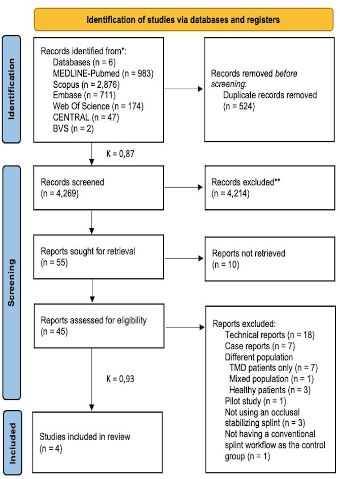
The literature identification and selection process was illustrated in the PRISMA [27] flowchart (Figure 1). In the selection process, Cohen’s kappa coefficient [29] was 0.87 for title and abstract and 0.93 for full-text selection. The search yielded 4,269 unique records, of which 4 studies were included in the qualitative synthesis.
Details of the included articles, consisting of 4 RCTs [15,28,32,33], are provided in the data extraction sheet (Tables II and III). Although all articles address PROMs, each author utilized different methodologies as different interocclusal record methods, splint materials (PMMA, PEEK, and 3D-printed resin), manufacture methods (3D-printing and milling), and minimum splint thickness parameters (0.5-2mm) in both the intervention and comparison (hard acrylic resin, PET-G rebased with autopolymerizing resin and soft splints using a vacuum-forming machine) groups, along with varying scales and evaluation techniques to measure PROMs, which complicates the analysis and clustering of the results.
Primary outcomes
MILLED PEEK SPLINTS [0.5mm milled PEEK vs. 2mm hard transparent acrylic resin [32] and 1mm milled PEEK vs. 2mm soft splints [28].
Two studies [28,32] found that the test group had statistically significantly higher comfort scores. One study [28] showed significant differences in appearance between the two splints, with higher scores for the digital one.
MILLED PMMA SPLINTS (1mm milled PMMA vs. 1mm prosthetic resin) [33]
On the other hand, one study [33] showed that ease of handling was significantly favored in digital splints. Also, regarding the impression technique, there was a significant preference for intraoral scanning over traditional impressions [33].
3D-PRINTED SPLINTS (2mm 3D-printed resin vs. PET-G rebased with autopolymerizing resin 2.0mm) [15]
For quality of life, one study [15], reported improvements in digital splints with a significant improvement in lifestyle and perceived reduction of their parafunction.
Secondary outcomes
Secondary outcomes, including time spent, T-scan analysis of occlusal contacts, retention, and stability as assessed by the operator, suggest that the digital approach may offer potential advantages compared to the conventional method. For time spent between devices, Wang et al. [32] evaluated time on impression, manufacture, and occlusal adjustment, and it was shown that less time was spent in the digital workflow. In contrast, Brandt et al. [33] evaluated the time needed for adjustments between devices and found no notable differences. Aiming stability occlusal contacts Wang et al. [28] demonstrated improved stability on T-scan occlusal in the digital group.
In the domain of SB and electromyography (EMG) activity, it was reported significant differences in tonic contractions between groups and between times, with conventional devices being more effective in reducing tonic contractions [15]. Significant differences between recording times were observed for general phasic contractions, but not between groups. During night recordings without splints, differences were observed for general phasic tonic contractions. For the total sMMA during night recordings without the use of splints, there was a significant difference between recording times. Lastly, no significant difference was observed in the overall SB index analysis [15].
Risk of bias assessment
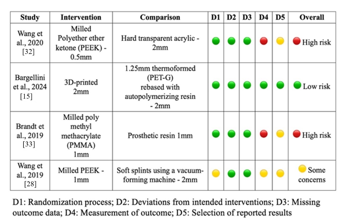
The assessment of the risk of bias using the RoB-2 tool is shown in Figure 2, with two RCTs assessed as high risk [32,33], one as low risk [15], and one with some concerns about bias [28]. The RCTs assessed as high risk [32,33] have deficiencies in the measurement of the outcome and selection of the reported result. The RCT [28] assessed as having some concerns about bias, presented the same deficiencies as the two reported as high risk, and had problems in the randomization process. A meta-analysis could not be performed due to the heterogeneity in the presentation of results across the studies.

The certainty of the evidence is reported in Table IV and was “high” for quality of life [15] and SB-related EMG activity [15]. The study that analyses these outcomes presents no risk of bias and has shown statistically significant differences between groups [15]. A “moderate” certainty of the evidence was found for appearance [28] and the SB overall index [15]. The reason for downgrading one level of certainty is that those studies [15,28] presented some type of risk of bias or did not show statistically significant differences between groups. The certainty of the evidence was “low” for comfort [28,32,33] and preference [33] as the methods used to measure the results were quite heterogeneous, and the comparator and intervention were different in each trial. The certainty of the evidence was “very low” for retention and fit [28,32,33]. The reason for lowering the level of evidence was that there were no statistically significant differences between the groups.
DISCUSSION
This systematic review aimed to investigate whether digitally manufactured occlusal stabilizing splints yield superior PROMs compared to conventional splints for patients with bruxism. The results showed that digital splints have the potential to improve patient satisfaction when compared to conventional OSS. These inquiries hold significant relevance for the dental healthcare system given the prevalence of bruxism in the general population [4]. Overall, OSS is a preferred intervention for both preventing teeth wear in bruxism and alleviating pain in myogenous TMD [7,14].
In this systematic review, only clinical trials [15,28,32,33] were included in which the test group used a fully digital workflow, including intraoral scanning and CAD/CAM. The analysis of the studies reveals some significant differences in favor of the test group across various parameters, however, there is difficulty in clustering these results since there is a difference between methodology workflow and splint material chosen for manufacturing. It is important to consider the different material choices used across various groups, as variations in material malleability could lead to bias when comparing clinical results [15,28,32,33]. These differences may account for the observed PROMs. Bargellini et al. [15] produced a 3D-printed splint with a virtual increase in the occlusal vertical dimension (OVD) of up to 2 mm between the molars but did not provide information about the interocclusal record method. Brandt et al. [33] chose PMMA (1mm) and registered the interocclusal relationship in the maximum intercuspal position. Two studies [28,32] utilized PEEK but with different approaches; one study [32] used 0.5mm disocclusion between molars and applied a leaf gauge for interocclusal record, while the other [28] minimum thickness was 1mm, and the interocclusal record methods were unclear.
For comfort, it is suggested that digital OSS outperforms traditional ones. This review encompasses studies that examine splints made from various thicknesses and materials, which can significantly affect comfort, fit, and clinical efficacy. The use of more resistant materials like milled PEEK splints allows the operator to do thinner splints [0.5-1mm] that may provide enhanced stability and durability without the expense of comfort. Conversely, if the material of choice does not present sufficient hardness, thinner splints could be manufactured to offer improved comfort but may be less durable. In this review, the two studies [28,32] using PEEK showed statistically significant differences in wearing comfort, whether no difference was noted in the study [32] that used PMMA (1mm). On the other hand, also the methodology of comparison groups, the choice of splint material, and the minimal thickness in the conventional workflow are matters of interest. For instance, Wang et al. [28] utilized soft splints (2 mm) created with a vacuum-forming machine to compare against milled PEEK splints, which is a concern as it did not include a control group with rigid splints. In the other two studies [32,33] the control group used hard resin, while Brandt et al. [33] compared prosthetic resin (1mm) to milled PMMA. Additionally, Wang et al. [32] compared hard acrylic resin (2mm) to milled PEEK splints. The difference in resilience could introduce a bias in comfort perception and improve the wearing comfort of the patient, as literature reported that the electrical activity of masticatory muscles in patients with bruxism using hard splints differed statistically from soft ones, which could cause an awareness of tiredness; and hard splint, as well as natural dentition, did not cause the same awareness [28]. Also, the interocclusal record method to obtain the splint may influence the splint comfort, as Brandt et al. [33] registered the interocclusal relationship in the maximum intercuspal position and Wang et al. [32] applied a leaf gauge for interocclusal record.
It is suggested that digital occlusal splints outperform traditional ones in terms of appearance, quality of life, and sleep-related EMG activity. However, each outcome was evaluated in only one study. Aesthetic considerations were higher for 1mm milled PEEK splints compared to vacuum-formed soft splints (2mm) [28]. Additionally, quality of life measures were superior for 3D-printed splints (2mm) compared to traditional splints made using a mixed technique with PET-G rebased with autopolymerizing resin (2mm) [15]. The digital group was highly effective in promoting better oral health outcomes, and the impression technique did not influence the analysis; in this study, [15] the traditional group deviated from a fully conventional workflow. Instead of using an analog impression, the author performed an intraoral scan to create 3D-printed casts, followed by the fabrication of conventional splints.
The only study that assessed an outcome directly related to bruxism management was Bargellini et al. [15], which reported significant differences between groups in SB and EMG activity. Conventional splints were more effective in reducing tonic contractions, while both splint types showed a similar impact on phasic contractions. These findings appear to be linked to flexural strength: 3D-printed splints made from a single rigid resin with higher flexural strength (75 MPa) may stabilize phasic contractions within one month but negatively affect tonic contractions, leading to their increase. Conversely, conventional splints produced from thermoformed PET-G and rebased with more elastic resins, despite their lower flexural strength (>50–69 MPa), seem to reduce tonic contractions but are associated with a progressive increase in phasic contractions. This suggests that digital splints are not invariably superior and that material properties are a key factor influencing clinical performance. When observing the total of sMMA during night recordings without the use of splints, significant differences between recording times were noted. This suggests that the absence of a splint affected sMMA, but it does not provide a direct comparison of effectiveness between the two groups. It is important to emphasize technique sensitivity when using 3D-printed resin as the manufacturing material choice. The 3D workflow involves data acquisition, digital design, pre-processing, printing, and post-processing. The combinations of these final three phases directly influence the mechanical properties, cost, accuracy, and clinical performance of the splint [16,17]. For instance, printing an object at a 90° angle takes longer than printing one at a 0° angle due to the horizontal inclination, resulting in fewer layers. The anisotropic property between layers also affects how the object responds to different mechanical forces [18,20]. Additionally, printing an object at 0° requires more support structures, which are crucial for accuracy and precision and influence printing results [21,22]. It's important to report information such as printer technology, material used, printing parameters, and post-processing methods in research papers [21-24]. Following the manufacturer's protocol for the printer and selected material during manufacturing is recommended [16]. The impact of combining each variable is still unclear [28].
When considering the outcomes related to patient experience in the tested workflow, it is important to consider the clinician's practice. For example, the impression process requires a learning curve that affects the accuracy of the cast (digital or analog) and patient satisfaction during the process. A clinician might not have the same ability to scan and make conventional impressions. Therefore, this learning curve is individual to each clinician. In a study performed by Brandt et al. [33], in the two groups, to guarantee blindness, both digital and conventional impressions were taken, and a range of PROMs were analyzed. In this analysis, the individual abilities in each workflow could impact the results. In the Wang et al. [32] study, it was informed that a senior clinician carried out the patient's clinical procedures, and two skilled technicians completed the splint fabrication. Meanwhile, in Wang et al. [28] study, there was no information about the clinician or technician experience in either workflow.
To diminish the impact of bias in the patient preference and assess the treatment effectiveness, both conventional and digital impressions would need to be conducted in both groups, regardless of the therapy administered. In the crossover study [33], included in this review, both impressions were taken. The randomization was done to determine the intervention order and there was no washout period between the interventions. This could indicate a residual effect from the first intervention applied to the second. In the study from Bargellini et al. [15], digital intraoral scanning was used instead of conventional impressions to manufacture conventional OSS (mixed workflow). Furthermore, studies involving blinded patients and evaluators could produce more unbiased results.
The findings indicate that utilizing digital workflows can enhance the patient experience, potentially leading to improved patient compliance and satisfaction. It is recommended in clinical practice to adopt digital workflows for splint production and to implement training programs that enhance proficiency in using these digital tools. However, the substantial heterogeneity across studies must be considered. PROMs were measured with different instruments, such as VAS, OHIP-14, and T-Scan, each targeting specific aspects of patient experience. This complicates direct comparisons and adds subjective bias. Comparator splints also varied in material and thickness, affecting reported comfort, retention, and adaptation. This methodological variability makes it hard to synthesize results and limits generalizability. Clinicians need standardized outcome measures and more uniform comparator designs in future studies so PROMs can reliably reflect patient experiences and support evidence-based decisions.
The review included only patients with bruxism assessment, according to the last consensus [36]. Bruxism can be assessed by self-report, clinical exams, or device-based tools. Specifically, the patient's self-report reflects personal experiences and beliefs. In contrast, clinical exams measure clinical signs of motor behavior, which may exist independently of patient beliefs and may be historical. Additionally, device-based tools are used to measure muscle activity. Regarding the studies included, Wang et al. [28], Brandt et al. [33], and Wang et al. [32] assessed bruxism through self-report and clinical exam, whereas Bargellini et al. [15] used device-based tools. It’s important to note that since all methods used to evaluate bruxism are valid, the differences between the assessment modes don’t impact the results.
Studies that included TMD patients with arthrogenous and mixed symptoms were excluded. However, in any of the included studies, the exclusion criteria were gold-standard questionnaires to track TMD. This could potentially have confounded the analysis. The conditions of myalgia and bruxism, although commonly related, require different treatments and response mechanisms. OSS can relieve myalgia symptoms, while it can reduce parafunctional activity and protect teeth from occlusal wear. As the review potentially includes patients with myalgia, this may obscure the true effect of OSS on bruxism-specific outcomes, such as reductions in SB index sMMA. Due to different baseline conditions, patients with non-bruxism myalgia may not experience the same EMG response to splints, and their levels of comfort or satisfaction may also differ.
CONCLUSION
This review indicates that the use of digital OSS results in potentially better PROMs in patients with bruxism compared to traditional methods and may offer advantages for the patient and clinical efficiency for operators. However, due to the lack of standardized outcome measures and manufacturing methodological variations in the available literature, further research involving standardized PROM assessments and workflow in each process is warranted to validate findings.
Data availability
The data supporting the findings of this study are available from the corresponding author upon reasonable request.
-
How to cite:
Oliveira JI, Kfouri M, Oliveira SSI, Silva EF, Mukai MK, Sesma N, et al. Patient-reported outcome measures between digital and conventional splints for bruxism: systematic review. Braz. Dent. Sci. 2026; 29:e4930. https://doi.org/10.4322/bds.2026.e4930
-
Funding
The authors declare that no financial support was received.
-
Regulatory Statement
Not applicable.
REFERENCES
-
1 Manfredini D, Ahlberg J, Aarab G, Bender S, Bracci A, Cistulli PA, et al. Standardized tool for the assessment of bruxism. J Oral Rehabil. 2024;51(1):29-58. https://doi.org/10.1111/joor.13411 PMid:36597658.
» https://doi.org/10.1111/joor.13411 -
2 Lobbezoo F, Ahlberg J, Raphael KG, Wetselaar P, Glaros AG, Kato T, et al. International consensus on the assessment of bruxism: report of a work in progress. J Oral Rehabil. 2018;45(11):837-44. https://doi.org/10.1111/joor.12663 PMid:29926505.
» https://doi.org/10.1111/joor.12663 -
3 Lobbezoo F, Ahlberg J, Aarab G, Manfredini D. Why using ‘harmless behaviour’, ‘risk factor’ and ‘protective factor’ as terms describing the various possible consequences of bruxism is still the best option. J Oral Rehabil. 2021;48(6):762-3. https://doi.org/10.1111/joor.13063 PMid:32812239.
» https://doi.org/10.1111/joor.13063 -
4 Manfredini D, Colonna A, Bracci A, Lobbezoo F. Bruxism: a summary of current knowledge on aetiology, assessment and management. Oral Surg. 2020;13(4):358-70. https://doi.org/10.1111/ors.12454
» https://doi.org/10.1111/ors.12454 -
5 Limrachtamorn T, Jorns TP, Ruangsri S, Chaichit R, Paphangkorakit J. Volumetric measurement of sleep bruxism-like indentations on acrylic surface using intraoral scanner and inlay wax. Braz Dent Sci. 2024;27(4):e4394. https://doi.org/10.4322/bds.2024.e4394
» https://doi.org/10.4322/bds.2024.e4394 - 6 Okeson JP. Management of temporomandibular disorders and occlusion. 3rd ed. St. Louis: Mosby-Year Book; 1993. p. 112-23.
-
7 Alkhutari AS, Alyahya A, Rodrigues Conti PC, Christidis N, Al-Moraissi EA. Is the therapeutic effect of occlusal stabilization appliances more than just placebo effect in the management of painful temporomandibular disorders? A network meta-analysis of randomized clinical trials. J Prosthet Dent. 2021;126(1):24-32. https://doi.org/10.1016/j.prosdent.2020.08.015 PMid:33070972.
» https://doi.org/10.1016/j.prosdent.2020.08.015 -
8 Oliveira SS, Oliveira JI, Frigerio ML, Laganá DC. Quality of life assessment in patients with temporomandibular disorder with stabilization splints and home therapeutic exercises: a randomized clinical trial. Braz Dent Sci. 2023;26(1):e3669. https://doi.org/10.4322/bds.2023.e3669
» https://doi.org/10.4322/bds.2023.e3669 -
9 Abdelsalam SE, Eskandar AE, Ghaly RM, Elkhashab MA. Relief of signs and symptoms in patients with temporomandibular disorders using occlusal stabilization splints equilibrated by T-scan versus articulating foil: a randomized clinical trial. Braz Dent Sci. 2025;28(2):e4521. https://doi.org/10.4322/bds.2025.e4521
» https://doi.org/10.4322/bds.2025.e4521 -
10 Albagieh H, Alomran I, Binakresh A, Alhatarisha N, Almeteb M, Khalaf Y, et al. Occlusal splints-types and effectiveness in temporomandibular disorder management. Saudi Dent J. 2023;35(1):70-9. https://doi.org/10.1016/j.sdentj.2022.12.013 PMid:36817028.
» https://doi.org/10.1016/j.sdentj.2022.12.013 -
11 Oliveira SSI, Pannuti CM, Paranhos KS, Tanganeli JPC, Laganá DC, Sesma N, et al. Effect of occlusal splint and therapeutic exercises on postural balance of patients with signs and symptoms of temporomandibular disorder. Clin Exp Dent Res. 2019;5(2):109-15. https://doi.org/10.1002/cre2.136 PMid:31049212.
» https://doi.org/10.1002/cre2.136 -
12 Benli M, Al-Haj Husain N, Ozcan M. Mechanical and chemical characterization of contemporary occlusal splint materials fabricated with different methods: a systematic review. Clin Oral Investig. 2023;27(12):7115-41. https://doi.org/10.1007/s00784-023-05360-0 PMid:37910242.
» https://doi.org/10.1007/s00784-023-05360-0 -
13 Gibreel M, Perea-Lowery L, Vallittu PK, Lassila L. Characterization of occlusal splint materials: CAD-CAM versus conventional resins. J Mech Behav Biomed Mater. 2021;124:104813. https://doi.org/10.1016/j.jmbbm.2021.104813 PMid:34530298.
» https://doi.org/10.1016/j.jmbbm.2021.104813 -
14 Ainoosah S, Farghal AE, Alzemei MS, Saini RS, Gurumurthy V, Quadri SA, et al. Comparative analysis of different types of occlusal splints for the management of sleep bruxism: a systematic review. BMC Oral Health. 2024;24(1):29. https://doi.org/10.1186/s12903-023-03782-6 PMid:38182999.
» https://doi.org/10.1186/s12903-023-03782-6 -
15 Bargellini A, Mannari E, Cugliari G, Deregibus A, Castroflorio T, Es Sebar L, et al. Short-term effects of 3D-printed occlusal splints and conventional splints on sleep bruxism activity: EMG-ECG night recordings of a sample of young adults. J Clin Med. 2024;13(3):776. https://doi.org/10.3390/jcm13030776 PMid:38337469.
» https://doi.org/10.3390/jcm13030776 -
16 Berli C, Thieringer FM, Sharma N, Müller JA, Dedem P, Fischer J, et al. Comparing the mechanical properties of pressed, milled, and 3D-printed resins for occlusal devices. J Prosthet Dent. 2020;124(6):780-6. https://doi.org/10.1016/j.prosdent.2019.10.024 PMid:31955837.
» https://doi.org/10.1016/j.prosdent.2019.10.024 -
17 Pereira ABN, Almeida RC, Marassi C, Quintão CCA, Carvalho FAR. Do low-cost 3-dimensional printers produce suitable dental models? Am J Orthod Dentofacial Orthop. 2022;161(6):858-65. https://doi.org/10.1016/j.ajodo.2021.06.018 PMid:35636876.
» https://doi.org/10.1016/j.ajodo.2021.06.018 -
18 Grymak A, Waddell JN, Aarts JM, Ma S, Choi JJE. Evaluation of wear behaviour of various occlusal splint materials and manufacturing processes. J Mech Behav Biomed Mater. 2022;126:105053. https://doi.org/10.1016/j.jmbbm.2021.105053 PMid:34998068.
» https://doi.org/10.1016/j.jmbbm.2021.105053 -
19 Pereira ER, Sichi LGB, Coelho MS, Lopes GC, Araújo RM. Dimensional accuracy of provisional complete crown made by the 3D printing method. Braz Dent Sci. 2024;27(2):e4366. https://doi.org/10.4322/bds.2024.e4366
» https://doi.org/10.4322/bds.2024.e4366 -
20 Grymak A, Aarts JM, Ma S, Waddell JN, Choi JJE. Comparison of hardness and polishability of various occlusal splint materials. J Mech Behav Biomed Mater. 2021;115:104270. https://doi.org/10.1016/j.jmbbm.2020.104270 PMid:33341739.
» https://doi.org/10.1016/j.jmbbm.2020.104270 -
21 Väyrynen VO, Tanner J, Vallittu PK. The anisotropicity of the flexural properties of an occlusal device material processed by stereolithography. J Prosthet Dent. 2016;116(5):811-7. https://doi.org/10.1016/j.prosdent.2016.03.018 PMid:27312654.
» https://doi.org/10.1016/j.prosdent.2016.03.018 -
22 de Paula Lopez V, Dias Corpa Tardelli J, Botelho AL, Marcondes Agnelli JA, Cândido Dos Reis A. Mechanical performance of 3-dimensionally printed resins compared with conventional and milled resins for the manufacture of occlusal devices: a systematic review. J Prosthet Dent. 2024;132(6):1262-9. https://doi.org/10.1016/j.prosdent.2022.12.006 PMid:36631367.
» https://doi.org/10.1016/j.prosdent.2022.12.006 -
23 Piedra-Cascón W, Krishnamurthy VR, Att W, Revilla-León M. 3D printing parameters, supporting structures, slicing, and post-processing procedures of vat-polymerization additive manufacturing technologies: a narrative review. J Dent. 2021;109:103630. https://doi.org/10.1016/j.jdent.2021.103630 PMid:33684463.
» https://doi.org/10.1016/j.jdent.2021.103630 -
24 Grymak A, Badarneh A, Ma S, Choi JJE. Effect of various printing parameters on the accuracy (trueness and precision) of 3D-printed partial denture framework. J Mech Behav Biomed Mater. 2023;140:105688. https://doi.org/10.1016/j.jmbbm.2023.105688 PMid:36753847.
» https://doi.org/10.1016/j.jmbbm.2023.105688 -
25 Muresanu SA, Hedesiu M, Dinu C, Roman R, Almasan O. Digital occlusal splints for temporomandibular joint disorders: a systematic review. Rev Rom Stomatol. 2022;68(3):97-105. https://doi.org/10.37897/RJS.2022.3.1
» https://doi.org/10.37897/RJS.2022.3.1 -
26 Algabri RS, Alqutaibi AY, Abo-Alrejal H, Al Adashi O, Abdulrahman F, Amr Elkhadem A. Effect of computer-aided design/computer-assisted manufacture versus conventional occlusal splints on the management of temporomandibular disorders: a systematic review and meta-analysis. Int Dent Med J Adv Res. 2017;3(1):1-9. https://doi.org/10.15713/ins.idmjar.72
» https://doi.org/10.15713/ins.idmjar.72 -
27 Page MJ, McKenzie JE, Bossuyt PM, Boutron I, Hoffmann TC, Mulrow CD, et al. The PRISMA 2020 statement: an updated guideline for reporting systematic reviews. BMJ. 2021;372:n71. https://doi.org/10.1136/bmj.n71 PMid:33782057.
» https://doi.org/10.1136/bmj.n71 -
28 Wang SM, Li Z, Wang GB, Ye HQ, Liu YS, Tong D, et al. Preliminary clinical application of complete digital workflow of design and manufacturing occlusal splint for sleep bruxism. Beijing Da Xue Xue Bao Yi Xue Ban. 2019;51(1):105-10. https://doi.org/10.19723/j.issn.1671-167X.2019.01.019 PMid:30773553.
» https://doi.org/10.19723/j.issn.1671-167X.2019.01.019 -
29 McHugh ML. Interrater reliability: the kappa statistic. Biochem Med. 2012;22(3):276-82. https://doi.org/10.11613/BM.2012.031 PMid:23092060.
» https://doi.org/10.11613/BM.2012.031 -
30 Sterne JAC, Savović J, Page MJ, Elbers RG, Blencowe NS, Boutron I, et al. RoB 2: a revised tool for assessing risk of bias in randomised trials. BMJ. 2019;366:l4898. https://doi.org/10.1136/bmj.l4898 PMid:31462531.
» https://doi.org/10.1136/bmj.l4898 -
31 Guyatt GH, Oxman AD, Vist GE, Kunz R, Falck-Ytter Y, Alonso-Coello P, et al. GRADE: an emerging consensus on rating quality of evidence and strength of recommendations. BMJ. 2008;336(7650):924-6. https://doi.org/10.1136/bmj.39489.470347.AD PMid:18436948.
» https://doi.org/10.1136/bmj.39489.470347.AD -
32 Wang S, Li Z, Ye H, Zhao W, Liu Y, Zhou Y. Preliminary clinical evaluation of traditional and a new digital PEEK occlusal splints for the management of sleep bruxism. J Oral Rehabil. 2020;47(12):1530-7. https://doi.org/10.1111/joor.13083 PMid:32841412.
» https://doi.org/10.1111/joor.13083 -
33 Brandt S, Brandt J, Lauer HC, Kunzmann A. Clinical evaluation of laboratory-made and CAD-CAM-fabricated occlusal devices to treat oral parafunction. J Prosthet Dent. 2019;122(2):123-8. https://doi.org/10.1016/j.prosdent.2018.11.017 PMid:30853140.
» https://doi.org/10.1016/j.prosdent.2018.11.017 -
34 Murad MH, Mustafa RA, Schünemann HJ, Sultan S, Santesso N. Rating the certainty in evidence in the absence of a single estimate of effect. Evid Based Med. 2017;22(3):85-7. https://doi.org/10.1136/ebmed-2017-110668 PMid:28320705.
» https://doi.org/10.1136/ebmed-2017-110668 -
35 Cochrane Handbook for Systematic Reviews of Interventions. Completing the 'Summary of findings' table and writing the abstract. Version 6.3 (updated February 2022) [Internet]. London: Cochrane; 2022. Chapter 14 [cited 2025 Jan 16]. Available from: https://training.cochrane.org/handbook/current/chapter-14
» https://training.cochrane.org/handbook/current/chapter-14 -
36 Verhoeff MC, Lobbezoo F, Ahlberg J, Bender S, Bracci A, Colonna A, et al. Updating the bruxism definitions: report of an international consensus meeting. J Oral Rehabil. 2025;52(9):1335-42. https://doi.org/10.1111/joor.13985 PMid:40312776.
» https://doi.org/10.1111/joor.13985
Edited by
-
Editor:
Sergio Eduardo de Paiva Gonçalves.