Different conditioning agents provide TGF-β1 release from root dentin: an in vitro study
ABSTRACT
Objective This study evaluated the effectiveness of different conditioning agents in promoting the release of Transforming Growth Factor Beta 1 (TGF-β1) from root dentin.
Material and Methods Sixty-eight third molars were distributed into four groups (n=17): 17% ethylenediaminetetraacetic acid (EDTA); 10% EDTA; 10% citric acid (CA); CA + 3% ferric chloride (10-3 solution). Slices of the cervical third of the root were obtained and irrigated with 2% chlorhexidine for 5 min. The slices were immersed in 220 μL of each conditioning agent for 10 min to expose TGF-β1 and analyzed by ELISA. Data normality was tested using the Shapiro-Wilk test. Kruskal-Wallis tests with Dunn-Bonferroni post hoc were used for comparison between groups (p<0.05).
Results Significant differences were observed among the treatments (p<0.001). The lowest TGF-β1 release was found in the group treated with 17% EDTA, which was significantly lower compared to the 10% EDTA, 10% CA and 10-3 solution groups.
Conclusion It could be concluded that different conditioning agents significantly affect the release of TGF-β1 from root dentin. Among the tested agents, 17% EDTA resulted in the lowest release, whereas 10% EDTA and the 10–3 solution promoted higher levels of growth factor release.
KEYWORDS:
Dentin; Endodontics; Growth factor beta 1; Regeneration; Regenerative endodontics
RESUMO
Objetivo Este estudo avaliou a eficácia de diferentes agentes condicionantes na liberação de Fator de Crescimento Transformador Beta 1 (TGF-β1) da dentina radicular.
Material e Métodos Sessenta e oito terceiros molares foram distribuídos em quatro grupos (n=17): ácido etilenodiamino tetra-acético (EDTA) 17%; EDTA 10%; ácido cítrico (AC) 10%; e AC com cloreto férrico 3% (solução 10-3). Fatias do terço cervical da raiz foram obtidas e irrigadas com clorexidina 2% por 5 minutos. Em seguida, foram imersas em 220 μL de cada agente condicionante por 10 minutos para exposição do TGF-β1, e analisadas pelo ensaio ELISA. A normalidade dos dados foi avaliada pelo teste de Shapiro-Wilk. Para comparação entre os grupos, foram utilizados os testes de Kruskal-Wallis com pós-teste de Dunn-Bonferroni (p<0,05).
Resultados Diferenças significativas foram observadas entre os tratamentos (p<0,001). A menor liberação de TGF-β1 foi observada no grupo tratado com EDTA 17%, sendo significativamente inferior em comparação aos grupos tratados com EDTA a 10%, AC a 10% e com a solução 10-3.
Conclusão Pode-se concluir que diferentes agentes condicionantes afetam significativamente a liberação de TGF-β1 da dentina radicular. Entre os agentes testados, o EDTA 17% resultou na menor liberação, enquanto o EDTA 10% e a solução 10–3 promoveram níveis mais elevados de liberação do fator de crescimento.
PALAVRAS-CHAVE:
Dentina; Endodontia; Fator de crescimento beta 1; Regeneração; Endodontia regenerativa
INTRODUCTION
Alternative approaches aimed at regenerating dental pulp have been gaining prominence in endodontics. Regenerative endodontic procedures are designed to restore the biological structure and original function of dental pulp, enhancing the root's resilience, length, and thickness compared to roots subjected to apexification [1]. The success of pulp regeneration relies on the presence of essential elements, including stem cells, scaffolds or support structures, and growth factors [2-4]. Among the growth factors, transforming growth factor beta 1 (TGF-β1) stands out due to its remarkable ability to recruit, to regulate cell proliferation, differentiation, and dentinogenesis, playing a crucial role in tissue repair and remodeling [5,6]. Furthermore, TGF-β1 demonstrates anti-inflammatory effects by regulating pro-inflammatory cytokines [7]. However, both TGF-β1 and other factors employed in the pulp regeneration process are confined within the dentin matrix, necessitating the use of dentin-conditioning agents for their release [7-9].
Conditioning agents, such as EDTA, in different concentrations and application times have been studied with the aim of increasing the release of bioactive molecules, such as TGF-β1, which are found within the dentin matrix. This phenomenon occurs during dentin demineralization, increasing migration, fixation and differentiation of stem cells from the periapical region [7-11]. Although EDTA is effective in removing the smear layer, dissolving organic material, and releasing growth factors, its prolonged use or use at high concentrations can cause detrimental effects on dentin. These effects include reduced microhardness, destruction of the dentin matrix, dentin erosions, and decreased fracture resistance, especially in immature teeth with thin dentin walls [12,13]. In view of the biological challenges in regenerative procedures, alternative conditioning agents to EDTA, such as citric acid (CA), have been investigated [14].
Research on the use of CA for dentin demineralization in pulp regeneration is justified in the search for more effective and less invasive dental treatments, highlighting its relatively mild action compared to other acids [15]. This agent, naturally found in citrus fruits such as lemons and oranges, can dissolve minerals like calcium in dentin. Being a weak acid, it can demineralize dentin in a controlled manner, increasing its permeability and facilitating pulp regeneration [16]. CA demonstrated the removal of the smear layer in vitro, exhibited lower cytotoxicity to fibroblasts, and showed greater biocompatibility compared to 17% EDTA [12,17]. Additionally, CA has been shown to be superior to EDTA concerning the recruitment, attachment, and survival of stem cells [18]. A systematic review reported that dentin conditioning with 10% CA promotes a greater release of TGF-β1 compared to EDTA, although both agents produced similar cellular responses [19]. Furthermore, a more recent systematic review noted methodological heterogeneity among studies but highlighted a tendency for increased TGF-β1 release with EDTA (10% or 17%) and with 10% CA [19]. Consequently, the researchers recommended conducting further studies with standardized protocols to validate these findings [19,20].
Considering research on new conditioning agents and the promising results observed in the dentin substrate using a solution containing 10% CA and 3% ferric chloride (10-3 solution) [21 –24], we hypothesize that this combination could enhance the release of growth factors and, in the future, improve the regenerative endodontic procedures. Although the 10-3 solution has been investigated mainly in the context of adhesive dentistry to evaluate resin–dentin bonding, it has not yet been assessed in endodontic or regenerative procedures, representing an important gap in the literature. Some studies suggest that dentin conditioners containing iron may offer a protective effect on collagen. This assumption is supported by paleontological findings showing that iron contributes to the preservation of collagen and elastin in fossils [25–26]. Additionally, ferric chloride binds to collagen, providing protection against structural degradation and allowing for greater depth of dentin demineralization [24]. Therefore, it is assumed that a chelating agent such as CA promotes controlled demineralization of the dentin structure, while ferric chloride acts as a stabilizing agent, helping protect the dentin against degradation. Thus, the 10-3 solution may represent an interesting alternative for dentin conditioning.
Therefore, this study aims to evaluate the effectiveness of different conditioning agents on TGF-β1 release. The study hypothesis is that different conditioning agents result in varying amounts of TGF-β1 release. The study hypothesis is that different conditioning agents result in variable rates of TGF-β1 release.
MATERIAL AND METHODS
Experimental design
The manuscript was prepared in accordance with the Preferred Reporting Items for Laboratory studies in Endodontology (PRILE) guidelines [27]. Figure 1 shows the PRILE flowchart with the study steps. The sample consisted of 68 third molars randomly distributed in 4 experimental groups (n=17) considering the conditioning agents as follows: 17% EDTA; 10% EDTA; 10% CA; 10-3 solution. The sample size was calculated with a significance level of 0.05, effect size of 0.35 and power of 0.85, using the G* Power v3.1 program (Heinrich Heine, Universitat Dusseldorf, Germany). The dependent variable was the release of TGF-β1. The irrigating and conditioning agents, composition and manufacturers used in the study are described in the Table I.
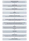
- Flowchart according to the Preferred Reporting Items for Laboratory Studies in Endodontology (PRILE) 2021 guidelines.
Teeth selection and root dentin preparation
The local Research Ethics Committee approved this study (#64296022.8.0000.5418). Sixty-eight extracted permanent third molars, aged between 18 and 35 years, were donated. The specimens were free of carious lesions, with at least two roots and no pathological or anatomical changes. With the help of periodontal curettes, bone, and periodontal tissue, when present, were removed. They were subsequently frozen at -80°C in 0.9% saline solution.
The teeth were sectioned in a perpendicular to the long axis, at the cementoenamel junction, using a straight handpiece and a water-cooled low-speed diamond disc. The roots were measured with a digital caliper (Mitutoyo, Suzano, SP, Brazil) and the crowns were discarded. The first cervical third was sectioned in a similar way, the remaining roots were discarded, and specimens of 3 mm thick root dentin were obtained. After that, the exposed dentin surface was polished with 600 silicon carbide grit to expose the pulp in occlusal and apical views. The remaining pulp was removed, and the specimens were stored in 0.5% chloramine for 24 h at 4°C and before the experiment, the solution was replaced with deionized water [28].
Radicular dentin treatment
The specimens were externally coated with an acid-resistant varnish and treated with 2 mL of 2% CHX irrigating solution for 5 min [11,28]. After the irrigation solution was used, the specimens were transferred to a 48-well plate and treated with 220 μL (protocol determined in a pilot study, unpublished data) of 17% EDTA (pH=7), 10% EDTA (pH=7), 10% CA (pH=3), and 10-3 solution (pH=0.5) as conditioning agents, for 10 min, on a shaker table (CT-165, Cientec, Belo Horizonte, MG, Brazil) to expose dentin growth factors. The samples were shaken to simulate the clinical dynamics of irrigation that occur during the chemical preparation of the root canal. This volume (220 μL), is sufficient to fully immerse the specimen, ensuring complete contact with the conditioning solution [11,28]. The solutions applied for each specimen after irrigation were stored and frozen for subsequent assessment and TGF-β1 quantification.
Quantification of TGF-β1 by ELISA assay
TGF-β1 levels were quantified using the Quantikine® ELISA kit (DuoSet Human TGF-b1, R&D Systems, Minneapolis, MN, USA, #P323635) specific for human, mouse, rat, porcine, and canine TGF-β1. This is a solid-phase sandwich enzyme immunoassay that employs monoclonal and polyclonal antibodies specific for TGF-β1. Samples were pre-activated to convert latent TGF-β1 into its immunoreactive form. For this, 100 μL of sample were acidified with 20 μL of 1 N HCl and incubated for 10 min at room temperature. Then, the samples were neutralized with 20 μL of 1.2 N NaOH/0.5 M HEPES solution. After neutralization, the activated samples were diluted according to the manufacturer's instructions using Calibrator Diluent RD6-11 (prediluted 1:2), and the final dilution factor was considered for data analysis. Optical density was measured at 450 nm with wavelength correction at 540 nm using a microplate reader. TGF-β1 concentrations were calculated by interpolation from a seven-point standard curve (31.3 to 2000 pg/mL) using a four-parameter logistic (4-PL) model and corrected for the dilution factor.
Statistical analysis
Data normality was tested using the Shapiro-Wilk test. Kruskal-Wallis tests with Dunn-Bonferroni post hoc were used for comparison between groups. P values with Bonferroni correction <0.05 were considered statistically significant. Statistical analyses were performed using JASP for Windows software (JASP Team, Version 0.19.1) and graphs were made using GraphPad Prism 8.0 for Windows (GraphPad Soft-ware, Boston, Massachusetts USA).
RESULTS
The results for the release of TGF- β1 were presented with statistic (H), p value (p) and effect size (Eta-squared) for Kruskall-Wallis, and statistic (z), p value considering Bonferroni correction (pbonf) and Rank-biserial correlation (rrb).
The Kruskal-Wallis test showed that there was a statistical difference in the release of TGF-β1 (H(3)=20.844; p<0.001; rank h2=0.279). In the comparison between groups, considering the Bonferroni correction, the 17% EDTA group showed lower values in release when compared to the 10% EDTA group (z=3.202; pbonf=0.008; rrb=0.661) and the 10-3 solution (z=4.416; pbonf<0.001; rrb= 0.768). There was no statistical difference when comparing the 10% EDTA group with the 10-3 solution group (z=1.215; pbonf=1.000; rrb=0.298), and furthermore, the CA group did not show a statistical difference with 10% EDTA (z=2.412; pbonf = 0.095; rrb = 0.571), 10% EDTA (z=-0.790; pbonf = 1.000; rrb = 0.190), and 10-3 Solution (z=2.004; pbonf=0.270; rrb=0.457). Table II shows the descriptive data of the groups considering the results in pg/mL.
DISCUSSION
Regenerative therapies rely on three core elements: stem cells, structures, and growth factors. However, achieving comprehensive success in regeneration requires the essential incorporation of materials, irrigants, conditioners, and medications throughout the regenerative endodontics procedures. Effectively addressing all pertinent aspects and variables related to regenerative success in young permanent teeth is a remarkable task [3,4].
Considering the importance of understanding the impact of agents used in regenerative endodontic procedures on growth factor release, the hypothesis of this study was confirmed, since significant differences were observed between root dentin conditioning agents. In the present study, we investigated 10% and 17% EDTA in 10 min, a shorter period compared to previous studies [11,28]. In addition, we evaluated two additional conditioning agents, 10% CA and a combination of CA with ferric chloride (10-3 solution). All of these conditioning agents provided TGF-β1 release.
We observed greater release of TGF-β1 when 10% EDTA was used compared to 17% EDTA. This finding supports the observations made by Galler et al. [11], who reported that conditioning with 10% EDTA resulted in the greatest release of TGF-β1, whereas 17% EDTA was less effective. In addition, the results of the present study indicated that 17% EDTA had the lowest levels of growth factor release when compared to 10% EDTA (pbonf=0.008) and 10-3 solution (pbonf<0.001), with strong effect sizes (rrb=0.661 and 0.768, respectively). This can be explained by considering the impact of EDTA on dentin. When the EDTA concentration increases, its osmolarity also increases, probably causing an acid accumulation during the exchange process of hydrogen and calcium ions present in hydroxyapatite. This higher concentration of acid can directly influence the EDTA action into calcium ions of root dentin and result in unwanted effects, such as dehydration of dentin and changes in the physical properties of cleaning solutions [29]. These findings highlight the importance of considering not only the demineralizing capacity of conditioning solutions but also their effects on the dentin matrix and the release of growth factors essential for regeneration.
In this context, CA especially when combined with ferric chloride, in our study, has proven promising, exhibiting a TGF-β1 release rate notably superior to that achieved with 17% EDTA. This combination stands out for its impressive results and the potential application of CA, given its proven non-cytotoxic nature in vitro [12]. These findings reinforce the assumption that CA is effective in TGF-β1 release, aligning consistently with results from comparable in vitro studies reported in two recent systematic reviews [19,20].
Furthermore, the release of TGF-β1 in the group subjected to CA as a conditioning agent showed superior results compared to that treated with 17% EDTA and comparable results to that treated with 10% EDTA. These findings align with studies, which similarly compared 17% EDTA and CA [2,18]. The consistent outcomes underscore that etching with CA attracts a significantly higher number of stem cells to dentin when compared to EDTA [17]. Therefore, conditioning with CA may be a viable alternative to current treatments, along with favorable biocompatibility in the context of regenerative endodontics.
Conditioning agents play an important role in regenerative endodontics by facilitating root surface cleaning and creating a substrate that promotes cell adhesion and migration, both essential for the regenerative process [11]. Additionally, many of these agents contain acids that can demineralize root dentin, thereby opening dentinal tubules and enhancing the penetration and migration of stem cells and other regenerative components. In our quest for novel conditioning agents aligning with these objectives, we have explored combinations displaying promise. In this context, we studied the combination of CA with ferric chloride in the present study. Rodrigues et al. [24] previously investigated this combination, aiming to assess the effects of conditioning solutions containing ferric chloride (FeCl3) on the resin-dentin bond strength, the safeguarding of dentin collagen against enzymatic degradation, and the activity of cathepsin-K [24]. Their study demonstrated that FeCl3 binds to collagen, providing protection against cathepsin K-mediated degradation. This supports the notion that the combination of citric acid and ferric chloride represents a favorable alternative for dentin conditioning, with the added advantage of preventing collagen degradation [24].
The 10–3 solution has emerged as a versatile and effective alternative for dentin conditioning. Saeki et al. [30] demonstrated that the addition of ferric chloride enhances the demineralizing effect of CA, intensifying dentin conditioning without causing irreversible collapse of the collagen matrix [30]. More recent studies indicate that the 10–3 solution can be a viable alternative to phosphoric acid, even on dry substrates, providing a stable adhesive interface without compromising the mechanical properties of dentin [31]. Additionally, the literature shows that this solution is highly effective in removing the smear layer without causing excessive demineralization, favoring controlled exposure of dentinal tubules and the release of bioactive signals important in regenerative endodontic procedures [32]. Its effectiveness even in the presence of blood contamination reinforces its clinical applicability in challenging scenarios such as reimplantation and regenerative therapies, promoting both durable adhesion and a microenvironment conducive to tissue regeneration [33]. Based on this assumption, one of the objectives of the present study was to investigate whether the favorable effects observed with FeCl3 on the bond strength between dentin and resin could potentially extend to one of the factors involved in the regenerative processes. Our findings indicated that the 10-3 solution significantly enhanced the release of TGF-β1 from root dentin, exceeding even the levels achieved with 17% EDTA. These results suggest that further exploration of this combination is warranted, especially concerning its implications for various procedures involving root dentin.
Although this study provides relevant contributions, some methodological limitations should be acknowledged. The in vitro design does not fully replicate the complex biological conditions of the clinical environment, particularly regarding the interaction of conditioning agents with vital tissues, inflammatory mediators, and blood components. Another limitation is the absence of a negative control group without treatment, which could have strengthened the interpretation of the results. However, it is important to note that a negative control would not be applicable to the clinical scenario, because even if no release occurred, or if it were lower than that of the test groups, such a condition could not be used in clinical practice, given the additional characteristics that a conditioning agent must present. Considering these limitations, future studies should incorporate biologically relevant models to evaluate stem cell viability, dentin surface alterations, and the clinical applicability of these conditioning protocols.
It is important to highlight that the use of conditioning agents during pulp regeneration protocols may affect the structure of root dentin, thereby influencing the release of growth factors, particularly TGF-β1. Our findings reinforce the importance of continually investigating the conditioning agents currently used, as other available solutions may also yield favorable outcomes. The selection of a conditioning agent for pulp regeneration should consider not only its antibacterial efficacy during root canal disinfection but also its effects on stem cells and on the release of dentin-derived growth factors. In addition to the present results, further studies are necessary to deepen the understanding of how different conditioning agents influence the regenerative potential of root dentin.
CONCLUSION
This study demonstrated that different conditioning agents significantly affect the release of TGF-β1 from root dentin. Among the tested agents, 17% EDTA resulted in the lowest release, whereas 10% EDTA and the 10–3 solution promoted higher levels of growth factor release.
Acknowledgements
We are grateful to the Coordenação de Aperfeiçoamento de Pessoal de Nível Superior (CAPES) for supporting this work.
Data availability
The data that support the findings of this study are available from the corresponding author upon reasonable request.
-
How to cite:
Theodoro AL, Gois CMB, dos Santos VAB, Puppin-Rontani RM, Pascon FM. Different conditioning agents provide TGF-β1 release from root dentin: an in vitro study. Braz. Dent. Sci. 2026;29:e4989. https://doi.org/10.4322/bds.2026.e4989
-
Funding
This work was partially supported by Coordenação de Aperfeiçoamento de Pessoal de Nível Superior (CAPES) - Código de Financiamento 001 and in the form of a master scholarship, under process number 88887.645797/2021-00.
-
Regulatory Statement
This study was conducted in accordance with all the provisions of the local human subjects oversight committee guidelines and policies of the Faculdade de Odontologia de Piracicaba (FOP), University of Campinas (UNICAMP). This study protocol was reviewed and approved by the local Research Ethics Committee, approval number 64296022.8.0000.5418.
REFERENCES
-
1 Hargreaves KM, Geisler T, Henry M, Wang Y. Regeneration potential of the young permanent tooth: what does the future hold? J Endod. 2008;34(7):S51-6. https://doi.org/10.1016/j.joen.2008.02.032 PMid:18565373.
» https://doi.org/10.1016/j.joen.2008.02.032 -
2 Chae Y, Yang M, Kim J. Release of TGF-β1 into root canals with various final irrigants in regenerative endodontics: an in vitro analysis. Int Endod J. 2018;51(12):1389-97. https://doi.org/10.1111/iej.12951 PMid:29774548.
» https://doi.org/10.1111/iej.12951 -
3 Diogenes A, Hargreaves KM. Microbial modulation of stem cells and future directions in regenerative endodontics. J Endod. 2017;43(9S):S95-101. https://doi.org/10.1016/j.joen.2017.07.012 PMid:28844309.
» https://doi.org/10.1016/j.joen.2017.07.012 -
4 Shalaby L, Shawkat S, Fathy I. Potential effect of platelet rich fibrin prepared under different centrifugation protocols on stem cells from the apical papilla. Braz Dent Sci. 2023;26(4):1-14. https://doi.org/10.4322/bds.2023.e3847
» https://doi.org/10.4322/bds.2023.e3847 -
5 Niwa T, Yamakoshi Y, Yamazaki H, Karakida T, Chiba R, Hu JC, et al. The dynamics of TGF-β in dental pulp, odontoblasts and dentin. Sci Rep. 2018;8(1):4450. https://doi.org/10.1038/s41598-018-22823-7 PMid:29535349.
» https://doi.org/10.1038/s41598-018-22823-7 -
6 Yongchaitrakul T, Pavasant P. Transforming growth factor-beta1 up-regulates the expression of nerve growth factor through mitogen-activated protein kinase signaling pathways in dental pulp cells. Eur J Oral Sci. 2007;115(1):57-63. https://doi.org/10.1111/j.1600-0722.2007.00420.x PMid:17305717.
» https://doi.org/10.1111/j.1600-0722.2007.00420.x -
7 Bracks IV, Espaladori MC, Barros P, de Brito LCN, Vieira LQ, Ribeiro Sobrinho AP. Effect of ethylenediaminetetraacetic acid irrigation on immune-inflammatory response in teeth submitted to regenerative endodontic therapy. Int Endod J. 2019;52(10):1457-65. https://doi.org/10.1111/iej.13136 PMid:31046128.
» https://doi.org/10.1111/iej.13136 -
8 Atesci AA, Avci CB, Tuglu MI, Ozates Ay NP, Eronat AC. Effect of different dentin conditioning agents on growth factor release, mesenchymal stem cell attachment and morphology. J Endod. 2020;46(2):200-8. https://doi.org/10.1016/j.joen.2019.10.033 PMid:31812361.
» https://doi.org/10.1016/j.joen.2019.10.033 - 9 Bucchi C, Valdivia-Gandur IM, Sánchez-Bizjak R, Tallón-Walton V. Manzanares- Céspedes MC. Regenerative endodontic therapy: a systematic review of clinical protocols. Int J Clin Exp Med. 2017;10(2):2006-15.
-
10 Dos Reis-Prado AH, Abreu LG, Fagundes RR, Oliveira SC, Bottino MC, Ribeiro-Sobrinho AP, et al. Influence of ethylenediaminetetraacetic acid on regenerative endodontics: A systematic review. Int Endod J. 2022;55(6):579-612. https://doi.org/10.1111/iej.13728 PMid:35305029.
» https://doi.org/10.1111/iej.13728 -
11 Galler KM, Buchalla W, Hiller KA, Federlin M, Eidt A, Schiefersteiner M, et al. Influence of root canal disinfectants on growth factor release from dentin. J Endod. 2015;41(3):363-8. https://doi.org/10.1016/j.joen.2014.11.021 PMid:25595468.
» https://doi.org/10.1016/j.joen.2014.11.021 -
12 Malheiros CF, Marques MM, Gavini G. In vitro evaluation of the cytotoxic effects of acid solutions used as canal irrigants. J Endod. 2005;31(10):746-8. https://doi.org/10.1097/01.don.0000157994.49432.67 PMid:16186755.
» https://doi.org/10.1097/01.don.0000157994.49432.67 -
13 Ozdemir HO, Buzoglu HD, Calt S, Cehreli ZC, Varol E, Temel A. Chemical and ultramorphologic effects of ethylenediaminetetraacetic acid and sodium hypochlorite in young and old root canal dentin. J Endod. 2012;38(2):204-8. https://doi.org/10.1016/j.joen.2011.10.024 PMid:22244637.
» https://doi.org/10.1016/j.joen.2011.10.024 -
14 Hanafy MS, Abdella Ahmed AK, Salem RG. Impact of using XP-endo finisher and nanobubble water during EDTA dentin conditioning on TGF-β1 release in regenerative endodontic procedures. BMC Oral Health. 2024;24(1):595. https://doi.org/10.1186/s12903-024-04355-x PMid:38778321.
» https://doi.org/10.1186/s12903-024-04355-x -
15 Tidmarsh BG. Acid-cleansed and resin-sealed root canals. J Endod. 1978;4(4):117-21. https://doi.org/10.1016/S0099-2399(78)80201-5 PMid:370323.
» https://doi.org/10.1016/S0099-2399(78)80201-5 -
16 Gómez-Delgado M, Camps-Font O, Luz L, Sanz D, Mercade M. Update on citric acid use in endodontic treatment: a systematic review. Odontology. 2023;111(1):1-19. https://doi.org/10.1007/s10266-022-00744-2 PMid:36220913.
» https://doi.org/10.1007/s10266-022-00744-2 -
17 Scelza MF, Pierro V, Scelza P, Pereira M. Effect of three different time periods of irrigation with EDTA-T, EDTA, and citric acid on smear layer removal. Oral Surg Oral Med Oral Pathol Oral Radiol Endod. 2004;98(4):499-503. https://doi.org/10.1016/j.tripleo.2004.03.027 PMid:15472668.
» https://doi.org/10.1016/j.tripleo.2004.03.027 -
18 Ivica A, Zehnder M, Mateos JM, Ghayor C, Weber FE. Biomimetic conditioning of human dentin using citric acid. J Endod. 2019;45(1):45-50. https://doi.org/10.1016/j.joen.2018.09.015 PMid:30448020.
» https://doi.org/10.1016/j.joen.2018.09.015 -
19 Reis-Prado AHD, Toledo PTA, Nunes GP, Ferreira PAV, Rahimnejad M, Dal-Fabbro R, et al. Citric acid conditioning as an alternative to EDTA for growth factors release and stem cell response in regenerative endodontics: A systematic review of in vitro studies. J Endod. 2024;50(2):129-43. https://doi.org/10.1016/j.joen.2023.11.006 PMid:37984798.
» https://doi.org/10.1016/j.joen.2023.11.006 -
20 Oliveira CLL, Ferreira FM, Puppin-Rontani J, Puppin-Rontani RM, Pascon FM. Potential of irrigants and medicaments in regenerative endodontics: insights from a systematic review on dentin growth factor release. Odontology. 2025. https://doi.org/10.1007/s10266-025-01128-y PMid:40517172.
» https://doi.org/10.1007/s10266-025-01128-y -
21 Li X, Ma W, Chen X, Zhao J, Nakata T, Tanaka K, et al. Bonding to NaOCl-treated dentin: effect of pretreatment with sodium toluene sulfinic acid. J Adhes Dent. 2012;14(2):129-36. https://doi.org/10.3290/j.jad.a22090 PMid:21935518.
» https://doi.org/10.3290/j.jad.a22090 - 22 Marshall GW, Saeki K, Gansky SA, Marshall SJ. AFM study of citric acid-ferric chloride etching characteristics of dentin. Am J Dent. 1999;12(6):271-6. PMid:10850245.
-
23 Nakabayashi N, Takarada K. Effect of HEMA on bonding to dentin. Dent Mater. 1992;8(2):125-30. https://doi.org/10.1016/0109-5641(92)90067-M PMid:1521692.
» https://doi.org/10.1016/0109-5641(92)90067-M -
24 Rodrigues RV, Giannini M, Pascon FM, Panwar P, Brömme D, Manso AP, et al. Effect of conditioning solutions containing ferric chloride on dentin bond strength and collagen degradation. Dent Mater. 2017;33(10):1093-102. https://doi.org/10.1016/j.dental.2017.07.004 PMid:28779890.
» https://doi.org/10.1016/j.dental.2017.07.004 -
25 San Antonio JD, Schweitzer MH, Jensen ST, Kalluri R, Buckley M, Orgel JP. Dinosaur peptides suggest mechanisms of protein survival. PLoS One. 2011;6(6):e20381. https://doi.org/10.1371/journal.pone.0020381 PMid:21687667.
» https://doi.org/10.1371/journal.pone.0020381 -
26 Schweitzer MH, Zheng W, Cleland TP, Goodwin MB, Boatman E, Theil E, et al. A role for iron and oxygen chemistry in preserving soft tissues, cells and molecules from deep time. Proc Biol Sci. 2013;281(1775):20132741. https://doi.org/10.1098/rspb.2013.2741 PMid:24285202.
» https://doi.org/10.1098/rspb.2013.2741 -
27 Nagendrababu V, Murray PE, Ordinola-Zapata R, Peters OA, Rôças IN, Siqueira JF Jr, et al. PRILE 2021 guidelines for reporting laboratory studies in Endodontology: A consensus-based development. Int Endod J. 2021;54(9):1482-90. https://doi.org/10.1111/iej.13542 PMid:33938010.
» https://doi.org/10.1111/iej.13542 -
28 Ferreira LN, Puppin-Rontani RM, Pascon FM. Effect of intracanal medicaments and irrigants on the release of transforming growth factor Beta 1 and vascular endothelial growth factor from cervical root dentin. J Endod. 2020;46(11):1616-22. https://doi.org/10.1016/j.joen.2020.07.034 PMid:32795548.
» https://doi.org/10.1016/j.joen.2020.07.034 -
29 Calvo Pérez V, Medina Cárdenas ME, Sánchez Planells U. The possible role of pH changes during EDTA demineralization of teeth. Oral Surg Oral Med Oral Pathol. 1989;68(2):220-2. https://doi.org/10.1016/0030-4220(89)90196-5 PMid:2506500.
» https://doi.org/10.1016/0030-4220(89)90196-5 -
30 Saeki K, Marshall SJ, Gansky SA, Marshall GW. Etching characteristics of dentin: effect of ferric chloride in citric acid. J Oral Rehabil. 2001;28(4):301-8. https://doi.org/10.1046/j.1365-2842.2001.00715.x PMid:11350581.
» https://doi.org/10.1046/j.1365-2842.2001.00715.x -
31 Sebold M, André CB, Carvalho RM, Giannini M. Dry-bonding to dentin using alternative conditioners based on iron-containing solutions or nitric acid. J Mech Behav Biomed Mater. 2019;94:238-48. https://doi.org/10.1016/j.jmbbm.2019.03.015 PMid:30927591.
» https://doi.org/10.1016/j.jmbbm.2019.03.015 -
32 Shimizu K, Satoh T, Shinkai K. Evaluation of restorative techniques for vertically fractured roots. Materials. 2021;14(9):2099. https://doi.org/10.3390/ma14092099 PMid:33919304.
» https://doi.org/10.3390/ma14092099 -
33 Takefu H, Shimoji S, Sugaya T, Kawanami M. Influence of blood contamination before or after surface treatment on adhesion of 4-META/MMA-TBB resin to root dentin. Dent Mater J. 2012;31(1):131-8. https://doi.org/10.4012/dmj.2011-142 PMid:22277617.
» https://doi.org/10.4012/dmj.2011-142
Edited by
-
Editor-in-chief:
Sergio Eduardo de Paiva Gonçalves
-
Section Editor:
Letícia Chaves de Souza