
 Pathological alterations associated with mandibular third molars: a clinical, radiographic and histopathological study
ABSTRACT
Objective Radiographic examination is routinely performed to detect pathoses associated with impacted teeth; however, evidence suggests that pathological alterations may occur in asymptomatic dental follicles exhibiting normal radiographic features. This study aimed to evaluate the presence of pathological changes in pericoronal follicles of unerupted mandibular third molars without radiographic evidence of associated lesions.
Material and Methods Patients indicated for surgical removal of mandibular third molars without evident clinical or radiographic abnormalities were included. Clinical and intraoperative data regarding sex, age, pain, gingival coverage, and distal bone loss of adjacent second molars were recorded. Radiographs were assessed for tooth position, impaction pattern, and root resorption. All pericoronal follicles were submitted to histopathological examination to identify cystic changes, epithelial lining characteristics, and inflammatory infiltrate.
Results The sample comprised 49 patients and 73 unerupted or partially erupted mandibular third molars. Histopathological analysis revealed 45.2% normal follicles, 28.8% dentigerous cysts, and 26.0% paradental cysts. An epithelial lining was identified in 56.2% of specimens, predominantly stratified squamous epithelium (82.9%). Inflammatory infiltrate was present in 64.4% of cases and was significantly more frequent in teeth partially covered by gingival tissue. No significant associations were observed with sex or age.
Conclusion A substantial proportion of pericoronal follicles exhibited pathological alterations despite the absence of clinical or radiographic signs of disease. These findings underscore the value of histopathological evaluation in supporting clinical decision-making regarding monitoring or removal of impacted mandibular third molars in selected cases.
KEYWORDS:
Histology; Mandibular third molar impacted; Pathology; Radiography
RESUMO
Objetivo O exame radiográfico é rotineiramente realizado para detectar patologias associadas a dentes impactados; entretanto, evidências indicam que alterações patológicas podem ocorrer em folículos dentários assintomáticos com características radiográficas normais. Este estudo teve como objetivo avaliar a presença de alterações patológicas em folículos pericoronários de terceiros molares inferiores não irrompidos, sem evidência radiográfica de lesões associadas.
Material e Métodos Foram incluídos pacientes com indicação para remoção cirúrgica de terceiros molares inferiores sem alterações clínicas ou radiográficas evidentes. Dados clínicos e transoperatórios referentes a sexo, idade, dor, cobertura gengival e perda óssea distal do segundo molar adjacente foram registrados. As radiografias foram avaliadas quanto à posição dentária, padrão de impactação e reabsorção radicular. Todos os folículos pericoronários foram submetidos à análise histopatológica para identificação de alterações císticas, características do revestimento epitelial e infiltrado inflamatório.
Resultados A amostra foi composta por 49 pacientes e 73 terceiros molares inferiores não irrompidos ou parcialmente irrompidos. A análise histopatológica revelou 45,2% de folículos normais, 28,8% de cistos dentígeros e 26,0% de cistos paradentários. Revestimento epitelial foi identificado em 56,2% das amostras, predominantemente epitélio escamoso estratificado (82,9%). Infiltrado inflamatório esteve presente em 64,4% dos casos, sendo significativamente mais frequente em dentes parcialmente cobertos por tecido gengival. Não foram observadas associações significativas com sexo ou idade.
Conclusão Uma proporção substancial dos folículos pericoronários apresentou alterações patológicas, mesmo na ausência de sinais clínicos ou radiográficos de doença. Esses achados ressaltam o valor da avaliação histopatológica no suporte à tomada de decisão clínica quanto ao acompanhamento ou à remoção de terceiros molares inferiores impactados em casos selecionados.
PALAVRAS-CHAVE:
Histologia; Terceiro molar incluso; Patologia; Radiografia
INTRODUCTION
The surgical extraction of unerupted or partially erupted mandibular third molars (MTM), whether symptomatic or asymptomatic, is among the most frequently performed procedures by general dentists and oral and maxillofacial surgeons [1-3]. Radiographic examination is routinely employed to assess the presence or absence of pathologic conditions associated with impacted teeth. However, several studies have demonstrated that pathological alterations may be present in asymptomatic dental follicles exhibiting normal radiographic appearance, with reported prevalences higher than previously anticipated [4-7].
One of the main arguments supporting prophylactic extraction is the potential for impacted MTM follicles to undergo cystic degeneration and, less frequently, neoplastic transformation [8]. Nevertheless, there is no consensus regarding the routine removal of asymptomatic impacted third molars, as pathological processes may develop even in the absence of clinical signs and/or radiographic abnormalities [8-13]. In recent years, research has increasingly focused on improving early detection of pericoronal pathologies in order to prevent disease progression that could ultimately require more extensive surgical intervention and be associated with greater morbidity [13,14].
The present study aimed to evaluate the presence of pathological alterations in pericoronal follicles of partially erupted and unerupted MTM lacking radiographic evidence of associated lesions.
MATERIALS AND METHODS
This study is in accordance with the Declaration of Helsinki, and the protocol was approved by the local Ethical Committee (registration number 14670819.6.0000.5374). All procedures were carried out with the full understanding and written consent of the subjects. The study follows the Enhancing the Quality and Transparency of Health Research” (EQUATOR Network) and on The Strengthening the Reporting of Observational Studies in Epidemiology (STROBE) statement [15].
This was an observational prospective study conducted on a cohort of 49 Brazilian patients who underwent MTM extraction between November 2019 and August 2021. Inclusion criteria comprised volunteers aged 18 years or older presenting with pain, difficulty maintaining adequate oral hygiene, and or occlusal alterations, without clinical, radiographic, or tomographic evidence of lesions associated with the impacted MTM follicle. Patients diagnosed clinically and or radiographically with maxillary bone lesions, odontogenic or non-odontogenic cysts or tumors in the MTM region, as well as those with systemic bone diseases affecting the jaws, were excluded. Cases presenting a pericoronal space greater than 3 mm were also excluded. The final sample consisted of 73 MTMs.
All extractions were performed under local anesthesia by a single operator, following established surgical and biosafety protocols. During clinical examination, the principal investigator recorded the presence of pain and gingival coverage status, classified as partially erupted or unerupted. Distal bone resorption affecting the adjacent second molar was assessed intraoperatively.
Radiographic assessment was performed by an experienced radiologist and included periapical radiographs, panoramic radiography, cone beam computed tomography, or computed tomography when indicated. The evaluated variables included pericoronal space limits, categorized as well defined or poorly defined, measurement of the pericoronal space, presence or absence of a radiopaque cortical halo, radicular resorption, adjacent bone loss, and stage of root development, classified as open or closed apex. MTM position was classified according to eruption status, categorized as fully impacted or partially impacted, angulation according to Winter’s classification [16], and depth of impaction according to the Pell and Gregory classification [17]. The frequency of each impaction pattern was also recorded (Figure 1 and 2)

Radiographic images illustrating different conditions associated with mandibular third molars. (A) Pericoronal follicle of tooth 38, showing a pericoronal space of normal thickness with well-defined limits; (B) Dentigerous cyst associated with tooth 38, characterized by a unilocular radiolucent image surrounding the dental crown, with well-defined corticated borders and enlargement of the pericoronal space; (C) Paradental cyst associated with tooth 48, evidenced by a radiolucent image located on the distal aspect of the tooth, with preservation of the pericoronal space.

Detail of the radiographic images above illustrating different conditions associated with mandibular third molars. (A) Pericoronal follicle of tooth 38; (B) Dentigerous cyst associated with tooth 38; (C) Paradental cyst associated with tooth 48.
During extraction, pericoronal tissues were collected and fixed in 10 percent buffered formalin, stained with hematoxylin and eosin, and submitted for histopathological examination. The analysis was performed independently by two oral pathologists at three section levels, and disagreements were resolved by a third pathologist. When epithelial lining was present, it was classified as cuboidal, stratified squamous, or pseudostratified ciliated columnar. Additional histopathological features evaluated included the presence of Rushton bodies, mucous cells, connective tissue pattern classified as loose, dense, or myxomatous, inflammatory infiltrate categorized as mononuclear or mixed, calcifications, odontogenic epithelial remnants, and cholesterol clefts.
Descriptive statistics was performed, in which absolute and relative frequencies were used for categorical variables and mean, standard deviation, median, minimum, and maximum value for quantitative variables. The Chi-square and Fisher's exact tests were used to analyze the associations between the presence of pathological alterations, gingival coverage, and inflammation with clinical, radiographic, and pathological variables. Data normality was evaluated using the Shapiro-Wilk test. The Kruskal-Wallis test was used to assess the relationship between pathological changes and patient age. The Man-Witney U test was applied to assess the association between gingival coverage and inflammation. Analyzes were performed using the SPSS 29 program (IMB, USA), with a significance level of 5%.
RESULTS
All clinical, radiographic, and histopathological characteristics are presented in Table I. Among the included patients, 65.3% were female and 34.7% were male, with a mean age of 23 years, ranging from 18 to 32 years. Pain was reported in 29 cases (59.2%). Regarding gingival coverage, 42 teeth (57.5%) were partially erupted and 31 (42.5%) were unerupted. Distal bone loss affecting the second molar was observed in 42.5% of cases.
Radiographic evaluation revealed partially impacted MTMs in 60.3% of cases and fully impacted teeth in 39.7%. The mean pericoronal space width was 1.54 mm, ranging from 0.48 to 2.58 mm. A radiopaque cortical boundary delimiting the pericoronal space was observed in 39.7% of cases. Regarding tooth inclination, 34.2% were vertical, 35.6% mesioangular, 12.3% distoangular, and 17.8% horizontal. Most cases were classified as Class II and Position B according to Pell and Gregory. Incomplete root formation was observed in 34.2% of cases, and no radicular resorption of the adjacent second molar was identified. However, distal bone loss of the second molar occurred in 42.5% of cases.
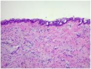
Histopathological examination identified alterations compatible with dentigerous cysts in 28.8% and paradental cysts in 26.0% of cases (Figure 3, 4 and 5), totaling 40 altered follicles, while 45.2% exhibited normal histological features. An epithelial lining was present in 56.2% of specimens, predominantly stratified squamous epithelium (82.9%). Cuboidal and pseudostratified ciliated columnar epithelium were each observed in 4.9% of cases. Mixed epithelial patterns were identified in three cases. Rushton bodies were observed in one case, and mucous cells in four cases (5.5%).

Dental Follicle (Cap) – Loose connective tissue containing islands of odontogenic epithelium (10× magnification).
Dentigerous Cyst – Cystic capsule composed of dense fibrous connective tissue lined by odontogenic cuboidal epithelium with more than one cell layer and superficial ciliated cells (10× magnification).

Fragment of a cystic capsule partially lined internally by stratified squamous epithelium with elongated projections, spongiosis, and exocytosis. The cystic capsule is composed of dense connective tissue and exhibits a mild mononuclear inflammatory infiltrate.
Loose connective tissue was observed in 20.5% of cases, dense in 49.3%, and myxomatous in 30.1%. Inflammatory infiltrate was present in 64.4% of cases, predominantly mononuclear (94.5%), with mixed infiltrate observed in 5.5%. Calcifications were identified in 64.4% of specimens, odontogenic epithelial remnants in 53.4%, and cholesterol clefts in 8.2%.
Pain was significantly more frequent in follicles diagnosed as paradental cysts (40.5%) compared with dentigerous cysts and normal follicles (both 29.7%) (p = 0.007) (Table II). Paradental cysts were more frequently associated with vertically positioned teeth, whereas dentigerous cysts and normal follicles were more commonly associated with mesioangular inclination (p = 0.039). No significant association was found between histological diagnosis and other clinical or radiographic variables.
Pain and distal bone loss were significantly more frequent in partially erupted teeth. The absence of a cortical boundary was also more common in partially erupted teeth. Mesioangular inclination was more frequent in unerupted teeth, whereas vertical inclination predominated in partially erupted teeth (p < 0.001). Follicle size was not associated with gingival coverage.
Inflammation was significantly more frequent in partially erupted teeth (p = 0.004). All paradental cysts and most dentigerous cysts exhibited inflammatory infiltrate. However, inflammation was not significantly associated with pain or other clinical and radiographic variables (Table III).
Association between gingival coverage and presence of inflammation with clinical, radiographic and histopathological variables
DISCUSSION
This study evaluated pathological alterations in pericoronal follicles of partially erupted and unerupted MTMs without radiographic evidence of associated lesions. Although a pericoronal space ≤ 3 mm is traditionally considered within normal limits, cystic or neoplastic changes have been reported in follicles measuring less than 2.5 to 3 mm [3,6,12,14]. The reported prevalence of pathological alterations in radiographically normal follicles ranges from 34% to 76.4% [12-14]. In the present study, the majority of pericoronal follicles (54.8%) exhibited pathological alterations, including dentigerous (28.8%) and inflammatory paradental cysts (26.0%), indicating that disease may occur despite radiographic parameters considered normal.
Dentigerous cysts are the most common cystic lesions associated with impacted third molars, accounting for approximately 18% to 20% of developmental odontogenic cysts [18-20]. Consistently, dentigerous cysts were the most frequent diagnosis, followed by paradental cysts, whereas a smaller proportion of follicles were histologically normal. These findings reinforce the clinical relevance of submitting pericoronal tissues for histopathological examination after extraction [1,5,6]. The prevalence observed here is higher than that reported in some studies of radiographically normal follicles [2,7].
It is important to emphasize that the histopathological differentiation between normal dental follicles and early odontogenic cystic lesions can be challenging, particularly in cases with subtle epithelial proliferation or minimal cystic change. To ensure methodological rigor and diagnostic reliability, all specimens in the present study were independently evaluated by two experienced oral pathologists. In cases of disagreement, a third senior pathologist reviewed the slides, and a consensus diagnosis was established.
Squamous metaplasia was identified in several specimens. Previous studies have reported squamous metaplasia in approximately 32% of follicles with pericoronal radiolucency smaller than 2.5 mm [13]. However, whether focal metaplasia alone is sufficient to establish cystic transformation remains unclear [1,2]. The relatively high frequency of pathological alterations may be influenced by the patients’ age range of 18 to 32 years, as increased cystic incidence with advancing age has been described [12,13,21].
Histologically, cyst capsules typically consist of loose connective tissue lined by stratified squamous epithelium and may present inflammatory infiltrate. In this study, most specimens exhibited stratified squamous epithelium associated with inflammation, whereas odontogenic epithelial remnants were less frequent. Reduced enamel epithelium and focal squamous metaplasia are common findings in dental follicles [22]. Metaplasia is considered an adaptive response to stress or injury, and eruption-related pressure or ischemia between 18 and 25 years of age may contribute to these changes [12,13,22]. This association, however, was not clearly demonstrated in the present sample.
No significant association was observed between histopathological alterations and radiographic features such as follicular limits, cortical halo, or pericoronal width, corroborating previous findings [5]. Early pathological changes may therefore be undetectable on routine imaging. Pericoronal spaces measuring between 3 and 4 mm may still represent normal follicles [22].
Management of asymptomatic impacted third molars remains controversial. Current evidence does not support routine removal or retention in the absence of clinical or radiographic disease [8]. However, retention may increase the risk of periodontal involvement of adjacent second molars [10]. In this study, distal alveolar bone loss affecting the second molar was observed more frequently when the adjacent third molar was partially erupted/partially impacted. These findings suggest that clinical decision-making regarding third molar management should take into account individual patient factors, professional judgment, and local clinical guideline [23].
Most teeth were partially erupted, and pain was frequently reported. Pain was present in all cases diagnosed as paradental cyst. Although surgical indication and symptomatology may have influenced the sample, this reflects clinical practice. Partially erupted molars are commonly associated with pain and inflammation [23], and periodontal disease or occlusal caries may occur even in asymptomatic cases [10]. Inflammation was the most frequent alteration, consistent with previous studies [11]. Inflammatory infiltrate was also observed in asymptomatic impacted teeth, possibly related to periodontal contamination or eruption dynamics [1]. Inflammatory follicles have been reported to be more frequently associated with pathological alterations than non-inflammatory follicles [5]. These findings may aid in the differential diagnosis of orofacial pain related to impacted third molars.
This study has some limitations that should be acknowledged. First, the sample size derived from a single center, which may limit the generalizability of the findings. Second, the inclusion of patients already indicated for extraction, many of whom reported symptoms may have overestimated the prevalence of pathological alterations compared with an asymptomatic population. Finally, the cross-sectional design precludes assessment of the natural progression of radiographically normal follicles over time.
CONCLUSION
Pathological alterations were identified in a substantial proportion of pericoronal follicles associated with unerupted and partially erupted MTM lacking radiographic evidence of lesions. Histopathological examination of pericoronal tissues therefore provides relevant diagnostic information and may support more informed clinical decision-making regarding the management of impacted MTM.
Data availability
The data that support the findings of this study are available from the corresponding author upon reasonable request.
-
How to cite:
Silveira RJ, Cascaes PSL, Oenning AC, Dorta R, Botelho T, Junqueira JL, Soares MQS. Pathological alterations associated with mandibular third molars: a clinical, radiographic and histopathological study. Braz. Dent. Sci. 2026;29:e4966. https://doi.org/10.4322/bds.2026.e4966
-
Funding
This research did not receive any specific grant from funding agencies in the public, commercial, or not-for-profit sectors.
-
Regulatory Statement
This study was conducted in accordance with all the provisions of the local human subjects oversight committee guidelines and policies. The study is in accordance with the Declaration of Helsinki, and the protocol was reviewed and approved by the Research Ethics Committee of Faculdade São Leopoldo Mandic, Campinas, São Paulo, Brazil (registration number 14670819.6.0000.5374). All procedures were carried out with the full understanding and written informed consent of the subjects prior to their inclusion in the study.Additionally, the study follows the recommendations of the Enhancing the Quality and Transparency of Health Research (EQUATOR Network) and adheres to the Strengthening the Reporting of Observational Studies in Epidemiology (STROBE) statement.
REFERENCES
-
1 Baykul T, Saglam AA, Aydin U, Başak K. Incidence of cystic changes in radiographically normal impacted lower third molar follicles. Oral Surg Oral Med Oral Pathol Oral Radiol Endod. 2005 May;99(5):542-5. https://doi.org/10.1016/j.tripleo.2004.08.010 PMid:15829874.
» https://doi.org/10.1016/j.tripleo.2004.08.010 -
2 Costa FW, Viana TS, Cavalcante GM, de Barros Silva PG, Cavalcante RB, Nogueira AS, et al. A clinicoradiographic and pathological study of PFs associated to mandibular third molars. J Craniofac Surg. 2014;25:e283-7. https://doi.org/10.1097/SCS.0000000000000712 PMid:24777014.
» https://doi.org/10.1097/SCS.0000000000000712 -
3 Esen A, Isik K, Findik S, Suren D. Histopathological evaluation of dental follicles of clinically symptomatic and asymptomatic impacted third molars. Niger J Clin Pract. 2016;19(5):616-21. https://doi.org/10.4103/1119-3077.188700 PMid:27538550.
» https://doi.org/10.4103/1119-3077.188700 -
4 Yildirim G, Ataoğlu H, Mihmanli A, Kiziloğlu D, Avunduk MC. Pathologic changes in soft tissues associated with asymptomatic impacted third molars. Oral Surg Oral Med Oral Pathol Oral Radiol Endod. 2008;106(1):14-8. https://doi.org/10.1016/j.tripleo.2007.11.021 PMid:18439856.
» https://doi.org/10.1016/j.tripleo.2007.11.021 -
5 Saravana GH, Subhashraj K. Cystic changes in dental follicle associated with radiographically normal impacted mandibular third molar. Br J Oral Maxillofac Surg. 2008;46(7):552-3. https://doi.org/10.1016/j.bjoms.2008.02.008 PMid:18406023.
» https://doi.org/10.1016/j.bjoms.2008.02.008 -
6 Adelsperger J, Campbell JH, Coates DB, Summerlin DJ, Tomich CE. Early soft tissue pathosis associated with impacted third molars without pericoronal radiolucency. Oral Surg Oral Med Oral Pathol Oral Radiol Endod. 2000;89(4):402-6. https://doi.org/10.1016/S1079-2104(00)70119-3 PMid:10760721.
» https://doi.org/10.1016/S1079-2104(00)70119-3 -
7 Ghaeminia H, Nienhuijs ME, Toedtling V, Perry J, Tummers M, Hoppenreijs TJ, et al. Surgical removal versus retention for the management of asymptomatic disease-free impacted wisdom teeth. Cochrane Database Syst Rev. 2020;5(5):CD003879. https://doi.org/10.1002/14651858.CD003879.pub5 PMid:32368796.
» https://doi.org/10.1002/14651858.CD003879.pub5 -
8 Güven O, Keskin A, Akal UK. The incidence of cysts and tumors around impacted third molars. Int J Oral Maxillofac Surg. 2000;29(2):131-5. https://doi.org/10.1016/S0901-5027(00)80011-9 PMid:10833151.
» https://doi.org/10.1016/S0901-5027(00)80011-9 -
9 Blakey GH, Marciani RD, Haug RH, Phillips C, Offenbacher S, Pabla T, et al. Periodontal pathology associated with asymptomatic third molars. J Oral Maxillofac Surg. 2002;60(11):1227-33. https://doi.org/10.1053/joms.2002.35717 PMid:12420253.
» https://doi.org/10.1053/joms.2002.35717 - 10 Rakprasitkul S. Pathologic changes in the pericoronal tissues of unerupted third molars. Quintessence Int. 2001 Sep;32(8):633-8. PMid:11526892.
-
11 Adelsperger J, Campbell JH, Coates DB, Summerlin DJ, Tomich CE. Early soft tissue pathosis associated with impacted third molars without pericoronal radiolucency. Oral Surg Oral Med Oral Pathol Oral Radiol Endod. 2000;89(4):402-6. https://doi.org/10.1016/S1079-2104(00)70119-3 PMid:10760721.
» https://doi.org/10.1016/S1079-2104(00)70119-3 -
12 Glosser JW, Campbell JH. Pathologic change in soft tissues associated with radiographically “normal” third molar impactions. Br J Oral Maxillofac Surg. 1999;37(4):259-60. https://doi.org/10.1054/bjom.1999.0061 PMid:10475645.
» https://doi.org/10.1054/bjom.1999.0061 -
13 Dongol A, Sagtani A, Jaisani MR, Singh A, Shrestha A, Pradhan A, et al. Dentigerous cystic changes in the follicles associated with radiographically normal impacted mandibular third molars. Int J Dent. 2018;2018(1):2645878. https://doi.org/10.1155/2018/2645878 PMid:29755527.
» https://doi.org/10.1155/2018/2645878 -
14 Shin SM, Choi EJ, Moon SY. Prevalence of pathologies related to impacted mandibular third molars. Springerplus. 2016;5(1):915. https://doi.org/10.1186/s40064-016-2640-4 PMid:27386359.
» https://doi.org/10.1186/s40064-016-2640-4 -
15 von Elm E, Altman DG, Egger M, Pocock SJ, Gøtzsche PC, Vandenbroucke JP, STROBE Initiative. The Strengthening the Reporting of Observational Studies in Epidemiology (STROBE) statement: guidelines for reporting observational studies. Lancet. 2007;370(9596):1453-7. https://doi.org/10.1016/S0140-6736(07)61602-X PMid:18064739.
» https://doi.org/10.1016/S0140-6736(07)61602-X - 16 Winter GB. Principles of exodontia as applied to the impacted mandibular third molar. St Louis: American Medical Book Co; 1926.
- 17 Pell GJ, Gregory BT. Impacted mandibular third molars: classification and modified techniques for removal. Dent Dig. 1933;39:330-8.
-
18 Satheesan E, Tamgadge S, Tamgadge A, Bhalerao S, Periera T. Histopathological and Radiographic Analysis of Dental Follicle of Impacted Teeth Using Modified Gallego’s Stain. J Clin Diagn Res. 2016;10(5):ZC106-11. https://doi.org/10.7860/JCDR/2016/16707.7838 PMid:27437341.
» https://doi.org/10.7860/JCDR/2016/16707.7838 -
19 Bello SA, Adeyemo WL, Bamgbose BO, Obi EV, Adeyinka AA. Effect of age, impaction types and operative time on inflammatory tissue reactions following lower third molar surgery. Head Face Med. 2011;7:8. https://doi.org/10.1186/1746-160X-7-8 PMid:21527036.
» https://doi.org/10.1186/1746-160X-7-8 -
20 Dodson TB. The management of the asymptomatic, disease-free wisdom tooth: removal versus retention. Atlas Oral Maxillofac Surg Clin North Am. 2012;20(2):169-76. https://doi.org/10.1016/j.cxom.2012.06.005 PMid:23021394.
» https://doi.org/10.1016/j.cxom.2012.06.005 -
21 Damante JH, Fleury RN. A contribution to the diagnosis of the small dentigerous cyst or the paradental cyst. Pesqui Odontol Bras. 2001;15(3):238-46. https://doi.org/10.1590/S1517-74912001000300010 PMid:11705271.
» https://doi.org/10.1590/S1517-74912001000300010 -
22 Jones AV, Craig GT, Franklin CD. Range and demographics of odontogenic cysts diagnosed in a UK population over a 30-year period. J Oral Pathol Med. 2006;35(8):500-7. https://doi.org/10.1111/j.1600-0714.2006.00455.x PMid:16918602.
» https://doi.org/10.1111/j.1600-0714.2006.00455.x -
23 Poszytek D, Górski B. Relationship between the status of third molars and the occurrence of dental and periodontal lesions in adjacent second molars in the Polish population: a radiological retrospective observational study. J Clin Med. 2024;13(1):20. https://doi.org/10.3390/jcm13010020 PMid:38202026.
» https://doi.org/10.3390/jcm13010020
Edited by
-
Editor-in-chief:
Sergio Eduardo de Paiva Gonçalves
-
Editor:
Renata Falchete do Prado


