Computer assisted zygomatic implants in atrophic maxilla: a clinical report
ABSTRACT
Different bone anchorage techniques allow the rehabilitation of atrophic maxillae without the need for bone reconstructions techniques, the zygomatic implant is one of the therapeutic options. The present case reports the use of computer assisted zygomatic implants to rehabilitate an atrophic maxilla. The surgery was planned using a planning software, with the use of .stl and .dicom archives obtained from the patient mouth scanning and cone beam tomography. Four zygomatic implants were planned, along with a drill guide and a prototype, which were printed. Surgery was trained in the prototype, then was performed with local sedation of the patient. A protocol-type prosthesis was installed within 24 hours. The chosen technique proved to be effective in an 12-month follow-up, during which the implants showed no mobility, the prosthesis showed no prosthetic compensations and the patient had no complaints.
KEYWORDS:
Atrophic maxilla; Case reports; Computer-assisted implant surgery; Implant; Zygomatic implant
RESUMO
Diferentes técnicas de ancoragem óssea permitem a reabilitação de maxilas atróficas sem a necessidade de técnicas de reconstrução óssea, sendo o implante zigomático uma das opções terapêuticas. O presente caso relata o uso de implantes zigomáticos guiados por computador para reabilitar uma maxila atrófica. A cirurgia foi planejada utilizando um software de planejamento, com o uso de arquivos .stl e .dicom obtidos a partir da digitalização da boca do paciente e da tomografia computadorizada de feixe cônico. Quatro implantes zigomáticos foram planejados, juntamente com um guia de perfuração e um protótipo, que foram impressos. O treinamento cirúrgico foi realizado com o protótipo e, em seguida, o paciente foi submetido à sedação local. Uma prótese do tipo protocolo foi instalada em 24 horas. A técnica escolhida mostrou-se eficaz em um acompanhamento de 12 meses, durante o qual os implantes não apresentaram mobilidade, a prótese não apresentou compensações protéticas e o paciente não apresentou queixas.
PALAVRAS-CHAVE:
Maxila atrófica; Relatos de casos; Cirurgia de implante assistida por computador; Implante; Implante zigomático
INTRODUCTION
Implant-supported prosthetic rehabilitation in atrophic jaws is a complex process and limited by the lack of bone height and thickness necessary for the installation of conventional implants [1]. Many techniques have been developed aiming the bone reconstruction prior to the installation of implants, the most used being guided bone regeneration (GBR) and alveolar sinus lifting with bone graft [1,2]. These techniques have their effectiveness and safety well-founded in the literature and have been developed in recent years, but they result in longer surgery and recovery time for the patient after the procedure, in addition to prolonging the treatment time [1,2].
Seeking to solve these problems, over the years other techniques were developed, based on different positions and geometries of the implants, to enable prosthetic rehabilitation: All-on-four technique [3]; short implants in posterior regions [4]; zygomatic implants [5] and custom subperiosteal implants [6]. These techniques reduce treatment time, as well as improve patients' quality of life [7,8].
The use of the zygomatic bone as anchorage for implants was first explored by Branemark in 1988 and published by Branemark and Aparicio in 1993 [5], with the use of customized conventional implants with elongated lengths. They sought to overcome the obstacles faced by conventional treatment at this time, such as: the need for bone grafts, often taken from areas outside the mouth (such as the iliac crest) and accompanied by greater risks to the patient; the longer surgical and prosthetic treatment time, due to the need for 2 or 3 surgical stages (graft + implant installation with immediate loading or graft + implant installation + delayed loading); high cost, mainly related to the value of biomaterials, and a hospital stay [9-12].
Zygomatic implants can be used in association with conventional implants, when conventional implants are used in the anterior region of the maxilla and zygomatic implants in the posterior region (All-on-four hybrid) or the called Quad Zygoma, when 4 zygomatic implants are used [13]. Zygomatic implant shows high survival rate (90-100%), however, the osteotomy sinus technique (OST) is associated with higher complication rates compared with the anatomy-guided approach (AGA), including increased incidence of sinusitis, soft tissue infection, paresthesia, oroantral fistulas, and direct surgical complications, particularly in short-term follow-ups [13,14]. On the other hand, randomized clinical trial data indicate that immediately loaded zygomatic implants result in fewer prosthetic and implant failures and reduced time to functional loading compared with conventional implants placed in augmented bone [13,14]. Moreover, zygomatic implant rehabilitation allows satisfactory functional and anatomical prosthetic reconstruction through a less invasive surgical approach [13,14].
Zygomatic implants occupy a substantial portion of the zygomatic bone and therefore require precise preoperative evaluation to determine the optimal apex position [13]. Their long-term success depends on adequate bone–implant contact while avoiding damage to adjacent maxillofacial structures [13,14]. Particular caution is necessary due to the proximity of the infratemporal fossa, which contains critical neurovascular elements, injury to these structures may lead to severe complications such as hematoma, neurosensory deficits, and other serious postoperative outcomes [13,14]. Careful planning and accurate placement are thus essential to ensure safety and predictability [13,14].
Contemporary implant rehabilitation is increasingly supported by digital technologies that enhance diagnostic precision, surgical accuracy, and prosthetic predictability [15]. Cone-beam computed tomography (CBCT) is used to obtain three-dimensional volumetric data, allowing detailed assessment of bone quantity and quality, anatomical limitations, and spatial relationships with critical structures [15]. In parallel, intraoral scanning provides high-resolution digital impressions of the dental arches and surrounding soft tissues, eliminating conventional impression-related distortions and improving patient comfort [15]. The integration of DICOM data from CBCT with STL files from the intraoral scan enables comprehensive computer-assisted planning, in which virtual implant positioning could be prosthetically driven and biomechanically optimized [15]. This digital workflow facilitates precise evaluation of implant angulation, depth, and emergence profile prior to surgery, contributing to enhanced treatment predictability and more efficient interdisciplinary communication [15].
The aim of this work is to report a clinical case of implant-supported prosthetic rehabilitation in a patient with atrophic maxilla, using zygomatic implant technique associated to digital workflow.
CASE REPORT
This manuscript follows CARE guidelines and the patient provided written informed consent for treatment and publication of clinical information/images.
A male, 63 years old, was referred to the Arcus Institute in São José dos Campos, Brazil for dental treatment. The patient reported having suffered the loss of dental elements over the last 20 years, due to increasing mobility resulting from periodontal disease. His main complaint at that moment was both esthetic and functional. The intra-oral clinical examination revealed the total absence of teeth in the upper arch, as well as the absence of teeth and the presence of a protocol-type implant-supported prosthesis in his lower arch.
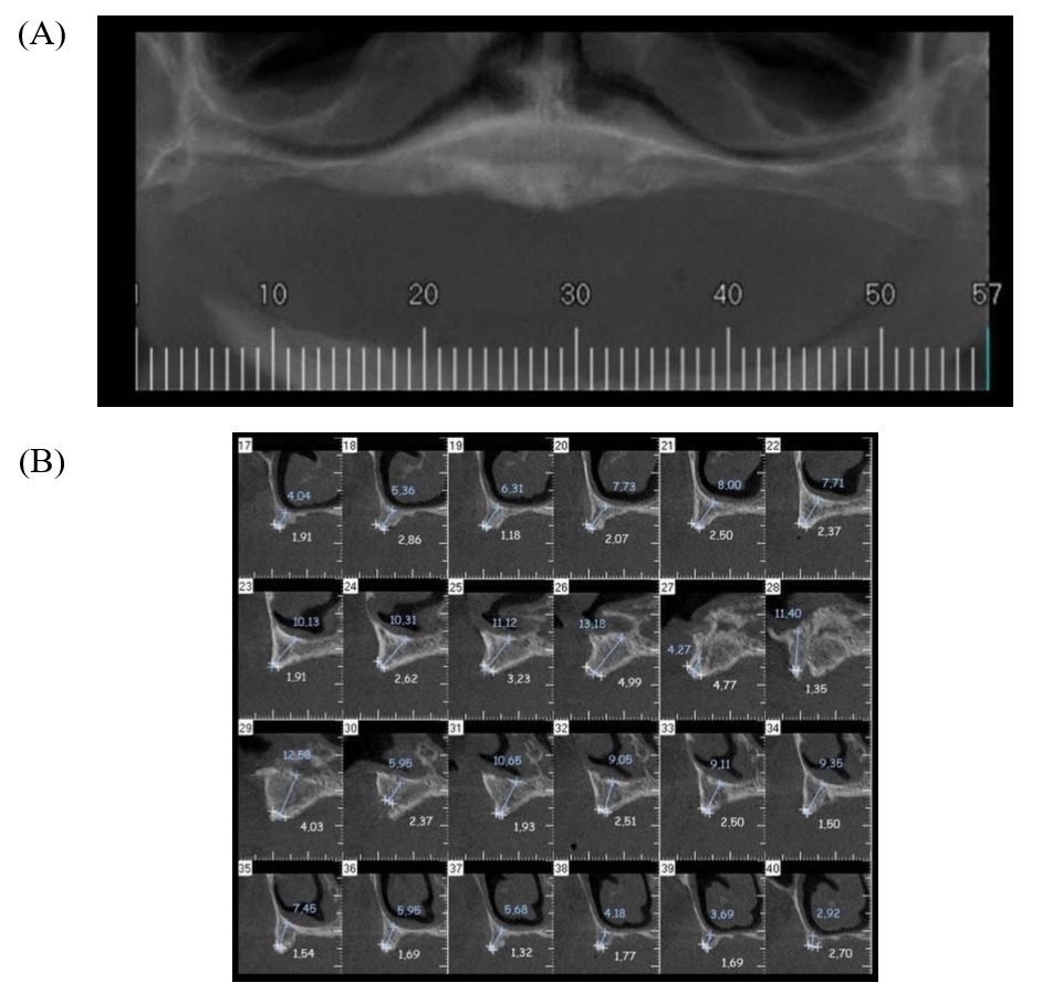
A cone bean computed tomography (CBCT) was requested, which revealed severe maxillary bone loss, in height and thickness (Figure 1), making it difficult to install convenient implants in both the anterior and posterior regions of the patient. The patient was also scanned to obtain the stereolithography (STL) archive required for planning.
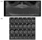
Patient’s imaging exams. (A) panoramic image reconstruction of the CBCT, showing height loss; (B) trans-axial slices from the CBCT showing thickness and height loss.
An evaluation by an anesthesiologist was requested, where the patient's ideal health conditions were assured after anamnesis, physical examination and laboratory tests (complete blood count, coagulogram and glycated hemoglobin), declaring the patient suitable for surgery.
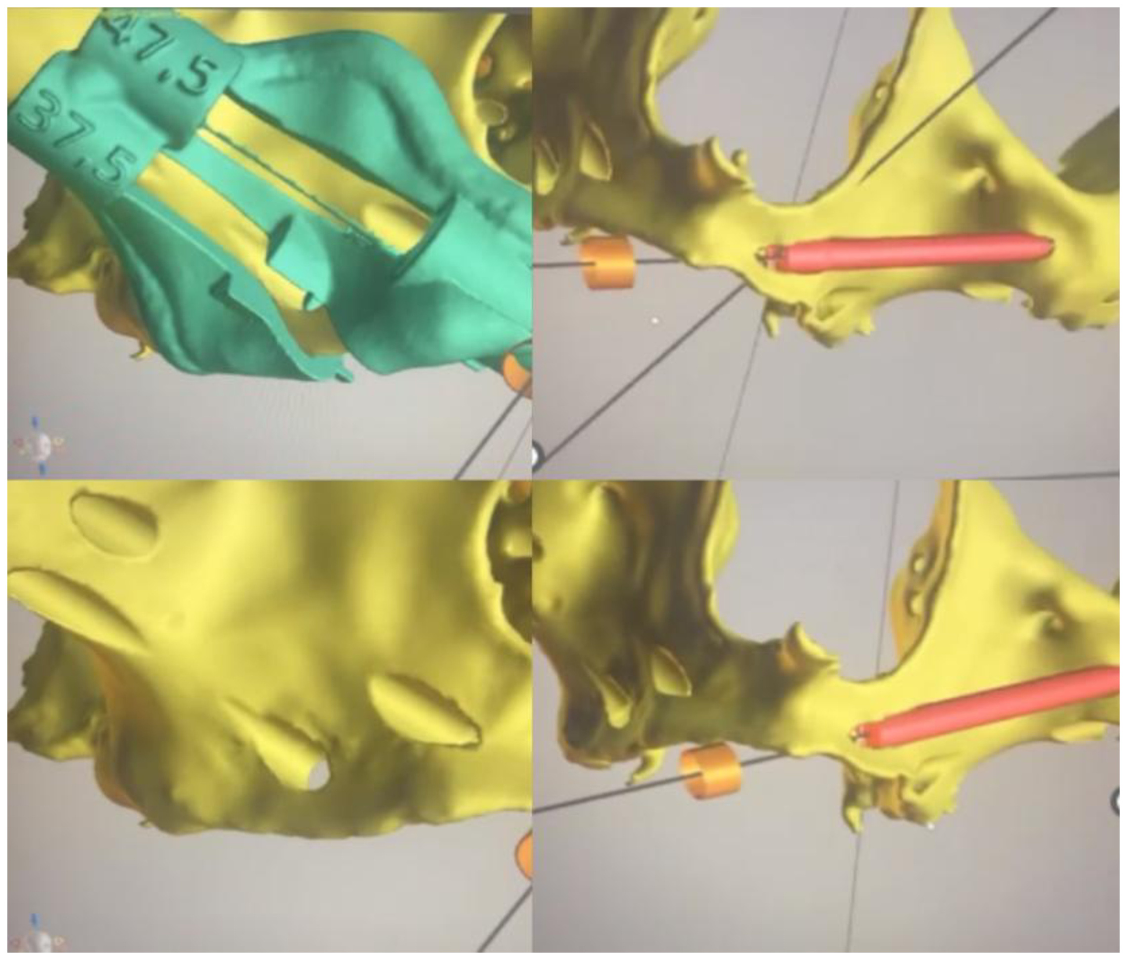
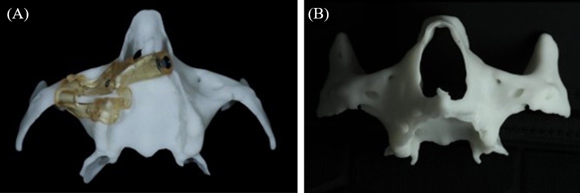
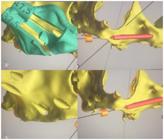
The DICOM (Digital Imaging and Communications in Medicine) and the STL (Stereolithography) archive from the patient were uploaded to the digital planning software Blue Sky Plan® (Blue Sky Bio, Illinois, USA). Four zygomatic implants (Neodent® GM Zygoma-S Implant) were planned, two 4.0mm × 37.5mm (posterior implant, in both sides) and two 4.0mm × 47.5mm (anterior implant, in both sides) and the drill guide was designed (Figure 2). Due to the length and inclination of the implants, the design of a single guide for all the four implants wasn’t possible, because the right and left drills would overlap each other, so two guides were planned and printed, one for each side (Figure 3A).
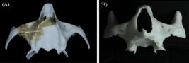
(A) Printed implant guide, separated by hemiarch; (B) Maxillae prototype with zygomatic drillings for implant placement.
A prototype of patient’s maxilla was printed and the procedure was performed in it (Figure 3B), using the same implant surgical kit (Neodent® GM Zygomatic Surgical Kit) that was planned to be used in the surgery.
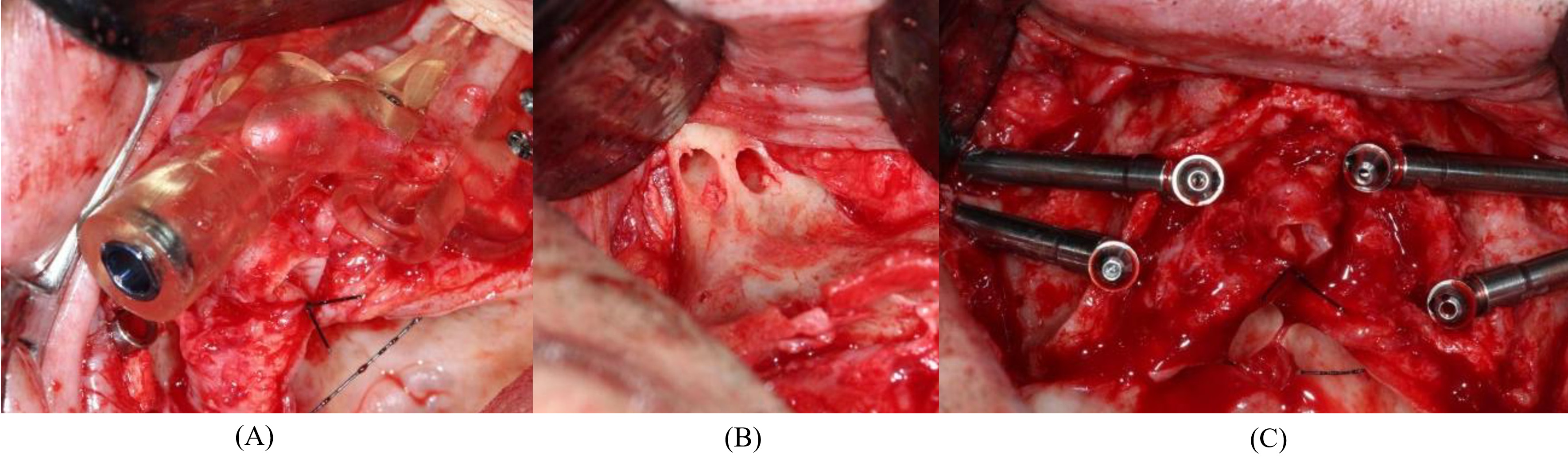
The procedure was performed under local anesthesia (Articaine Hydrochloride 4% Epinephrine 1:100,000) with intravenous conscious sedation, performed by an anesthesiologist. The main incision was made on the crest of the alveolar ridge with two posterior relaxing incisions, followed by a mucoperiosteal detachment. The surgical guide was placed and fixed with anchor pins (Figure 4A). Finally, drilling (Figure 4B) and installation of the implants were carried out, whose immediate torque was found to be above 60N. The path of the zygomatic implants was extrasinusal.
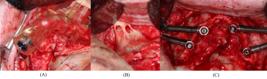
Surgical steps. (A) Guide in position fixed with anchor pins; (B) Bone aspect after drilling, showing the extrasinusal implant pathway; (C) Occlusal view of implants with abutments placed.
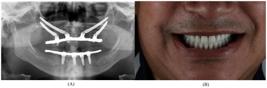
Four angled abutments (Neodent® GM Exact Mini Conical Abutment 60°) were inserted (Figure 4C) to enable the prosthetic rehabilitation with a protocol type prosthesis made with acrylic resin. Soft tissue was sutured with Nylon monofilament thread. 24 hours after the surgical procedure, a protocol type prosthesis made with Cobalt-Chromium (Co-Cr) metal framework and acrylic resin was installed, restoring the vertical dimension of occlusion and patient’s smile esthetic. A panoramic radiography was taken immediately after all the procedures (Figure 5A), showing implants, abutments and the metal framework in position. Patient’s final smile is shown in Figure 5B.
After a 12-month period, the patient was clinically evaluated and it was possible to verify the absence of edema and peri-implant inflammation, no significant differences in peri-implant bone level or soft tissue were observed. The concave shape of the prosthesis enabled effective oral hygiene, which could improve the prognosis. The patient reported aesthetic satisfaction and improvement in chewing capacity.
DISCUSSION
The digital planning used in this case report was a fundamental step for the placement of the zygomatic implants, given the anatomical complexity of the atrophic maxilla and the proximity of critical structures such as the orbit and maxillary sinus. The integration of cone-beam computed tomography (CBCT) data with intraoral scanning and virtual planning software allowed precise three-dimensional assessment of the zygomatic bone trajectory, identification of the optimal implant entry and apex points, and evaluation of bone-implant contact. This digital workflow enhanced prosthetically driven planning, ensuring proper implant angulation, emergence profile, and biomechanical distribution, particularly in this quad zygomatic approach. Moreover, computer-aided design and manufacturing (CAD/CAM) of surgical guides improved the accuracy and predictability of the implant positioning, reducing intraoperative risks and surgical time.
The choice of the zygomatic anchorage over a guided bone regeneration (GBR) in the posterior site was made by the patient due to the possibility of the immediate loading and short treatment time. Sinus lift and GBR technique are predictable, present a high success rate and are well established in the literature [16,17], however it requires more surgery procedures and there is a risk of graft loss, requiring reintervention before implant installation [1,2,18].
The ideal desired path of zygomatic implants has changed over the years. Initially, with its introduction by Brånemark, the trajectory was mostly intrasinusal, and his 5-year to 10-year follow-up revealed a 94% survival rate in 28 patients [7]. Aiming to reduce sinus complications, the vertical slot technique was introduced, where the body of the implant is allocated through the lateral wall of the maxilla [19]. In cases with a high concavity of maxillary anterior wall, the exteriorized technique was developed with the anterior wall of the maxillary sinus, introducing the exteriorized technique, where no prior sinus window or slot is required [20,21].
In this case report, the exteriorized technique was possible, which following the ZAGA (Zygoma anatomy-guided approach) classification developed by Aparicio [22,23] is a ZAGA Type 3 case. Because the maxillary wall is very concave, the implant head is located at the level of alveolar crest and the implant body has an anterior extrasinus path. This position of implant is possible in only 9% of the patients, and is related to the decrease of complications, especially those associated with the maxillary sinus [24,25]. Past studies showed a cumulative success rate of 94.4% at 7 years for extrasinusal implants, strengthening the indication of this technique whenever possible [21].
Regarding the prosthetic aspect, the alveolar position of the implant head and the angled abutments made it possible for the screw access holes to be positioned on the occlusal of molars and palatin of anterior teeth, which was covered with composite resin. The possible difference in color between the acrylic resin of the prosthesis and the restorative material won’t be seen due to its position.
CONCLUSION
In the present case, the prosthetic rehabilitation of an atrophic maxilla using the zygomatic implant technique proved to be effective. Zygomatic bone anchorage allowed treatment without the need for bone reconstruction procedures for vertical and horizontal maxillary augmentation. A digital workflow was used for planning and guiding the implant osteotomy. Implant insertion was performed manually, following the planned trajectory. The prosthetic rehabilitation was immediate, restoring masticatory function and appearance of the teeth, leaving the patient completely satisfied. The 12-month follow-up showed the stability and security of the zygomatic implants in this patient, no complications or adverse events were observed in the case report or reported by the patient.
LIMITATIONS
Despite the successful outcome observed in this report, it is important to acknowledge the inherent limitations of this study design, like small sample size, the lack of a control group and the selection bias, which prevents statistical analysis. Furthermore, this study presents a short follow-up period, which may not account for long-term peri-implant stability or late complications.
Acknowledgements
Authors should not acknowledge their own institution when it is already listed in the affiliations.
Data availability
Not applicable.
-
How to cite:
Miranda PRP, Campos JF, Leite SMG, Ribeiro NCR, Silva RR, Nishioka RS. Computer assisted zygomatic implants in atrophic maxilla: a clinical report. Braz. Dent. Sci. 2026;29:e5075. https://doi.org/10.4322/bds.2026.e5075
-
Regulatory Statement
Written informed consent was obtained from the patient for the publication of this case report and any accompanying images.This study was conducted in accordance with the ethical principles of the Declaration of Helsinki. The research protocol was reviewed and approved by the Research Ethics Committee from the Institute of Science and Technology, São Paulo State University (UNESP), under the approval number (CAAE: 88017625.5.0000.0077). All procedures involving human participants were carried out in compliance with the committee's guidelines and applicable regulations.
-
Funding
The authors declare that no financial support was received.
-
Ethics statement
This study was conducted in accordance with the ethical principles of the Declaration of Helsinki. The research protocol was reviewed and approved by the Research Ethics Committee from the Institute of Science and Technology, São Paulo State University (UNESP), under the approval number (CAAE: 88017625.5.0000.0077). All procedures involving human participants were carried out in compliance with the committee’s guidelines and applicable regulations.
REFERENCES
- 1 Miranda PRP, Leite SMG, Silva RR, Campos JF, Nishioka RS. Técnicas alternativas ao uso de enxertos na reabilitação de maxilas atróficas. Braz J Implantol Health Sci. 2026;8(2):277-96. https://doi.org/10.36557/2674-8169.2026v8n2p277-296.
-
2 Aghaloo T, Misch C, Lin GH, Iacono VJ, Wang HL. Bone augmentation of the edentulous maxilla for implant placement: a systematic review. Int J Oral Maxillofac Implants. 2016;31(Suppl):s19-30. https://doi.org/10.11607/jomi.16suppl.g1 PMid:27228250.
» https://doi.org/10.11607/jomi.16suppl.g1 - 3 Grangeiro MTV, Leite SMG, Miranda PRP, Oliveira CMA, Rodrigues CS, Nossi NR,et al. Use of cone beam tomography for imeddiate loading planning in upper protocol: a case report. REMUNOM. 2026;13(3):1-17. https://doi.org/10.66104/cnwp3v87.
-
4 Bechara S, Kubilius R, Veronesi G, Pires JT, Shibli JA, Mangano FG. Short (6-mm) dental implants versus sinus floor elevation and placement of longer (>/=10-mm) dental implants: a randomized controlled trial with a 3-year follow-up. Clin Oral Implants Res. 2017;28(9):1097-107. https://doi.org/10.1111/clr.12923 PMid:27402427.
» https://doi.org/10.1111/clr.12923 -
5 Gaur V, Doshi A, Palka L. Zygomatic approach with single-piece implants: a technical note. Natl J Maxillofac Surg. 2022;13(1):153-61. https://doi.org/10.4103/njms.NJMS_274_20 PMid:35911801.
» https://doi.org/10.4103/njms.NJMS_274_20 -
6 Herce-López J, Pingarrón MC, Tofé-Povedano Á, García-Arana L, Espino-Segura-Illa M, Sieira-Gil R, et al. Customized subperiosteal implants for the rehabilitation of atrophic jaws: a consensus report and literature review. Biomimetics. 2024;9(1):61. https://doi.org/10.3390/biomimetics9010061 PMid:38275459.
» https://doi.org/10.3390/biomimetics9010061 -
7 Brånemark PI, Gröndahl K, Ohrnell LO, Nilsson P, Petruson B, Svensson B, et al. Zygoma fixture in the management of advanced atrophy of the maxilla: technique and long-term results. Scand J Plast Reconstr Surg Hand Surg. 2004;38(2):70-85. https://doi.org/10.1080/02844310310023918 PMid:15202664.
» https://doi.org/10.1080/02844310310023918 -
8 Soto-Penaloza D, Zaragozí-Alonso R, Penarrocha-Diago M, Penarrocha-Diago M. The all-on-four treatment concept: systematic review. J Clin Exp Dent. 2017;9(3):e474-88. https://doi.org/10.4317/jced.53613 PMid:28298995.
» https://doi.org/10.4317/jced.53613 -
9 Roper MB, Vissink A, Dudding T, Pollard A, Gareb B, Malevez C, et al. Long-term treatment outcomes with zygomatic implants: a systematic review and meta-analysis. Int J Implant Dent. 2023;9(1):21. https://doi.org/10.1186/s40729-023-00479-x PMid:37405545.
» https://doi.org/10.1186/s40729-023-00479-x - 10 Migliorança RM, Ilg JP, Mayo TM, Serrano AS, Souza RP. Função imediata em fixações zigomáticas: relato de dois casos com 18 e 30 meses de acompanhamento clínico. Implant News. 2006;3(3):243-7.
-
11 Nkenke E, Hahn M, Lell M, Wiltfang J, Schultze-Mosgau S, Stech B, et al. Anatomic site evaluation of the zygomatic bone for dental implant placement. Clin Oral Implants Res. 2003;14(1):72-9. https://doi.org/10.1034/j.1600-0501.2003.140110.x PMid:12562368.
» https://doi.org/10.1034/j.1600-0501.2003.140110.x -
12 Bedrossian E, Brunski J, Al-Nawas B, Kämmerer PW. Zygoma implant under function: biomechanical principles clarified. Int J Implant Dent. 2023;9(1):15. https://doi.org/10.1186/s40729-023-00483-1 PMid:37347335.
» https://doi.org/10.1186/s40729-023-00483-1 -
13 Davó R, Fan S, Wang F, Wu Y. Long-term survival and complications of Quad Zygoma Protocol with Anatomy-Guided Approach in severely atrophic maxilla: a retrospective follow-up analysis of up to 17 years. Clin Implant Dent Relat Res. 2024;26(2):343-55. https://doi.org/10.1111/cid.13296 PMid:38084831.
» https://doi.org/10.1111/cid.13296 -
14 Wang J, Chen B, Zhao Y, Pan S, Xu X. The optional apex sites for quad zygomatic implant placement in edentulous patients with severe alveolar bone resorption: a CBCT anatomical analysis. BMC Oral Health. 2024;24(1):1393. https://doi.org/10.1186/s12903-024-05140-6 PMid:39550587.
» https://doi.org/10.1186/s12903-024-05140-6 -
15 Vrielinck L, Politis C, Schepers S, Pauwels M, Naert I. Image-based planning and clinical validation of zygoma and pterygoid implant placement in patients with severe bone atrophy using customized drill guides. Preliminary results from a prospective clinical follow-up study. Int J Oral Maxillofac Surg. 2003;32(1):7-14. https://doi.org/10.1054/ijom.2002.0337 PMid:12653226.
» https://doi.org/10.1054/ijom.2002.0337 -
16 Danesh-Sani SA, Loomer PM, Wallace SS. A comprehensive clinical review of maxillary sinus floor elevation: anatomy, techniques, biomaterials and complications. Br J Oral Maxillofac Surg. 2016;54(7):724-30. https://doi.org/10.1016/j.bjoms.2016.05.008 PMid:27235382.
» https://doi.org/10.1016/j.bjoms.2016.05.008 -
17 Del Fabbro M, Rosano G, Taschieri S. Implant survival rates after maxillary sinus augmentation. Eur J Oral Sci. 2008;116(6):497-506. https://doi.org/10.1111/j.1600-0722.2008.00571.x PMid:19049518.
» https://doi.org/10.1111/j.1600-0722.2008.00571.x -
18 Molina A, Sanz-Sánchez I, Sanz-Martín I, Ortiz-Vigón A, Sanz M. Complications in sinus lifting procedures: classification and management. Periodontol 2000. 2022;88(1):103-15. https://doi.org/10.1111/prd.12414 PMid:35103321.
» https://doi.org/10.1111/prd.12414 - 19 Stella JP, Warner MR. Sinus slot technique for simplification and improved orientation of zygomaticus dental implants: a technical note. Int J Oral Maxillofac Implants. 2000;15(6):889-93. PMid:11151591.
-
20 Aparicio C, Ouazzani W, Aparicio A, Fortes V, Muela R, Pascual A, et al. Extrasinus zygomatic implants: three year experience from a new surgical approach for patients with pronounced buccal concavities in the edentulous maxilla. Clin Implant Dent Relat Res. 2010;12(1):55-61. https://doi.org/10.1111/j.1708-8208.2008.00130.x PMid:19076181.
» https://doi.org/10.1111/j.1708-8208.2008.00130.x -
21 Maló P, de Araújo Nobre M, Lopes A, Ferro A, Moss S. Extramaxillary surgical technique: clinical outcome of 352 patients rehabilitated with 747 zygomatic implants with a follow-up between 6 months and 7 years. Clin Implant Dent Relat Res. 2015;17(Suppl 1):e153-62. https://doi.org/10.1111/cid.12147 PMid:24004279.
» https://doi.org/10.1111/cid.12147 - 22 Aparicio C. A proposed classification for zygomatic implant patient based on the zygoma anatomy guided approach (ZAGA): a cross-sectional survey. Eur J Oral Implantology. 2011;4(3):269-75. PMid:22043470.
-
23 Aparicio C, Olivo A, de Paz V, Kraus D, Luque MM, Crooke E, et al. The zygoma anatomy-guided approach (ZAGA) for rehabilitation of the atrophic maxilla. Clin Dent Rev. 2022;6(1):2. https://doi.org/10.1007/s41894-022-00116-7
» https://doi.org/10.1007/s41894-022-00116-7 -
24 Aleksandrowicz P, Kusa-Podkańska M, Grabowska K, Kotuła L, Szkatuła-Łupina A, Wysokińska-Miszczuk J. Extra-sinus zygomatic implants to avoid chronic sinusitis and prosthetic arch malposition: 12 years of experience. J Oral Implantol. 2019;45(1):21-30. https://doi.org/10.1563/aaid-joi-D-18-00034 PMid:30074849.
» https://doi.org/10.1563/aaid-joi-D-18-00034 -
25 Borgonovo A, Grandi T, Vassallo S, Signorini L. Extrasinus zygomatic implants for the immediate rehabilitation of the atrophic maxilla: 1-year postloading results from a multicenter prospective cohort study. J Oral Maxillofac Surg. 2021;79(2):356-65. https://doi.org/10.1016/j.joms.2020.10.003 PMid:33160924.
» https://doi.org/10.1016/j.joms.2020.10.003
Edited by
-
Editor-in-chief:
Sergio Eduardo de Paiva Gonçalves
-
Editor:
João Paulo Mendes Tribst