AI in early oral cancer detection: a systematic review of technologies and clinical impact
ABSTRACT
Objective To evaluate the effectiveness and clinical applicability of artificial intelligence (AI) in the early diagnosis of oral cancer compared to conventional methods.
Material and Methods This systematic review was prospectively registered in the PROSPERO database under the number CRD420251083609. A descriptive systematic review was conducted based on the PICO question: “In patients with suspected oral cancer, is AI more effective than traditional methods for early diagnosis?” Searches were performed between April and June 2025 in PubMed, LILACS, SciELO, Scopus, Web of Science, Embase, and Google Scholar using the keywords “artificial intelligence,” “oral neoplasm,” and “early diagnosis.” Comparative studies reporting diagnostic metrics such as sensitivity, specificity, and accuracy were included. Methodological quality was assessed using the Quality Assessment of Diagnostic Accuracy Studies – Artificial Intelligence extension (QUADAS-AI) and the Grading of Recommendations, Assessment, Development and Evaluation (GRADE) system.
Results Of 1,704 identified studies, 12 met eligibility criteria. Most studies employed retrospective observational designs, primarily using convolutional neural networks (CNNs) or hybrid models on clinical and histopathological images. Reported diagnostic accuracy was generally above 80%, and some lightweight models demonstrated potential for remote screening. Common limitations included lack of external validation, methodological heterogeneity, and dependence on image quality.
Conclusion AI shows promising potential to support early diagnosis of oral cancer, improving diagnostic speed and accuracy. Broader clinical implementation will require multicenter validation, standardized datasets, and integration with clinical and histopathological evaluation.
KEYWORDS:
Artificial Intelligence; Computer-aided diagnosis; Early diagnosis; Mouth neoplasms; Squamous Cell Carcinoma of Head and Neck
RESUMO
Objetivo Avaliar a eficácia e a aplicabilidade clínica da inteligência artificial (IA) no diagnóstico precoce do câncer bucal em comparação com métodos convencionais.
Material e Métodos Esta revisão sistemática foi registrada prospectivamente na base de dados PROSPERO sob o número CRD420251083609. Foi realizada uma revisão sistemática descritiva com base na questão PICO: “Em pacientes com suspeita de câncer bucal, a IA é mais eficaz do que os métodos tradicionais para o diagnóstico precoce?” As buscas foram realizadas entre abril e junho de 2025 nas bases PubMed, LILACS, SciELO, Scopus, Web of Science, Embase e Google Scholar, utilizando os termos “inteligência artificial”, “neoplasia bucal” e “diagnóstico precoce”. Estudos comparativos que relataram métricas de diagnóstico, como sensibilidade, especificidade e acurácia, foram incluídos. A qualidade metodológica foi avaliada utilizando os sistemas QUADAS-AI e GRADE.
Resultados De 1.704 estudos identificados, 12 atenderam aos critérios de elegibilidade. A maioria dos estudos utilizou desenhos observacionais retrospectivos, empregando principalmente redes neurais convolucionais (CNNs) ou modelos híbridos em imagens clínicas e histopatológicas. A acurácia diagnóstica relatada foi geralmente acima de 80%, e alguns modelos leves demonstraram potencial para triagem remota. Limitações comuns incluíram falta de validação externa, heterogeneidade metodológica e dependência da qualidade das imagens.
Conclusão A IA apresenta potencial promissor para apoiar o diagnóstico precoce do câncer bucal, melhorando a rapidez e a precisão diagnóstica. A implementação clínica mais ampla exigirá validação multicêntrica, conjuntos de dados padronizados e integração com avaliação clínica e histopatológica.
PALAVRAS-CHAVE:
Inteligência Artificial; Diagnóstico auxiliado por computador; Diagnóstico precoce; Neoplasias bucais; Carcinoma de células escamosas de cabeça e pescoço
INTRODUCTION
Oral cancer represents a significant public health challenge worldwide [1,2]. Global epidemiological data from the Global Burden of Disease indicate a marked increase in mortality from this neoplasm, with a 98.7% rise between 1990 and 2017, from 97,492 to 193,696 deaths [1,2]. In many countries, the average five-year survival rate remains below 50% [2,3]. Notable examples include India, with rates near 37%; Uganda and Egypt, around 20%; and Poland and Thailand, ranging from 36% to 39% [2,3]. These figures reflect challenges in early diagnosis and limited access to effective treatment [2,3]. In Brazil, five-year survival varies by region and health service but often remains below 50% [4]. While referral centers such as A.C. Camargo in São Paulo report survival rates close to 51.7%, other locations, such as Florianópolis, report significantly lower rates, with survival at 33.3% [4]. Factors contributing to these unfavorable outcomes include late diagnosis, unequal access to care, and structural limitations of the public health system [4].
The majority of oral cancer cases are oral squamous cell carcinoma (OSCC), accounting for over 90% of malignant neoplasms in the oral cavity [3,5]. OSCC is often preceded by oral potentially malignant disorders (OPMDs), such as leukoplakia, erythroplakia, and oral lichen planus, which carry variable risks of malignant transformation [2,6,7]. Early detection significantly improves prognosis, with cure rates up to 80% for early-stage diagnoses, compared to 20-30% in advanced stages [2,3]. However, timely diagnosis is often hindered by a shortage of experienced pathologists, interobserver and intraobserver variability in histopathological analysis, limited access to specialized services, and the low sensitivity of visual screenings performed by primary care professionals [5,8-11].
In this context, artificial intelligence (AI) has emerged as a promising tool to support the diagnosis of malignant and potentially malignant oral lesions [12,13]. Deep convolutional neural networks (CNNs), a type of AI inspired by human brain function, have been successfully applied to classification, segmentation, and analysis of both clinical and histopathological images [2,3,9,12]. Recent studies report diagnostic accuracy ranging from 72.1% to 98.6%, depending on the network architecture and dataset [2,8,11]. AI has also been applied to images obtained via smartphones in remote areas and to automated interpretation of digitized histological slides, demonstrating utility across diverse clinical scenarios [7,9,12].
Despite these advances, concerns remain regarding the diagnostic accuracy of AI compared to conventional methods, such as expert clinical examination and standard histopathological analysis [13,14]. Key limitations include variability in training datasets, risk of overfitting, limited generalizability to different populations, and the lack of robust external validation [6,10,11,13,15]. Moreover, many studies still involve small sample sizes or lack standardized methodological designs, which hinders direct comparison between algorithms [3,4,8].
METHODS
The strength of this review lies in its inclusion of the most recent (2023-2025) AI applications for oral cancer detection, the adoption of QUADAS-AI for methodological appraisal, and the comparison of diverse imaging modalities, offering a more holistic assessment of diagnostic potential. This systematic review was prospectively registered in the PROSPERO database under the number CRD420251083609
Research question
The research question was formulated according to the PICO strategy (P: population/patients; I: intervention; C: comparison/control; O: outcome), defined as follows:
-
P: Patients with suspected oral cancer
-
I: Artificial intelligence (AI)-assisted diagnosis
-
C: Traditional diagnostic methods (clinical examination, conventional complementary tests)
-
O: Higher diagnostic effectiveness in early detection (accuracy, sensitivity, specificity, and detection time)
The guiding question was:
“In patients with suspected oral cancer, does the use of artificial intelligence result in greater diagnostic accuracy than conventional methods, in terms of sensitivity, specificity, and accuracy?”
Search strategy
A comprehensive search was performed across the following electronic databases: PubMed, SciELO, LILACS, Scopus, Web of Science, Embase, and Google Scholar. The strategy included both indexed and gray literature to minimize publication bias due to non-indexed or unpublished studies.
Keywords were selected based on Medical Subject Headings (MeSH), using the National Library of Medicine database. Boolean operators (“AND”, “OR”), quotation marks, and parentheses were applied to enhance search precision and specificity.
To align the strategy with the PICO framework, the following descriptors were used:
-
P (Population): “Mouth Neoplasms”, “Early Detection of Cancer”, “Diagnosis”, “Precancerous Conditions”
-
I (Intervention): “Artificial Intelligence”, “Machine Learning”, “Deep Learning”, “Computer-Assisted Diagnosis”
-
C (Comparison): “Clinical Examination”, “Clinical Image”
-
O (Outcome): “Early Diagnosis”, “Sensitivity and Specificity”, “Diagnostic Accuracy”
In Embase, the strategy was refined using controlled vocabulary from the Emtree thesaurus, including the terms “oral cancer,” “early diagnosis,” and “artificial intelligence,” as well as their relevant variations and synonyms, to maximize retrieval sensitivity and specificity.
Study selection and eligibility criteria
Titles and abstracts were screened independently and in duplicate by ten reviewers using the Rayyan QCRI platform [16], which supports blinded screening, tagging of inclusion and exclusion decisions, and conflict resolution. Discrepancies were resolved through discussion and consensus after reviewing the full text when necessary.
Eligible studies met the following inclusion criteria:
-
Application of AI in the early diagnosis of oral cancer;
-
Comparison with conventional diagnostic methods;
-
Reporting of quantitative diagnostic metrics (example: sensitivity, specificity, or accuracy).
The selection process adhered to the PRISMA 2020 (Preferred Reporting Items for Systematic Reviews and Meta-Analyses) guidelines. The detailed inclusion and exclusion criteria are summarized in Table 1. Although the primary objective of this review was to evaluate AI in early oral cancer detection, the inclusion criteria also encompassed studies on precursor oral lesions (OPMDs), as these entities are biologically and clinically linked to malignant transformation and are routinely part of early diagnostic pathways.
Studies that lacked clinical evaluation, or those that did not compare AI performance with standard diagnostic approaches were excluded.
Methodological quality assessment
The methodological quality of the included studies was evaluated using the QUADAS-AI tool, complemented by the GRADE system for assessing the certainty of evidence (Tables 2 and 3).
The QUADAS-AI framework extends the original QUADAS-2 domains to include AI-specific aspects such as dataset representativeness, model training and validation protocols, algorithm transparency, and reproducibility (Table 2). The GRADE approach was applied to classify the overall certainty of the evidence based on five domains: risk of bias, inconsistency, indirectness, imprecision, and publication bias.
To ensure accessibility and color-perception neutrality across all figures, the Coblis – Color Blindness Simulator was used to evaluate whether contrasts, heatmaps, and categorical colors remained distinguishable for individuals with common forms of color vision deficiency (protanopia, deuteranopia, and tritanopia). This step followed current recommendations for inclusive scientific visualization.
RESULTS
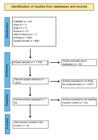
The PRISMA flow diagram is presented in Figure 1. A total of 1,704 records were retrieved from seven databases. After removing 32 duplicates, 1,672 studies were screened by title and abstract, resulting in 1,637 exclusions for not meeting the thematic criteria. Thirty-five full-text articles were assessed for eligibility, and 23 were excluded. Finally, twelve studies [3,5-8,10,11,13,17,18] fulfilled the eligibility criteria and were included in the qualitative synthesis. Table 1 presents the characteristics of the included studies, AI models, and datasets.
The included studies primarily employed retrospective designs to evaluate AI performance in diagnosing oral squamous cell carcinoma (OSCC) or oral epithelial dysplasia (OED). Sample sizes ranged widely, from 30 to 5,192 images/patients, with diverse modalities including clinical photographs, histopathological slides, and microRNA profiles. Convolutional neural networks (CNNs) were the most commonly used architectures, applied in eight of the twelve studies [3,5,7,8,10,11,13,17], while hybrid CNN models and Transformer-based models demonstrated the highest diagnostic performance. Artificial neural networks (ANNs) and traditional machine learning models generally showed lower accuracy and limited generalizability. Externally validated models, such as ODYN and DenseNet201, exhibited higher generalizability (AUC = 0.88-0.94), whereas single-center models often reported inflated accuracies without reproducibility.
Performance metrics varied across studies. Hybrid CNN models achieved accuracies between 94-99% [11,13], while Transformer-based models showed superior predictive performance for malignant transformation [10]. Smartphone-based models demonstrated diagnostic accuracy above 85%, highlighting feasibility for remote screening in underserved populations [7,12]. In contrast, ANN and traditional ML models achieved lower sensitivity and specificity [17,18]. Table 4 summarizes the sample, AI model, and outcomes of each study.
The risk of bias, assessed using QUADAS-AI (Table 2), ranged from low to moderate for most studies. Shephard et al. [10], Ahmad et al. [11], Warin et al. [8], Alabi et al. [17] and Talwar et al. [7] were classified as low risk, whereas Bashir et al. [3], Fati et al. [13], Jubair et al. [5], Pruthi et al. [6] and Alhazmi et al. [18] showed moderate risk, mainly due to limited sample sizes, lack of external validation, or incomplete reporting. Liyanage et al. [2] was the only study rated as high risk due to unclear reference verification and limited generalizability.
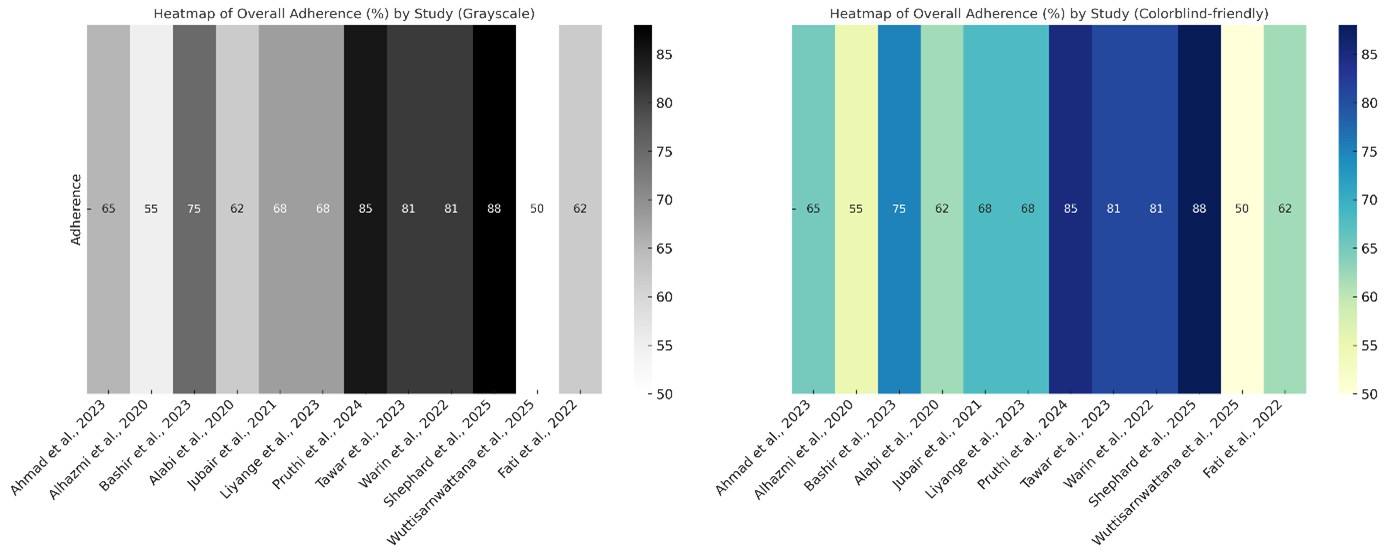
Reporting adherence according to STARD-AI criteria (Table 5 and Figure 2) varied from 50% to 90%. Shephard et al. [10] achieved the highest adherence by implementing a complete end-to-end diagnostic pipeline, whereas Wuttisarnwattana et al. [12] demonstrated the lowest adherence due to limited methodological transparency. While most studies reported accuracy, AUC, sensitivity, and specificity, few included explainability analyses such as Grad-CAM.
The certainty of evidence, evaluated using GRADE (Table 3), ranged from low to high. High certainty was achieved by Shephard et al. [10] and Alabi et al. [17], supported by robust validation and consistent results. Moderate certainty was assigned to Ahmad et al. [11], Talwar et al. [7], and Warin et al. [8]. The remaining studies [2,3,5,6,13,18] were rated as low due to methodological limitations, indirectness, or imprecision related to small sample sizes or incomplete reporting. Overall, the evidence suggests that AI is a promising tool for early oral cancer diagnosis, but broader clinical implementation requires multicenter validation, standardized datasets, and integration with clinical evaluation.
DISCUSSION
Unlike previous systematic reviews, this study incorporates research published up to mid-2025, including both histopathological and mobile-based diagnostic applications, and applies the QUADAS-AI framework. This represents the first structured methodological assessment of AI-based diagnostic tools specifically in oral oncology. Although methodological heterogeneity and the absence of quantitative synthesis limit the strength of conclusions, the absence of language restrictions reduces the risk of language bias. By providing a comprehensive evaluation of study quality and reporting, these findings can guide future research toward more robust, reproducible, and clinically translatable AI applications.
Variations in accuracy across studies can be attributed to differences in dataset size, heterogeneity of image acquisition (clinical vs. histopathological), annotation quality, preprocessing strategies, and the presence or absence of external validation. Lightweight CNNs trained on smartphone images often show lower sensitivity for benign lesions due to visual noise, while hybrid or Transformer-based architectures trained on large histopathological datasets tend to achieve higher accuracy due to richer feature representation and more stable patterns. Additionally, imbalanced datasets, common in OSCC research, can inflate performance metrics when minority malignant classes are underrepresented.
Artificial intelligence (AI) is increasingly recognized as a valuable tool for early detection of oral cancer, addressing limitations of conventional diagnostics such as specialist scarcity, subjectivity, and restricted access in remote areas [13-21]. Several studies have demonstrated that AI can enhance screening accuracy and prognostic assessment of malignant and potentially malignant lesions [13,19]. CNN models, including EfficientNetV2 and MobileNetV3, achieved accuracy above 80% for malignant and premalignant lesions, while the IDaRS histological model reached an AUROC of 0.78 [2,3]. By analyzing large datasets, AI systems can detect complex patterns with consistency, minimizing interobserver variability [2,3] and expediting the diagnostic process [13,14]. These advantages are crucial for early detection, which is directly linked to improved treatment outcomes [4,18].
Nonetheless, several limitations persist. Model performance depends heavily on image quality, and many algorithms display reduced sensitivity for benign lesions or require multicenter validation (13-14). The Transformer-based ODYN model, for example, achieved an F1-score of 0.96 and outperformed the WHO grading system for oral epithelial dysplasia [10]. Yet, its reliance on manual annotations indicates that subjectivity remains a challenge (20). The lack of publicly annotated databases for OSCC also restricts model robustness and reproducibility [22,23].
Recent advances in technological approaches for oral lesion assessment have shown promising potential for improving early diagnosis and prognosis. Computational models and deep learning architectures have been applied to oral tongue lesions, enabling accurate classification and risk prediction [23,24]. Other imaging techniques have also been explored for detecting oral malignancies. Ex vivo fluorescence confocal microscopy has shown high sensitivity and specificity compared with histopathology [25].
Building on these foundations, mobile-based AI applications have achieved high diagnostic accuracy (>85%) [7], representing a significant step toward accessible screening in resource-limited settings. Lightweight convolutional neural networks, such as EfficientNet-B0, have reached expert-level performance in analyzing tongue lesion images [5], while DeepLab v3+ combined with ResNet-50 has demonstrated 87.57% accuracy in lesion segmentation tasks [12,26].
Despite these encouraging results, limitations such as lack of clinical validation and imbalanced datasets continue to affect the generalizability of these models, highlighting the importance of integrating AI tools into standardized diagnostic workflows and multicenter validation studies [27-29].
Beyond imaging, boosted decision trees (BDT) have surpassed conventional histopathological parameters, such as depth of invasion, in predicting recurrence of early tongue squamous cell carcinoma [16]. Yet, classical histopathological variables remain essential [18,22].
ANN models based on clinical and behavioral data reached sensitivity up to 85% but suffered from limited interpretability [9]. AI-driven analysis of microRNA profiles showed 89.4% accuracy [6], demonstrating potential for molecular-level prediction, though small sample sizes and invasive sampling restrict applicability [20,24]. In contrast, smartphone-based intraoral imaging, even when performed by non-specialists, produced encouraging F1-scores (~0.86), suggesting utility for population-level screening [7].
Hybrid AI models combining CNNs with classical feature extraction techniques such as FCH, DWT, LBP, and GLCM achieved some of the best performances. Fati et al. [13] reported 99.1% accuracy, while Ahmad et al. [11] obtained 97% accuracy, 90.9% sensitivity, and 98.9% specificity using DenseNet201 and Xception architectures. However, their use of controlled datasets limits direct clinical translation [11,13]. Real-world performance is influenced by variable image quality and lack of standardized acquisition [23].
Artificial intelligence (AI) represents a valuable complementary tool for early oral cancer diagnosis and can help expand access to care [16,27,28]. Broader clinical implementation will require investment in digital infrastructure, standardized public databases [23,24], multicenter validation, and integration of clinical, histological, and molecular data [6,20]. Hybrid models that combine medical expertise with AI’s analytical capabilities appear to offer the most promising route for clinical translation [5,11,19].
Despite encouraging results, heterogeneity in datasets, annotation protocols, and validation methods remain considerable. Only three studies (Shephard et al., Ahmad et al., Alabi et al.) included multicenter or external validation, which is essential for ensuring generalizability. Inconsistent reporting of metrics (AUC, F1-score, accuracy) further complicates comparison and underscores the need for standardized frameworks such as STARD-AI and QUADAS-AI. None of the included studies reported integration into clinical workflows or prospective validation, indicating that AI currently serves best as an adjunctive diagnostic aid rather than a replacement for conventional methods.
The STARD-AI assessment, together with visualization procedures such as heatmap generation using the Coblis color-blindness simulator [30,31], highlighted substantial variability in reporting quality. While most studies detailed datasets and model architecture, few provided public access to data or code, limiting reproducibility. Validation strategies were inconsistent, with some studies using cross-validation or external datasets and others providing minimal information on data partitioning. Accuracy, AUC, sensitivity, and specificity were frequently reported, but explainability analyses were rare. Only one study implemented a fully integrated diagnostic pipeline, emphasizing that most AI tools remain experimental and not yet deployed in clinical practice. These findings reinforce the need for standardized reporting to enhance transparency, support replication, and facilitate safe clinical application of AI in oral oncology.
Early diagnosis plays a critical role in improving the prognosis of patients with oral cancer. Research by Bandeira et al. [32] highlights that a substantial proportion of individuals receive their diagnosis at advanced stages, which often complicates treatment and diminishes survival outcomes. This underscores the significant potential of artificial intelligence (AI) in facilitating timely detection, thereby enabling more favorable prognoses. AI applications have already demonstrated promising results in related areas, such as the Oral ID device, a handheld tool that utilizes autofluorescence technology to delineate safe surgical margins in patients with oral squamous cell carcinoma [33]. Comparative analyses indicate that using Oral ID improves margin assessment accuracy, surpassing the 52.2% achieved by the traditional 1 cm margin method [33].
Future research should prioritize prospective multicenter trials, open-access annotated datasets, and consistent use of AI-specific quality frameworks. Integration of multimodal data clinical, histopathological, and molecular may further enhance predictive accuracy and facilitate clinical translation.
CONCLUSION
Artificial intelligence shows strong potential as a complementary tool for early oral cancer diagnosis, with performance comparable to or exceeding conventional methods. Convolutional neural networks and hybrid models facilitate efficient analysis of clinical and histopathological images, improving access in resource-limited settings. Implementation challenges remain, including limited external validation, dataset heterogeneity, and generalizability. Careful integration with standard clinical and histopathological evaluation, along with standardized methodologies, well-annotated public datasets, and multicenter validation, is essential to establish AI as a reliable and clinically applicable diagnostic tool.
List of abbreviations
OSCC – Oral Squamous Cell Carcinoma
OPMDs – Oral Potentially Malignant Disorders
CNN – Convolutional Neural Network
ANN – Artificial Neural Network
WSI – Whole Slide Image
AUC – Area Under the Curve
AUROC – Area Under the Receiver Operating Characteristic Curve
miRNA – microRNA
BDT – Boosted Decision Tree
QUADAS-AI – Quality Assessment of Diagnostic Accuracy Studies – Artificial Intelligence extension
GRADE – Grading of Recommendations, Assessment, Development and Evaluation
OED – Oral Epithelial Dysplasia
Acknowledgements
The authors would like to thank the staff and colleagues at the Universidade Federal de Juiz de Fora (UFJF) for their support and valuable contributions during the preparation of this manuscript.
Data availability
The data is already available within the text.
-
How to cite:
Albertoni LA, Martins LSS, Rodrigues CH, Cunha LC, Magalhães HS, Ferreira BA, et al. AI in early oral cancer detection: a systematic review of technologies and clinical impact. Braz. Dent. Sci. 2026;29:e5017. https://doi.org/10.4322/bds.2026.e5017
-
Funding
This research did not receive any specific grant from funding agencies in the public, commercial, or not-for-profit sectors.
-
Regulatory Statement
This review was conducted in accordance with the PRISMA 2020 guidelines for systematic reviews and meta-analyses, as recommended by the EQUATOR Network.The protocol for this systematic review was prospectively registered in the PROSPERO database under registration number CRD420251083609.
-
Disclosure
All data extraction forms, analytic code, and datasets used in this review are available upon request from the corresponding author.
-
Ethics statement
Ethical approval and informed consent were obtained for all primary studies included in this review where reported. If such information was not reported in the original study, it was noted accordingly.
REFERENCES
- 1 Ó SF, Lima JRM, Oliveira SMD, Cruz RAN, Medeiros CGL. Oral cancer and public health. Braz J Implant Health Sci. 2024;4(8):1-7.
-
2 Liyanage V, Tao M, Park JS, Wang KN, Azimi S. Malignant and premalignant oral lesions classification and diagnosis with deep neural networks. J Dent. 2023;137:104657. https://doi.org/10.1016/j.jdent.2023.104657 PMid:37574105.
» https://doi.org/10.1016/j.jdent.2023.104657 -
3 Bashir RMS, Shephard AJ, Mahmood H, Azarmehr N, Raza SEA, Khurram SA, et al. A digital score of peri-epithelial lymphocytic activity predicts malignant transformation in oral epithelial dysplasia. J Pathol. 2023;260(4):431-42. https://doi.org/10.1002/path.6094 PMid:37294162.
» https://doi.org/10.1002/path.6094 -
4 Montoro JRMC, Hicz HA, Souza L, Livingstone D, Melo DH, Tiveron RC, et al. Prognostic factors in squamous cell carcinoma of the oral cavity. Braz J Otorhinolaryngol. 2008;74(6):861-6. https://doi.org/10.1016/S1808-8694(15)30146-4 PMid:19582342.
» https://doi.org/10.1016/S1808-8694(15)30146-4 -
5 Jubair F, Al Karadsheh O, Malamos D, Al Mahdi S, Saad Y, Hassona Y. A novel lightweight deep convolutional neural network for early detection of oral cancer. Oral Dis. 2022;28(4):1123-30. https://doi.org/10.1111/odi.13825 PMid:33636041.
» https://doi.org/10.1111/odi.13825 -
6 Pruthi N, Singh R, Kumar A, Sharma P, Gupta M, Yadav S. Applying machine learning for enhanced microRNA analysis: a companion risk tool for oral squamous cell carcinoma in standard care incisional biopsy. Biomolecules. 2024;14(4):458. https://doi.org/10.3390/biom14040458 PMid:38672474.
» https://doi.org/10.3390/biom14040458 -
7 Talwar V, Singh P, Mukhia N, Vinod PK, Rathore S, Singh A, et al. AI-assisted screening of oral potentially malignant disorders using smartphone-based photographic images. Cancers. 2023;15(16):4120. https://doi.org/10.3390/cancers15164120 PMid:37627148.
» https://doi.org/10.3390/cancers15164120 -
8 Warin K, Limprasert W, Suebnukarn S, Jinaporntham S, Jantana P, Vicharueang S. AI-based analysis of oral lesions using novel deep convolutional neural networks for early detection of oral cancer. PLoS One. 2022;17(8):e0273508. https://doi.org/10.1371/journal.pone.0273508 PMid:36001628.
» https://doi.org/10.1371/journal.pone.0273508 -
9 Chaudhary N, Rai A, Rao AM, Faizan MI, Augustine J, Chaurasia A, et al. High-resolution AI image dataset for diagnosing oral submucous fibrosis and squamous cell carcinoma. Sci Data. 2024;11(1):1050. https://doi.org/10.1038/s41597-024-03836-6 PMid:39333529.
» https://doi.org/10.1038/s41597-024-03836-6 -
10 Shephard AJ, Mahmood H, Raza SEA, Araújo ALD, Santos Silva AR, Lopes MA, et al. Development and validation of an artificial intelligence-based pipeline for predicting oral epithelial dysplasia malignant transformation. Commun Med. 2025;5(1):186. https://doi.org/10.1038/s43856-025-00873-z PMid:40394272.
» https://doi.org/10.1038/s43856-025-00873-z -
11 Ahmad M, Irfan MA, Sadique U, Haq IU, Jan A, Khattak MI, et al. Multi-method analysis of histopathological images for early diagnosis of oral squamous cell carcinoma using deep learning and hybrid techniques. Cancers (Basel). 2023;15(21):5247. https://doi.org/10.3390/cancers15215247 PMid:37958422.
» https://doi.org/10.3390/cancers15215247 -
12 Wuttisarnwattana P, Limprasert W, Suebnukarn S, Jinaporntham S, Jantana P, Vicharueang S, et al. Precise identification of oral cancer lesions using artificial intelligence. Stud Health Technol Inform. 2024;316:1096-7. https://doi.org/10.3233/SHTI240601 PMid:39176572.
» https://doi.org/10.3233/SHTI240601 -
13 Fati SM, Senan EM, Javed Y. Early diagnosis of oral squamous cell carcinoma based on histopathological images using deep and hybrid learning approaches. Diagnostics. 2022;12(8):1899. https://doi.org/10.3390/diagnostics12081899 PMid:36010249.
» https://doi.org/10.3390/diagnostics12081899 -
14 He J, Baxter SL, Xu J, Xu J, Zhou X, Zhang K. The practical implementation of artificial intelligence technologies in medicine. Nat Med. 2019;25(1):30-6. https://doi.org/10.1038/s41591-018-0307-0 PMid:30617336.
» https://doi.org/10.1038/s41591-018-0307-0 - 15 Sounderajah V, Ashrafian H, Aggarwal R, De Fauw J, Denniston AK, Greaves F, et al. Developing specific reporting guidelines for diagnostic accuracy studies assessing AI interventions: the QUADAS-AI extension. Nat Med. 2021;27(2):185-7.
-
16 Ouzzani M, Hammady H, Fedorowicz Z, Elmagarmid A. Rayyan: a web and mobile app for systematic reviews. Syst Rev. 2016;5(1):210. https://doi.org/10.1186/s13643-016-0384-4 PMid:27919275.
» https://doi.org/10.1186/s13643-016-0384-4 -
17 Alabi RO, Elmusrati M, Sawazaki Calone I, Kowalski LP, Haglund C, Coletta RD, et al. Comparison of supervised machine learning classification techniques in prediction of locoregional recurrences in early oral tongue cancer. Int J Med Inform. 2020;136:104068. https://doi.org/10.1016/j.ijmedinf.2019.104068 PMid:31923822.
» https://doi.org/10.1016/j.ijmedinf.2019.104068 -
18 Alhazmi FH, Alhazmi Y, Makrami A, Masmali A, Salawi N, Masmali K, et al. Application of artificial intelligence and machine learning for prediction of oral cancer risk. J Oral Pathol Med. 2021;50(5):444-50. https://doi.org/10.1111/jop.13157 PMid:33394536.
» https://doi.org/10.1111/jop.13157 - 19 Kaur S, Mupparapu M. Artificial intelligence in oral cancer diagnosis and prognosis: current trends and future directions. Oral Oncol Rep. 2023 In press.
-
20 Marque L, Souza FN, Silva LM, Silva BMR, Oliveira FB, Santos LCC. Artificial intelligence in oral cancer diagnosis and prognosis: current perspectives. J Oral Pathol Med. 2024;53(2):123-35. https://doi.org/10.1111/jop.13467
» https://doi.org/10.1111/jop.13467 - 21 El-Naggar AK. The 2017 WHO classification of head and neck tumours. Int J Head Neck Surg. 2017;48(6):30-7.
-
22 Dimitriou N, Arandjelović O, Caie PD. Deep learning for whole-slide image analysis: an overview. Front Med. 2019;6:264. https://doi.org/10.3389/fmed.2019.00264 PMid:31824952.
» https://doi.org/10.3389/fmed.2019.00264 - 23 Sengupta S. Challenges in AI-based diagnosis of oral cancer: data, validation, and deployment. Diagnostics. 2022;12(8):1903.
-
24 Bello IO, Soini Y, Salo T. Prognostic evaluation of oral tongue cancer: means, markers and perspectives (II). Oral Oncol. 2010;46(9):636-43. https://doi.org/10.1016/j.oraloncology.2010.06.008 PMid:20637679.
» https://doi.org/10.1016/j.oraloncology.2010.06.008 -
25 Shavlokhova V, Sandhu S, Flechtenmacher C, Koveshazi I, Neumeier F, Padrón-Laso V, et al. MobileNet-based detection of oral squamous cell carcinoma using ex vivo confocal images. J Clin Med. 2021;10(22):5326. https://doi.org/10.3390/jcm10225326
» https://doi.org/10.3390/jcm10225326 - 26 Wuttisarnwattana P, Limprasert W, Suebnukarn S, Jinaporntham S, Jantana P, Vicharueang S, et al. DeepLab v3+ for segmentation of oral potentially malignant and malignant lesions in photographic images. J Imaging. 2024;10(1):45.
- 27 Mahmood H. Use of artificial intelligence for the diagnosis of oral cancer: a systematic review. Oral Oncol. 2020;106:104741.
- 28 Pruthi P. A machine learning model based on miRNA expression for early detection of oral squamous cell carcinoma. J Oral Pathol Med. 2024;53(1):65-72.
- 29 Rahman MM. A review of artificial intelligence applications in oral cancer diagnosis and prognosis. Front Oncol. 2023;13:1057421.
- 30 Sounderajah V, Ashrafian H, Aggarwal R, De Fauw J, Denniston AK, Greaves F, et al. Developing specific reporting guidelines for diagnostic accuracy studies assessing AI interventions: the STARD-AI extension. Nat Med. 2021;27(2):185-7.
-
31 Coblis. Color Blindness Simulator [Internet]. 2025 [cited 2025 Oct 13]. Available from: https://www.color-blindness.com/coblis-color-blindness-simulator/
» https://www.color-blindness.com/coblis-color-blindness-simulator/ -
32 Bandeira CM, Almeida AA, Carta CFL, Almeida JD, Kaminagakura E. Como melhorar o diagnóstico precoce do câncer bucal? Braz Dent Sci. 2017;20(4):25-31. https://doi.org/10.14295/bds.2017.v20i4.1439
» https://doi.org/10.14295/bds.2017.v20i4.1439 -
33 Mohamed Y, Mohammed SA, Sulaiman AM. Oral ID® as an adjunctive tool for surgical margin assessment in patients with oral squamous cell carcinoma: a comparative study: Oral ID® as an Adjunctive Tool for Surgical Margin Assessment. Braz Dent Sci. 2022;24(4, Suppl):e2868. https://doi.org/10.4322/bds.2021.e2868
» https://doi.org/10.4322/bds.2021.e2868
Edited by
-
Editor-in-chief:
Sergio Eduardo de Paiva Gonçalves
-
Editor:
Renata Falchete do Prado