Digital palatal anthropometry in children with and without congenital zika syndrome-associated microcephaly
ABSTRACT
Objective To digitally compare palatal linear and angular anthropometry in children with and without congenital zika syndrome-associated microcephaly.
Material and Methods Thirty-eight digitized dental molds were divided into two groups: congenital zika syndrome-associated microcephaly (CZS) group and control group (no associated craniofacial anomaly). Linear and angular measurements were performed, as follow: left (I-C’) and right (I-C) anterior intersegment distance, left (I-T’) and right (I-T) anterior-posterior intersegment length, intercanine distance (C-C’), intertuberosity distance (T-T’); left (T’C’I) and right (ICT) deciduous canine angle, and interincisive angle (C’IC). Intraclass Correlation Coefficient, Mann-Whitney test, independent T test, and Pearson Correlation were used for statistical analysis with α=5%.
Results In CZS group, the intertuberosity measurement (T-T’) of boys were statistically higher than that of girls (p=0.0496). The CZS group showed strong correlations for I-C vs. C-C (r=0.919), while the control group showed strong correlations for I-C vs. C-C (r=0.846), I-C vs. C-C (r=0.847), and T-T vs. I-T (r=0.722). The means of angles T’C’I and ICT were greater in CZS group (p=0.0016 and p=0.0313, respectively), while measurement C’IC was greater in control group (p=0.0008). Whereas the linear measurements did not show a statistically significant difference (p>0.05).
Conclusion Children with congenital Zika syndrome–associated microcephaly showed a tendency toward anterior palatal narrowing compared with children without craniofacial anomalies, suggesting the need for early palatal expansion.
KEYWORDS:
Anthropometry; Dental arch; Imaging, three-dimensional; Microcephaly; Zika virus
RESUMO
Objetivo Comparar digitalmente a antropometria linear e angular palatina em crianças com e sem microcefalia congênita associada à síndrome do zika.
Material e Métodos Trinta e oito modelos dentários digitalizados foram divididos em dois grupos: grupo microcefalia congênita associada à síndrome do zika vírus (CZS) e grupo controle (sem anomalia craniofacial). Foram realizadas as seguintes medidas lineares e angulares: distância intersegmentar anterior esquerda (I-C') e direita (I-C), comprimento intersegmentar anterior-posterior esquerda (I-T') e direita (I-T), distância intercaninos (C-C'), distância intertuberosidade (T-T'); ângulo canino decíduo esquerdo (T'C'I) e direito (ICT) e ângulo interincisivo (C'IC). Coeficiente de correlação intraclasse, teste de Mann-Whitney, teste T independente e correlação de Pearson foram usados para análise estatística com α = 5%.
Resultados No grupo CZS, a medida da intertuberosidade (T-T’) dos meninos foi estatisticamente maior na comparação com as meninas (p = 0,0496). A correlação intergrupo mostrou forte correlação no grupo CZS para I-C' vs. C'-C (r = 0,919) e no grupo controle para I-C' vs. C'-C (r = 0,846), I-C vs. C'-C (r = 0,847) e T-T' vs. I-T (r = 0,722). As médias dos ângulos T'C'I e ICT foram maiores no grupo CZS (p = 0,0016 e p = 0,0313, respectivamente), enquanto a medida C'IC foi maior no grupo controle (p = 0,0008). Enquanto, as medidas lineares não apresentaram diferença estaticamente significante (p>0,05).
Conclusão Crianças com microcefalia associada à síndrome congênita do Zika apresentaram tendência ao estreitamento palatino anterior em comparação com crianças sem anomalias craniofaciais, sugerindo a necessidade de expansão palatina precoce.
PALAVRAS-CHAVE:
Antropometria; Arco dental; Imageamento tridimensional; Microcefalia; Zika vírus
INTRODUCTION
Zika virus (ZV) is an arbovirus transmitted by the Aedes Aegypti mosquito accounting for the global epidemic starting in 2015 [1]. This public health emergency affected thousands of people worldwide, reaching Asia, Africa, Pacific islands, and Latin America, including Brazil. The virus causes mild symptoms similar to dengue and Chikungunya fever, but in rare cases, it can lead to Congenital Zika Virus Syndrome (CZS) associated with microcephaly in newborns [2-5].
The rising incidence of microcephaly in newborns, both in Brazil and other countries, has become a matter of global concern [5-7]. In Brazil, Northeast region accounts for 28% of the Brazilian births and it had the highest incidence of the disease (97%) [5]. The relationship between the virus and microcephaly has been intensely studied by experts worldwide, as research indicates that the virus can cross the placental barrier and reach the fetus, where it can infect developing brain cells, resulting in irreversible damage. The infection can occur at any time during pregnancy, but the most critical period is the first trimester, during which the development of major organs, including bones, takes place [2,7,8].
A hypothesized mechanism for craniofacial anomalies in congenital Zika syndrome (CZS) involves the impact of Zika virus on cranial neural crest cells, which are essential for maxillary growth and bone formation. ZIKV infection in these cells blocks the epithelial-mesenchymal transition, activates autophagy, and induces secretion of cytokines (such as LIF and VEGF) that indirectly affect neurogenesis and facial development [9-11].
Although studies are still ongoing, it is possible to say that the presence of ZV during pregnancy poses a risk to the fetus's development. CZS can affect the child's craniofacial development, such as the presence of a narrow palate, short labial frenulum, enamel hypoplasia, agenesis, and delayed tooth eruption [7,8]. Therefore, it is of utmost importance that dentists are aware of the possible alterations that ZV can cause in the craniofacial development of children to provide accurate information about the potential effects of ZV and offer more predictable treatments with better prognosis for the individual. For this purpose, studies comparing children with and without CZS are necessary [12].
The analysis of the morphometry of dental arches in individuals with CZS can contribute to the rehabilitation process and establish criteria for future investigations. No studies have yet quantified palatal angular changes in CZS infants using 3D digital methods, despite reports of narrow palates in up to 80% of cases [13]. Thus, this study aimed to digitally compare the anthropometry of palatal linear and angular measurements in children with and without congenital zika syndrome-associated microcephaly. The null hypothesis is that the evaluated parameters would be different between groups.
MATERIAL AND METHODS
Inclusion criteria comprised children aged between 5 and 10 months with and without congenital zika syndrome-associated microcephaly. Controls from the pre-epidemic period [2010-2012] to avoid confounding due to Zika exposure, while participants with CZS were selected during the 2015-2016 period. The diagnosis of CZS was confirmed according to established criteria: microcephaly <32 cm at birth, associated with Zika virus exposure and characteristic neuroimaging findings (e.g., cerebral volume loss, calcifications, and cortical malformations) [14]. Exclusion criteria were dental molds with defects compromising the measurements. Sample size was obtained with a pilot study with standard deviation on 5.21º in interincisive angle measurement (Cohen's d = 1.06), with level of significance of 5%, test power of 80%, and a minimum difference to be detected of 5.5º. The minimum sample size was fifteen participants per group: CZS group – children with congenital zika syndrome-associated microcephaly; and control group – children without craniofacial anomalies.
All dental casts were obtained from the institutional files. The dental casts were digitized by a three-dimensional surface laser scanner (R700TM, 3Shape, Copenhagen, Denmark). The palatal morphometry was analyzed by software Mirror Imaging version 2.8.3 (Canfield Scientific Inc., Fairfield, NJ, USA) [12,15-18]. To evaluate the digital anthropometry, the following anatomic references were used: interincisive point (I); left (C’) and right (C) deciduous canine eruption point; left (T’) and right (T) tuberosity. Landmarks were placed at the palatal vault midpoint for interincisive, canines, and tuberosities measurements to ensure reproducibility.
Therefore, the following linear measurements were analyzed: left anterior intersegment length (I-C’, straight line from the interincisive point to the left deciduous canine eruption point); right anterior intersegment length (I-C, line from the interincisive point to the right deciduous canine eruption point); left anterior-posterior intersegment length (I-T’, straight line from the interincisive point to the left tuberosity); right anterior-posterior intersegment length (I-T, straight line from the interincisive point to the right tuberosity) (Figure 1A); intercanine distance (C-C’, line contouring the palatal surface between right and left deciduous canine eruption points); intertuberosity distance (T-T’, line contouring the palatal surface between right and left tuberosity) [12,15-18] (Figure 1B). The angular measurement were: left deciduous canine angle (T’C’I, angle between the left tuberosity, left deciduous canine eruption point, and the interincisive point) (Figure 1C); interincisive angle (C’IC, angle between the left deciduous canine eruption point, interincisive point, and right deciduous canine eruption point) (Figure 1D); right deciduous canine angle (ICT, angle between the right tuberosity, right deciduous canine eruption point, and the interincisive point) [19] (Figure 1E). The linear measurements were obtained in mm and the angular measurements in degrees (º) [18]. The obtained values were stored in electronic sheets (Microsoft Excel 2019, Microsoft Corporation, Albuquerque, USA).
- Parameters for palatal morphometry. (A) Straight linear measurements - left anterior intersegment length (I-C’), right anterior intersegment length (I-C), left anterior-posterior intersegment length (I-T’), and right anterior-posterior intersegment length (I-T). (B) Linear measurements contouring the palatal surface - intercanine distance (C’-C) and (T’-T) intertuberosity distance (T’T). (C) Angular measurement of the left deciduous canine (T’C’I). (D) interincisive angle measurement (C’IC). (E) Angular measurement of the right deciduous canine (ICT).
All linear and angular measurements were performed by two examiners. To determine the intra- and interexaminer error, 50% of the sample was randomly selected through a website. Next, the sample was measured twice, within 15-day interval, according to previous study [20]. The Intraclass Correlation Coeficient (ICC) was applied for intra- and interexaminer evaluation [21]. Data was analyzed by Shapiro-Wilk normality test, independent t-test, Mann-Whitney test, Cohen´s d, and Pearson Correlation. All tests were performed in Prism GraphPad Software (Prism 5 for Windows- Version 5.0- software GraphPad, NC, USA) with level of significance of 5%. The descriptive data were presented as mean/standard deviation and median/interquartile amplitude.
RESULTS
Sample description
Thirty-eight dental casts were selected (CZS group, n=15, male/female = 6/9; control group, n=23, male/female = 13/10). At the moment of the dental impression, the mean age of CZS group was 9.54 ±0.20 months and control group 6.57 ±0.18 months.
Intra- and interexaminer error
The intraexaminer analysis revealed that both examiners were calibrated (ICC = 0.98 and 0.99). The interexaminer error was of ICC=0.96. All these results indicated an excellent reliability [21].
Intragroup comparison (male vs female)
Concerning gender, in CZS group, the intragroup comparison showed a statistically greater intertuberosity distance (T-T’) for boys. In control groups, all measurements exhibited no statistically significant differences (p>0.05) (Table I).
Intragroup correlations
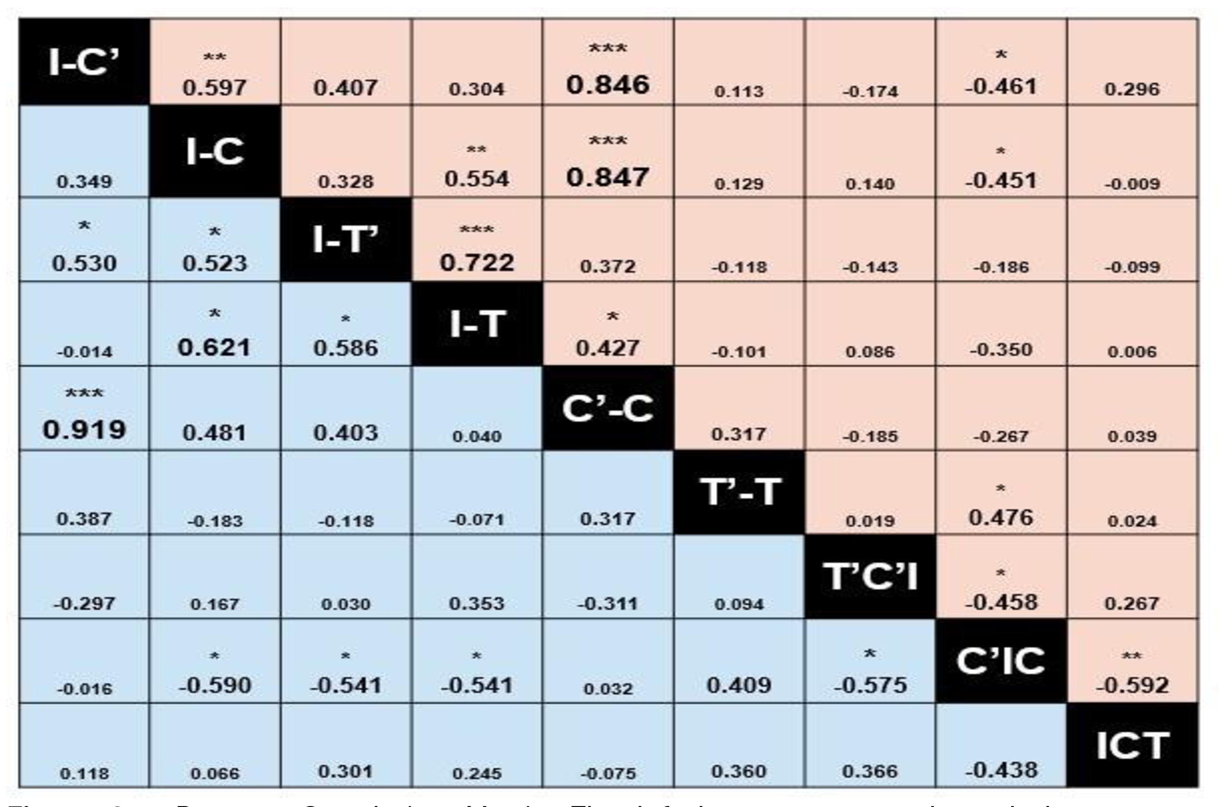
In CZS group, the correlation analyses of parameters I-C’ vs I-T’ (r=0.530 and p<0.05), I-C vs I-T’ (r=0.523 and p<0.05), I-C vs I-T (r=0.621 and p<0.05), and I-T’ vs I-T (r=0.586 and p<0.05) demonstrated moderate coefficients, while I-C’ vs C’-C (r=0.919 and p<0.001) demonstrated strong coefficient. All these analyses indicated positive linear relations (that is, the variables are directly proportional among each other). The correlations I-C vs C’IC (r=-0.590 and p<0.05), I-T’ vs C’IC (r=-0.541 and p<0.05), I-T vs C’IC (r=-0.541 and p<0.05), and T’C’I vs C’IC (r=-0.575 and p<0.05) showed moderate coefficients with negative linear relations (that is, the variables were inversely proportional among each other) (Figure 2).
- Pearson Correlation Matrix. The left bottom corner showed the coefficients of group CSZ (blue); the right upper conner showed the coefficients of control group (pink). This matrix indicates the values of the Pearson correlation coefficients with linear correlation among variables. Negative coefficients indicate variables are inversely proportional among each other (negative linear relation), while positive values indicate that variables are directly proportional among each other (positive linear relation). *p<0.05; **p<0.01; ***p<0.001.
In control group, the analyses of parameters I-C’ vs I-C (r=0.597 and p<0.01), I-C vs I-T (r=0.554 and p< 0.01), I-T vs C’-C (r=0.427 and p<0.05), and T’-T vs C’IC (r=0.476 and p<0.05) showed moderate coefficients, while I-C’ vs C’-C (r=0.846 and p<0.001), I-C vs C’-C (r=0.847 and p<0.001), and I-T’ vs I-T (r= 0.722 and p<0.001) showed strong coefficients. All these analyses indicated positive linear relations. The correlations I-C’ vs C’IC (r=-0.461 and p<0.05), I-C vs C’IC (r=-0.451 and p< 0.05), T’C’I vs C’IC (r=-0.458 and p<0.05), and C’IC vs ICT (r=-0.592 and p<0.01) showed moderate coefficients with negative linear relations (Figure 2).
Intergroup comparison
The linear measurement comparisons did not show statistically significant differences (p>0.05). For angular measurements, T’C’I and ICT were significantly greater in CZS group (p=0.0016 and p=0.0313, respectively), while C’IC was significantly greater in control group (p=0.0008) (Table II).
- Comparison between the means of linear and angular measurements in CZS group and control group (Independent t-test)
DISCUSSION
The literature has increasingly shown studies on the development and growth of the maxilla in edentulous individuals with and without anomalies, during early childhood, through quantitative morphometry. These studies enable the identification of anatomical differences among these individuals [15-18,20]. However, literature lacks studies on congenital zika syndrome-associated microcephaly due to the difficulty of obtaining models in the early months of life [12]. As far as we are concerned, this is the first study comparing linear and angular palatal measurements in children with and without CSZ.
The intragroup gender comparison, control group showed no statistically significant differences, while CZS group, boys showed greater intertuberosity measurements than that of girls. The intertuberosity measurement is the line between soft and hard palate. Many variables may have interfered with this difference, such as oral breathing, type of feeding (breast or formula), and post-delivery history. Even with a limited number of studies assessing growth differences in individuals with microcephaly associated with congenital Zika virus syndrome during early childhood, there are studies that demonstrate that these individuals are mouth breathers, have swallowing difficulties, and consequently, experience impaired bone growth due to differences in muscular tone within the stomatognathic system [12,21-24].
Both groups showed strong correlation for the measurements I-C’ vs. C’-C (directly proportional). In control group, the measurements I-C vs C’-C and T-T’ vs. I-T also exhibited strong correlation (directly proportional). The literature lacks studies on comparing these measurements in children with and without congenital zika syndrome-associated microcephaly.
The intergroup comparison revealed that CZS group had more atresic palate than that of control group, according to the angular measurements. According to authors [12,23] this occurs because children with congenital zika syndrome-associated microcephaly had hypertonic masticatory muscles and hypotonic swallowing muscles together with dysphagia. These deficiencies can lead to deficient craniofacial development, as the muscles of mastication guide the bone growth of the maxila [12,23,25].
In this study, angular metrics were more sensitive to the early effects of soft-tissue hypertonia, as they captured subtle conformational changes in the palate induced by uneven muscular traction on the developing maxilla, which guides bone growth. In contrast, linear measurements, although indicating a trend toward anterior narrowing, did not reach statistical significance (p>0.05), possibly due to the greater biological variability inherent to linear dimensions in small pediatric samples or to longitudinal compensation by tissue growth.
CZS group exhibited a narrower palate than that of control group, in angular measurements. Similar anterior narrowing seen in other craniofacial anomalies [15-20] suggests shared mechanotransduction deficits. Even with this expected result, further studies are necessary to evaluate possible dental malocclusions because recent studies have shown that individuals with congenital zika syndrome-associated microcephaly tend to have dental agenesia and malformation [25], reinforcing even more the need for the early monitoring [12,21,22].
Further studies on CZS and the consequences of the congenital defect in occlusion are necessary to guide the clinical management and to achieve a better prognosis. One limitation of this study was the cross-sectional measurement. It is important to emphasizing that in Brazil, the CZS epidemic mainly occurred in the Northeast region.
CONCLUSION
It is concluded that the results of this study provide evidence that children with microcephaly associated with congenital Zika virus syndrome have greater anterior palatal atresia, with differences in palatal angular measurements compared with children without craniofacial anomalies. Additionally, longitudinal studies are necessary to assess the craniofacial development of these children over time.
Acknowledgements
The authors thank the study participants.
-
How to cite:
Bisaia A, Ambrosio ECP, Moreira KMS, Benedito ECO, Oliveira TM, Machado MAAM. Digital palatal anthropometry in children with and without congenital zika syndrome-associated microcephaly. Braz. Dent. Sci. 2026;29:e4987. https://doi.org/10.4322/bds.2026.e4987
-
Funding
Coordination for the Improvement of Higher Education Personnel – Brazil (CAPES) – 88887.643071/2021-00.001.
-
Regulatory Statement
This study was reviewed and approved by the Institutional Review Board of the Bauru School of Dentistry, University of São Paulo (CAAE: 68825923.4.0000.5417), ensuring compliance with ethical standards for research involving human subjects.
Data availability
The dataset supporting the results of this study is not publicly available due to ethical restrictions related to the privacy of the participants. Data may be made available upon reasonable request to the corresponding author, subject to approval by the relevant Ethics Committee.
REFERENCES
-
1 Gubler DJ, Vasilakis N, Musso D. History and emergence of Zika virus. J Infect Dis. 2017;216(Suppl 10):S860-7. https://doi.org/10.1093/infdis/jix451 PMid:29267917.
» https://doi.org/10.1093/infdis/jix451 -
2 Calvet G, Aguiar RS, Melo ASO, Sampaio SA, de Filippis I, Fabri A, et al. Detection and sequencing of Zika virus from amniotic fluid of fetuses with microcephaly in Brazil: a case study. Lancet Infect Dis. 2016;16(6):653-60. https://doi.org/10.1016/S1473-3099(16)00095-5 PMid:26897108.
» https://doi.org/10.1016/S1473-3099(16)00095-5 -
3 Mlakar J, Korva M, Tul N, Popović M, Poljšak-Prijatelj M, Mraz J, et al. Zika virus associated with microcephaly. N Engl J Med. 2016;374(10):951-8. https://doi.org/10.1056/NEJMoa1600651 PMid:26862926.
» https://doi.org/10.1056/NEJMoa1600651 -
4 Stoyanova G, Jabeen S, Landazuri Vinueza J, Ghosh Roy S, Lockshin RA, Zakeri Z. Zika virus triggers autophagy to exploit host lipid metabolism and drive viral replication. Cell Commun Signal. 2023;21(1):114. https://doi.org/10.1186/s12964-022-01026-8 PMid:37208782.
» https://doi.org/10.1186/s12964-022-01026-8 -
5 Ayoub A, de Freitas Silva L, Mossey P, Al-Rudainy D, De Mattos AM, Garcia Júnior IR, et al. The characterisation of the craniofacial morphology of infants born with zika virus; innovative approach for public health surveillance and broad clinical applications. Front Med (Lausanne). 2021;8:612596. https://doi.org/10.3389/fmed.2021.612596 PMid:34249956.
» https://doi.org/10.3389/fmed.2021.612596 -
6 Brasil P, Calvet GA, Siqueira AM, Wakimoto M, de Sequeira PC, Nobre A, et al. Zika virus outbreak in Rio de Janeiro, Brazil: clinical characterization, epidemiological and virological aspects. PLoS Negl Trop Dis. 2016;10(4):e0004636. https://doi.org/10.1371/journal.pntd.0004636 PMid:27070912.
» https://doi.org/10.1371/journal.pntd.0004636 -
7 Do Rosário MS, De Jesus PAP, Vasilakis N, Farias DS, Novaes MAC, Rodrigues SG, et al. Guillain–Barré syndrome after Zika virus infection in Brazil. Am J Trop Med Hyg. 2016;95(5):1157-60. https://doi.org/10.4269/ajtmh.16-0306 PMid:27645785.
» https://doi.org/10.4269/ajtmh.16-0306 - 8 World Health Organization. WHO child growth standards: head circumference-for-age, arm circumference-for-age, triceps skinfold-for-age and subscapular skinfold-for-age: methods and development. Geneve: WHO; 2007.
-
9 Yan Y, Zhang XT, Wang G, Cheng X, Yan Y, Fu YJ, et al. Zika virus induces abnormal cranial osteogenesis by negatively affecting cranial neural crest development. Infect Genet Evol. 2019;69:176-89. https://doi.org/10.1016/j.meegid.2019.01.023 PMid:30665021.
» https://doi.org/10.1016/j.meegid.2019.01.023 -
10 Bayless NL, Greenberg RS, Swigut T, Wysocka J, Blish CA. Zika virus infection induces cranial neural crest cells to produce cytokines at levels detrimental for neurogenesis. Cell Host Microbe. 2016;20(4):423-8. https://doi.org/10.1016/j.chom.2016.09.006 PMid:27693308.
» https://doi.org/10.1016/j.chom.2016.09.006 -
11 Díaz C, Aragón N, Lopez-Medina E, Arango MC, Dávalos D, Contreras-Rengifo A. Craniofacial and dental features in children aged 3-5 years with congenital Zika syndrome. Clin Oral Investig. 2023;27(9):5181-8. https://doi.org/10.1007/s00784-023-05137-5 PMid:37578656.
» https://doi.org/10.1007/s00784-023-05137-5 -
12 Moreira Machado MAA, Passucci Ambrosio EC, Bringel M, Moffa EB, Siqueira WL, Oliveira TM. Comparative 3D study of dental arches in children with microcephaly associated with congenital Zika syndrome, orofacial cleft. Spec Care Dentist. 2022;18. https://doi.org/10.1111/scd.12800 PMid:36529535.
» https://doi.org/10.1111/scd.12800 -
13 Silva AR So, Ramos LFS, Maciel YL, Maurício HA, Cartaxo RO, Ferreira SJ, et al. Orofacial features in children with microcephaly associated with Zika virus: A scoping review. Oral Dis. 2022;28(4):1022-8. https://doi.org/10.1111/odi.13804 PMid:33590546.
» https://doi.org/10.1111/odi.13804 -
14 Aragao MDFV, van der Linden V, Brainer-Lima AM, Coeli RR, Rocha MA, Silva PS, et al. Clinical features and neuroimaging (CT and MRI) findings in presumed Zika virus related congenital infection and microcephaly: retrospective case series study. BMJ. 2016;353:i1901. https://doi.org/10.1136/bmj.i1901
» https://doi.org/10.1136/bmj.i1901 -
15 Ambrosio ECP, Pucciarelli MGR, Sforza C, Soares S, Menezes MD, Carrara CFC, et al. Correlation between cleft width and dental arch symmetry in cleft lip and palate: a longitudinal study. Braz Oral Res. 2023;37:e021. https://doi.org/10.1590/1807-3107bor-2023.vol37.0021 PMid:37018803.
» https://doi.org/10.1590/1807-3107bor-2023.vol37.0021 -
16 Ambrosio ECP, Sforza C, Carrara CFC, Machado MAAM, Oliveira TM. Innovative method to assess maxillary arch morphology in oral cleft: 3d-3d superimposition technique. Braz Dent J. 2021;32(2):37-44. https://doi.org/10.1590/0103-6440202104148 PMid:34614059.
» https://doi.org/10.1590/0103-6440202104148 -
17 Ambrosio ECP, Sforza C, De Menezes M, Carrara CFC, Machado MAAM, Oliveira TM. Post-surgical effects on the maxillary segments of children with oral clefts: new three-dimensional anthropometric analysis. J Craniomaxillofac Surg. 2018;46(9):1511-4. https://doi.org/10.1016/j.jcms.2018.06.017 PMid:30029840.
» https://doi.org/10.1016/j.jcms.2018.06.017 -
18 Ambrosio ECP, Sforza C, de Menezes M, Carrara CFC, Soares S, Machado MAAM, et al. Prospective cohort 3D study of dental arches in children with bilateral orofacial cleft: assessment of volume and superimposition. Int J Paediatr Dent. 2021;31(5):606-12. https://doi.org/10.1111/ipd.12731 PMid:32970887.
» https://doi.org/10.1111/ipd.12731 -
19 Hoffmannova E, Moslerová V, Dupej J, Borský J, Bejdová Š, Velemínská J. Three-dimensional development of the upper dental arch in unilateral cleft lip and palate patients after early neonatal cheiloplasty. Int J Pediatr Otorhinolaryngol. 2018;109:1-6. https://doi.org/10.1016/j.ijporl.2018.03.009 PMid:29728158.
» https://doi.org/10.1016/j.ijporl.2018.03.009 -
20 Rando GM, Ambrosio ECP, Jorge PK, Prado DZA, Falzoni MMM, Carrara CFC, et al. Anthropometric analysis of the dental arches of five-year-old children with cleft lip and palate. J Craniofac Surg. 2018;29(6):1657-60. https://doi.org/10.1097/SCS.0000000000004806 PMid:30028406.
» https://doi.org/10.1097/SCS.0000000000004806 -
21 Fleiss JL. Design and analysis of clinical experiments. Hoboken: John Wiley & Sons; 2011. https://doi.org/10.1002/9781118032923
» https://doi.org/10.1002/9781118032923 -
22 Silva LVO, Hermont AP, Magnani IQ, Martins CC, Borges-Oliveira AC. Oral alterations in children with microcephaly associated to congenital Zika syndrome: A systematic review and meta-analyses. Spec Care Dentist. 2023;43(2):184-98. https://doi.org/10.1111/scd.12761 PMid:35912588.
» https://doi.org/10.1111/scd.12761 -
23 Aragón N, Díaz C, Contreras A. Dental, occlusal, and craniofacial features of children with microcephaly due to congenital zika infection: 3 cases report from Valle del Cauca, Cali-Colombia-2020. Cleft Palate Craniofac J. 2021;58(10):1318-25. https://doi.org/10.1177/1055665621990978 PMid:33563005.
» https://doi.org/10.1177/1055665621990978 -
24 Rios D, Rios M, Nóbrega AC, Oliveira LB, Vaz D, Sales H, et al. Alterations in deglutition in children with congenital Zika virus syndrome. CoDAS. 2023;35(1):e20210270. https://doi.org/10.1590/2317-1782/20212021270 PMid:36629551.
» https://doi.org/10.1590/2317-1782/20212021270 -
25 Medina DT, Santos APPD, Rodrigues FMDF, Oliveira BH. Oral manifestations of congenital Zika virus infection in children with microcephaly: 18-month follow-up case series. Spec Care Dentist. 2022;42(4):343-51. https://doi.org/10.1111/scd.12681 PMid:34811766.
» https://doi.org/10.1111/scd.12681
Edited by
-
Editor-in-chief:
Sergio Eduardo de Paiva Gonçalves
-
Editor:
Cristiane Meira Assunção