A new alternative for finishing the surface of dental preparations with ultrasonic instruments and CVD technology
ABSTRACT
Objective This study aimed to evaluate surface roughness and surface finishing quality of bovine dentin prepared using different instrumentation protocols. Conventional high-speed diamond burs were compared with ultrasonic CVD diamond tips to assess their effectiveness in optimizing prosthodontic tooth preparations while preserving dental structure.
Material and Methods Forty bovine teeth were randomly assigned to four experimental groups (n = 10) according to the dentin finishing protocol. The Control group (DT) used a conventional diamond bur at High-speed. Group DT+CR4 employed a conventional bur followed by a CVD diamond tip (CR4) coupled to an ultrasonic device. Group DT+CR4U used a conventional bur followed by two ultrasonic CVD diamond tips (CR4 and ultrafine CR4U). Group DT+FF+RT consisted of a conventional bur followed by a multilaminated carbide bur, an ultrafine diamond tip, and rubber polishing tips. After preparation, surface roughness (Ra) was measured using a contact profilometer, and surface topography was qualitatively analyzed using scanning electron microscopy (SEM). Data were submitted to descriptive and variance analyses with a 95% confidence interval.
Results The Control group showed significantly higher surface roughness values (Ra = 3.46 ± 0.89; p < 0.001) compared with all experimental groups. No statistically significant differences were observed among the experimental protocols (Ra = 1.94 ± 0.80; 1.87 ± 0.43; 1.83 ± 0.66; p > 0.05). SEM analysis revealed that the DT+CR4 and DT+CR4U groups exhibited more regular and homogeneous dentin surfaces than the Control group, while DT+FF+RT presented the smoothest surface pattern.
Conclusion Tooth preparations performed with ultrasonic CVD diamond tips resulted in more regular and homogeneous dentin surfaces and significantly reduced surface roughness compared with conventional diamond burs alone. These findings support the use of CVD diamond tips as a viable and conservative alternative for the finishing of prosthodontic tooth preparation.
KEYWORDS:
Dental cavity preparation; Prosthodontic tooth preparation; Scanning electron microscopy; Surface properties; Ultrasonic
RESUMO
Objetivo Este estudo teve como objetivo avaliar a rugosidade e a qualidade do acabamento superficial da dentina bovina preparada utilizando diferentes protocolos de instrumentação. Pontas diamantadas convencionais de alta rotação foram comparadas com pontas diamantadas CVD ultrassônicas para avaliar sua eficácia na otimização do preparo de dentes para prótese, preservando a estrutura dental.
Material e Métodos Quarenta dentes bovinos foram aleatoriamente distribuídos em quatro grupos experimentais (n = 10) de acordo com o protocolo de acabamento dentinário. O grupo Controle (DT) utilizou uma ponta diamantada convencional em alta rotação. O grupo DT+CR4 empregou uma ponta convencional seguida de uma ponta diamantada CVD (CR4) acoplada a um dispositivo ultrassônico. O grupo DT+CR4U utilizou uma ponta convencional seguida de duas pontas diamantadas CVD ultrassônicas (CR4 e CR4U ultrafina). O grupo DT+FF+RT consistiu em uma ponta convencional seguida de uma broca de carboneto multilaminada, uma ponta diamantada ultrafina e pontas de polimento de borracha. Após o preparo, a rugosidade superficial (Ra) foi medida utilizando um perfilômetro de contato, e a topografia da superfície foi analisada qualitativamente por microscopia eletrônica de varredura (MEV). Os dados foram submetidos a análises descritivas e de variância com intervalo de confiança de 95%.
Resultados o grupo Controle apresentou valores de rugosidade superficial significativamente maiores (Ra = 3,46 ± 0,89; p < 0,001) em comparação com todos os grupos experimentais. Não foram observadas diferenças estatisticamente significativas entre os protocolos experimentais Ra = 1,94 ± 0,80; 1,87 ± 0,43; 1,83 ± 0,66 (p > 0,05). A análise por MEV revelou que os grupos DT+CR4 e DT+CR4U exibiram superfícies de dentina mais regulares e homogêneas do que o grupo Controle, enquanto o grupo DT+FF+RT apresentou o padrão de superfície mais liso.
Conclusão O preparo dentário realizado com pontas diamantadas CVD ultrassônicas resultou em superfícies de dentina regulares e homogêneas, além de uma redução significativa na rugosidade superficial, em comparação com o uso isolado de brocas diamantadas convencionais. Esses achados corroboram o uso de pontas diamantadas CVD como uma alternativa viável e conservadora para o acabamento do preparo dentário protético.
PALAVRAS-CHAVE:
Preparo de cavidade dentária; Preparo dentário para prótese; Microscopia eletrônica de varredura; Propriedades de superfície; Ultrassom
INTRODUCTION
Dental preparation is a determining factor for achieving mechanical, biological, and aesthetic success in adhesive oral rehabilitation [1-3], whether with indirect restorative materials, such as dental ceramics, or direct materials, such as composite resins [4]. This preparation can be performed using different devices, such as High-Speed (HS) or Ultrasonic (US) tools to which different diamond tips (DT) are attached [5-7].
The diamond tip was the first cutting rotary instrument established in the dental market and has been in use since 1932. It is considered the most widely used abrasive instrument in Restorative Dentistry and Prosthodontics [8]. The tip consists of a highly resistant metal shaft, such as steel or stainless steel, in which small cavities are filled with diamond particle [4]. The method used to bond the diamond particles to the metal matrix of the cavities is through the electrolytic deposition of natural or synthetic diamond; alternative methods such as welding, agglomeration, and adhesives can also be used. During manufacturing, there may be heterogeneity in the shape of the diamond particles and challenges in automation, factors that contribute to low durability [4,5,9]. Additionally, repeated sterilizations of these tips reduce their cutting effectiveness, as they affect the component that binds the diamond particles to the metal shaft, leading to the loss of these particles [5,6]. Its use requires professional clinical skill to ensure that its high cutting power does not produce excessive removal of healthy dental tissues or excessive trauma to the dental pulp [10].
In 1988, the National Institute for Space Research (INPE – São José dos Campos, SP, Brazil), with the support of the São Paulo Research Foundation (FAPESP), developed a new tip called CVD (Chemical Vapor Deposition). The CVD technology for diamond production uses gases such as methane, which, in the presence of a high concentration of hydrogen within a specific reactor, undergo various physicochemical interactions, depositing a continuous layer of diamond onto a molybdenum metal shaft without the need for methods to promote adhesion [11]. Initially, these tips were associated with high-speed turbines and demonstrated greater durability due to their high wear resistance combined with high cutting effectiveness [12]. Given the strong adhesion of diamond to the metal shaft, the use of CVD diamond tips associated with ultrasonic devices was proposed, as the continuous layer of diamond formed would be able to withstand the oscillatory movements generated by the device [6,13]. The use of ultrasonic devices for performing cavity preparations through vibration has been studied since 1949 and has been shown not to cause significant damage to the dentin-pulp complex [14]. Ultrasonic technology offers advantages such as reduced heat, pressure, and noise, as well as a decreased need for anesthesia in patients. The tips used with this device are angled and fit directly onto the handpiece, allowing better access to certain areas of the tooth and, consequently, enabling the creation of conservative cavity preparations [15,16]. Ultrasonic vibration can be generated in two ways: magnetically or piezoelectrically [17]. In 1988, ultrasonic technology based on the piezoelectric principle was developed, which involves electrification under pressure, where an electric voltage is applied to a ceramic material (quartz crystals) within a transducer. When these crystals are subjected to an electric charge, they expand and contract alternately to produce ultrasonic waves. The tip of the insert is attached to this transducer, which vibrates in a low-amplitude, back-and-forth motion, causing disorganization and rupture of solid fragments [17,18]. A study compared cavity preparations made with US and AR instruments. Those performed with US showed more favorable characteristics in terms of smoothness, definition, rounded axial angles, and exposed dentinal tubules, which facilitate the adhesive process [6]. Additionally, the piezoelectric technology allows for the cutting of highly mineralized structures without causing damage to the soft tissues [17,18].
The prepared dental structure should have clear details and smooth marginal contours[1]. Achieving preparations with fewer roughnesses improves the accuracy of impressions and, consequently, leads to better fitting of the final prosthetic restoration, as well as optimizing the resin cementation process in dentin [19-21]. The finishing aims to improve texture, smoothness, luster, and anatomy, as well as to reduce the levels of bacterial plaque and gingival inflammation [4,22,23]. Furthermore, different types of finishing can also affect the accuracy of scanning with various intraoral scanners [23,24].
The potential improvement in the quality of cavity preparations could contribute to increased longevity of restorations and prostheses [25,26]. In this context, it is possible to use efficient alternatives with oscillatory instruments for finishing preparations, aiming to optimize the quality and precision of the prepared dentin. Therefore, the purpose of this study was to evaluate different protocols for prosthodontic tooth preparation and finishing, in order to quantitatively measure surface roughness and qualitatively assess surface topography. Null hypothesis (H0): The use of CVD diamond tips associated with ultrasonic technology does not significantly affect the quantitative surface roughness (Ra) of dentin preparations compared to conventional finishing methods. Alternative hypothesis (H1): The use of CVD diamond tips associated with ultrasonic technology significantly affects the quantitative surface roughness (Ra) of dentin preparations compared to conventional finishing methods.
In addition, the surface topography of the prepared dentin was qualitatively analyzed by scanning electron microscopy (SEM) as a complementary descriptive approach to support the interpretation of the quantitative roughness data, rather than as a variable subjected to statistical hypothesis testing.
MATERIAL AND METHODS
Specimen preparation
Forty bovine teeth were selected and cleaned with periodontal curettes and stored in distilled water in a freezer at a temperature between 1.7 and 3.3ºC. The roots were sectioned with a precision cutter (ISOMET 1000, Buehler, Illinois, USA) and discarded, using only the coronal portions. The crowns had their vestibular surfaces flattened, exposing the dentin surface, using a plaster cutting machine (Kohl Baco S.A. Ind. Bras), under water cooling to minimize frictional heating induced by the removal of tooth structure. To smooth the exposed dentin surfaces, the crowns were subjected to waterproof sandpaper with 240, 400 and 600 grits (3M ESPE®), always under refrigeration with water in a polisher (Ecomet 250, Buehler, Illinois, USA), so that the resulting smear layer was uniform over the dentin surface [27]. The action time of each sandpaper was standardized at 15s and carried out manually, with circular movements and trying to maintain uniform and constant pressure. The crowns were fixed to the base of a circular specimen cutter (Micromil) with the buccal side facing upwards. Cylindrical samples were obtained from the dentin layer of the bovine teeth using a trephine diamond drill with an internal diameter of 6mm.
To carry out the abrasions, the samples were positioned and fixed with silicone on a metal base with circular holes of 6mm in diameter, allowing the samples to remain stable during the abrasion sequence. The abrasions were carried out in the sequence defined for each of the 4 study groups (Control [DT], DT+CR4, DT+CR4U and DT+FF+RT) (Table 1). All the groups used the KG® #2135 diamond tip attached at High-speed under refrigeration as the first tip, which was applied 3 times to each specimen. A KG® #2135 diamond tip was used for each group.
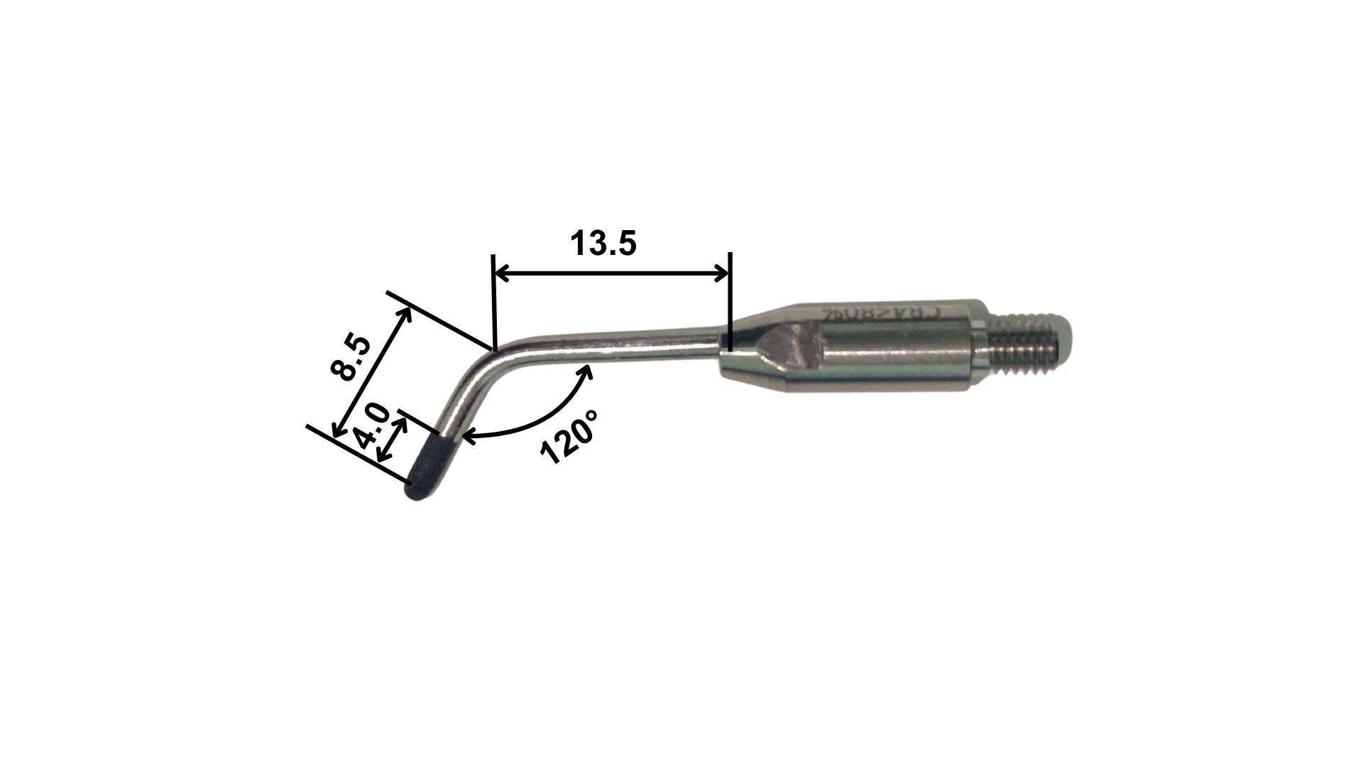
In groups DT+CR4, DT+CR4U CVDentus® ultrasonic (Figure 1) and High-speed diamond tips were used, each tip being applied 5 times to each specimen. The ultrasonic tips were attached to the Piezoelectric Clinical Plus DentSurg device (CVDentus®, Brazil) under refrigeration. Each tip was specifically programmed in the device. The experimental groups (n = 10), the specification and recommendation of the tooth preparation tips used are described in Table 1 and Table 2, respectively.
Ultrasonic tips developed for the refinement of dental preparations — dimensions of the CR4 and CR4U inserts. Both have the same shape but differ in diamond grit size. Source: Prepared by the author.
Surface roughness analysis
After making the prosthodontic tooth preparation, the average roughness (Ra) of the specimens (n = 10) was evaluated with a contact profilometer (SJ-400 Small). Three measurements were taken on the x-axis, and three measurements were taken on the y-axis for each sample [9,25,28].
Surface morphology analysis
The surface of each group was evaluated (n = 2) by scanning electron microscopy. The specimens were kept in a glass bell jar with silica for 7 days for complete dehydration. They were then mounted on stubs and a 4 nm layer of Au-Pd alloy was vacuum deposited on the surfaces of the samples (Emitech SC7620 Sputter Coater, Moorestown, NJ, USA). They were then examined using an SEM (Inspect S50, FEI, Hillsboro, Oregon, USA) with 15 kV of power. The images were collected using a computer coupled to the SEM [9].
Statistical analysis
Statistical analysis software (JAMOVI Statistics) was used to compare the differences between the groups. Roughness data (Ra) were submitted to the homogeneity of variance test (Levene's test) and the normality test (Shapiro-Wilk test). These values were evaluated using the One-way ANOVA test, followed by Tukey’s post hoc test for multiple comparisons, adopting a significance level of 95% (p=0.05).
RESULTS
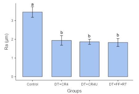
The average Ra values ± 95% CI and the normality results for the surface roughness analysis are shown in detail in Table 3. The control group showed the highest Ra values compared to the other groups (3.46±0.89), differing statistically from the others (p<0.001), with the DT+CR4 group: 1.94±0.81; DT+CR4U: 1.87±0.43; DT+FF+RT: 1.83±0.66. These experimental groups showed similar values, with no significant difference between them (p>0.05).
The data comparing the Ra values of the different methods used for each group and the roughness assessment scores are shown in Figures 2.
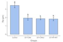
Scores ±95% CI values for Surface Roughness analysis between different methods for each group (p<.001). Source: Prepared by the author.
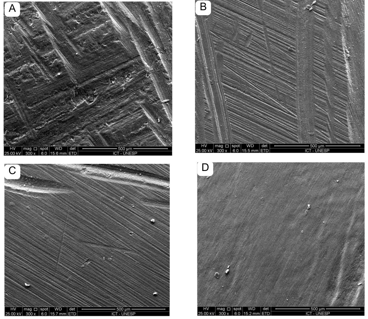
Analysis of the surface morphology by SEM is shown in Figure 3 and revealed clear differences. Specimens prepared by the method in the Control group showed discontinuous dentin topography with deep, irregular grooves.
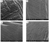
Topographical features of the test specimens from each experimental group. Images A represents Control group. A shows specimens prepared in the conventional way with a diamond tip only and larger surface defects can be seen which have given rise to grooves; B-C show specimens prepared with CVD tips and/or associated with piezoelectric ultrasonic equipment with more regular dentin surfaces, i.e. fewer grooves and greater surface smoothness; D shows a protocol for finishing and polishing preparations using multilaminate tips and rubber tips, with a smoother surface. Source: Prepared by the author.
The DT+CR4 and DT+CR4U groups showed dentin surfaces with greater homogeneity and regularity, with the latter showing grooves with greater uniformity compared to the former. The dentin of the DT+FF+RT group was smoother and more regular than the others.
DISCUSSION
Based on the quantitative roughness measurements, all experimental groups exhibited a statistically significant reduction in Ra values compared with the control. Therefore, the null hypothesis (H0), which stated that the use of CVD diamond tips associated with ultrasonic technology does not significantly affect the quantitative surface roughness (Ra) of dentin preparations, was rejected, while the alternative hypothesis (H1) was accepted. This reformulation explicitly states whether the hypotheses were rejected or accepted to improve scientific consistency.
In the qualitative SEM evaluation, more homogeneous and less grooved surface patterns were observed in the experimental groups, particularly in DT+FF+RT, indicating smoother and more uniform topographies. However, because SEM provides only descriptive, non-quantitative information, these observations are presented strictly as complementary evidence to support the quantitative roughness data, rather than as hypothesis-testing results.
Oscillating instruments with CVD diamond surfaces were developed to overcome the limitations of conventional preparation designs dictated by HS burs [7]. In previous studies in the literature, this device has proven to be an efficient and durable alternative, as it does not produce high-pitched noise, minimizes pulp damage by generating less heat, and offers less removal of tooth structure, resulting in well-defined and smooth preparation margins with less roughness [7,16]. In agreement with previous literature, the results of the present study demonstrated that ultrasonic-assisted CVD instrumentation produced significantly lower surface roughness compared with preparations performed solely with conventional high-speed diamond burs, supporting the acceptance of H1.
The quality of the dental surface is essential, as it enhances the fit and marginal seating of restorations, creates a homogeneous layer of remaining dentin, which optimizes adhesion to self-etching and self-adhesive resin cements, and improves the accuracy of impressions and intraoral scanning [9,21,22]. The dentin surface is uniformly instrumented when subjected to prosthodontic tooth preparation using CVD tips associated with piezoelectric ultrasonic instruments, as the vibration generated by ultrasonic waves produces a homogeneous segmentation of solid surfaces, thereby eliminating irregularities that may be caused by conventional tips used with rotary instruments [6]. This can be observed in the images of the present study showing the surface topography obtained by scanning electron microscopy (SEM) (Figure 3), which demonstrated that the DT+CR4 and DT+CR4U groups exhibited less deep surface grooves compared to the control groups, suggesting a less rough surface. The DT+FF+RT group showed the least amount of surface grooves and the most homogeneous surface due to the use of rubber tips at low speed, which aligns with the smoother surfaces expected from polishing protocols. A similar study demonstrated through surface roughness analysis, scanning electron microscopy, and micro-tensile testing that the use of ultrasonic equipment in surface finishing of preparations improves the quality and precision of this procedure in dentin of human teeth, producing a bond strength comparable to the conventional method; however, it did not reveal any statistical difference between these protocols [21]. In our study, the roughness data showed a statistical difference between the experimental groups and the control group. Furthermore, the SEM images qualitatively demonstrate the influence on the surface texture of the prepared dentin.
The effect of surface smoothness on retention varies according to the luting agent: conventional cements benefit from increased roughness, whereas adhesive resin cements perform better on smoother surfaces with thinner smear layers [23]. Some investigations demonstrate that surface roughness improves the retention of zinc phosphate cement, as a rougher area increases the contact surface, optimizes mechanical interlocking, and reduces the need for additional retention features, such as grooves [28]. Conversely, a rough preparation is not beneficial for crown retention when using adhesive cements, as it can affect bond strength due to the increased thickness and density of the dentin smear layer [21]. Dentin smear layers produced by ultrasonic abrasion are thinner compared to conventional techniques using diamond abrasives and rotary instruments, which suggests that they may optimize the bond strength of resin cement to the dental structure [29,30]. The results obtained from our investigation suggest that the surface characteristics achieved with CVD tips associated with the ultrasonic instrument may influence the adhesion of resin cements. These alternative devices are recommended for cavity preparation and finishing, in an effort to preserve dental structure and take advantage of the benefits of new adhesive systems. Additional studies will be necessary to further evaluate the adhesive and mechanical behavior of these surfaces, as this falls beyond the scope of the present investigation.
Another factor influenced by surface roughness is the fit and marginal seating of restorations. The cervical finish is considered a critical point in preparations intended for prosthetics, as it corresponds to the line that will define the adaptation of the prosthesis to the remaining tooth structure [8,31]. Therefore, it must exhibit precise, sharp, and regular characteristics, which reduce the risks of infiltration and secondary caries at the cement line and facilitate the maintenance of periodontal structures [20,29]. With CVD technology for the manufacture of diamond tips and their association with ultrasonic equipment, it is possible to achieve more regular and precise finishes, relying less on the operator's skill, as there is greater control over removal of tooth structure due to the oscillatory action. This also allows for a more precise internal finish line without damaging soft tissue, compared to conventional diamonds used with rotary instruments.
The different finishing procedures tested for dental preparations demonstrated that this is a factor that can influence surface regularity. A homogeneous and regular surface positively affects the accuracy of functional impressions and intraoral scanning, as it eliminates imperfections, allows better flow of the impression material over the preparation surface, and provides better reflectance properties for capturing images with the intraoral scanner [22,23,31]. The dental literature shows that achieving a smoother and more regular preparation surface, as obtained in the experimental groups of this study, can improve long-term prognosis by promoting better adaptation and longevity of restorations [3,23,32,33].
Although these findings may demonstrate the potential of using CVD technology with ultrasonic instruments for finishing dental preparations, the SEM images still showed small surface irregularities, as seen in the DT+CR4 and DT+CR4U groups, which could be improved to achieve a more homogeneous surface like that of the DT+FF+RT group. A possible solution to this issue may be the combination of CVD tips attached to ultrasonic instruments with rubber tips in a contra-angle at low speed, which could have the potential to compensate for the surface grooves of the diamonds. To the authors' current knowledge, there are no studies in the literature that specifically address this topic.
Even with the limited clinical inference of an in vitro study, the findings suggest the potential for an alternative material for finishing dental preparations using tips made with CVD technology associated with ultrasonic instruments, which could directly influence the quality of the final outcome, both aesthetically and physiologically. This could lead to greater resistance and adhesion of the material to the dental structure, as the Ra values of these groups are close to those of the group that used the established surface finishing method in the literature [9,19,21,25,26,32,33]. The dental preparation technique is sensitive to the operator in achieving ideal properties [34]. This limitation is inherent to the technique using rotary instruments. The clinical handling of oscillatory instruments may help reduce these limitations by producing more uniform preparations and minimizing technical errors. Ultimately, all these characteristics contribute to the provision of a successful prosthesis. Further studies are recommended to evaluate the effect of surface finishing with ultrasonic instruments on adhesion, accuracy of impressions, and scanning. In addition, clinical studies are needed to reinforce the conclusions of this in vitro analysis.
These findings confirm that finishing protocols involving CVD-coated oscillatory instruments produce measurable and clinically relevant modifications to dentin surface roughness. Such effects align with the alternative hypothesis (H1) and contradict the null hypothesis (H0), reinforcing that ultrasonic-assisted CVD instrumentation significantly alters the quantitative surface characteristics when compared with conventional finishing approaches.
CONCLUSION
Within the limitations of this in vitro study, it can be concluded that:
-
Dental preparations made with CVD diamond tips combined with piezoelectric ultrasonic produce a dentin surface that is significantly less rough compared to the use of conventional diamond tips associated with high-speed handpieces.
-
The topographic evaluation of the superficial dentin showed that dental preparations made with CVD diamond tips combined with piezoelectric ultrasonic yield smoother and more regular surfaces, with less pronounced superficial grooves.
All these characteristics demonstrate promising alternatives for the finishing of dental preparations aimed at producing long-lasting functional prostheses.
Acknowledgements
The authors would like to thank the dental company CVDentus®, São Paulo, Brazil for supplying materials. The authors would like to thank Prof. Dr. Milton Santamaria Júnior for his help in interpreting the statistical data.
Data availability
The data that support the findings of this study are available from the corresponding author upon reasonable request.
-
How to cite:
Silva MB, Muramatsu LCR, Silva LAS, Marques TO, Silva JMF, Uemura ES. A new alternative for finishing the surface of dental preparations with ultrasonic instruments and CVD technology. Braz. Dent. Sci. 2026;29:e4855. https://doi.org/10.4322/bds.2026.e4855
-
Funding
This research did not receive any specific grant from funding agencies in the public, commercial, or not-for-profit sectors.
-
Regulatory Statement
This study did not involve human participants or live animals. Bovine teeth were obtained from a licensed slaughterhouse as by-products of the food industry. Therefore, according to Brazilian regulations and institutional guidelines, approval from an Ethics Committee on Animal Use was not required.
REFERENCES
- 1 Dietschi D, Maeder M, Holz J. In vitro evaluation of marginal fit and morphology of fired ceramic inlays. Quintessence Int. 1992;23(4):271-8. PMid:1502303.
-
2 Goodacre CJ, Campagni WV, Aquilino SA. Tooth preparations for complete crowns: an art form based on scientific principles. J Prosthet Dent. 2001;85(4):363-76. https://doi.org/10.1067/mpr.2001.114685 PMid:11319534.
» https://doi.org/10.1067/mpr.2001.114685 -
3 Li SY, Duan XF, Cao Y. Evaluation of the effect of using ultrasonic instruments to improve the shoulder of the preparations. Beijing Da Xue Xue Bao Yi Xue Ban. 2020;53(1):88-94. https://doi.org/10.19723/j.issn.1671-167X.2021.01.014
» https://doi.org/10.19723/j.issn.1671-167X.2021.01.014 -
4 Machado AC, Tolentino AB, Spini PHR, Gonzaga RCQ, Zeola LF, Reis BR, et al. Analysis of standard dimensions of the macrogeometry of diamond burs from different manufacturers and models. Rev Odontol Bras Central. 2014;23(64):47-52. http://doi.org/10.36065/robrac.v23i64.844
» http://doi.org/10.36065/robrac.v23i64.844 -
5 Boldieri T, Bussaneli D, Rodrigues J, Cardoso K, Josgrilberg E, Cordeiro R. Influence of sterilization process on cutting effectiveness and durability of ultrasonic tips. Braz Dent Sci. 2015;18(3):10-6. https://doi.org/10.14295/bds.2015.v18i3.1143
» https://doi.org/10.14295/bds.2015.v18i3.1143 -
6 Cardoso MV, Coutinho E, Ermis RB, Poitevin A, Van Landuyt K, De Munck J, et al. Influence of dentin cavity surface finishing on micro-tensile bond strength of adhesives. Dent Mater. 2008;24(4):492-501. https://doi.org/10.1016/j.dental.2007.04.011 PMid:17675225.
» https://doi.org/10.1016/j.dental.2007.04.011 -
7 Horne P, Bennani V, Chandler N, Purton D. Ultrasonic margin preparation for fixed prosthodontics: a pilot study. J Esthet Restor Dent. 2012;24(3):201-10. https://doi.org/10.1111/j.1708-8240.2011.00477.x PMid:22691082.
» https://doi.org/10.1111/j.1708-8240.2011.00477.x - 8 Pegoraro LF, Lins do Valle A, Araújo CRP, Bonfante G, Conti PCR. Fixed prosthodontics: foundations for oral rehabilitation planning. 2nd ed. Porto Alegre: Grupo A; 2009.
-
9 Rocha SC, Barcellos DC, Pucci CR, Pagani C, Lima DR. CVD and diamond tips: can they influence the bond strength of a total-etch adhesive to dentin? Braz Dent Sci. 2016;19(2):89-95. https://doi.org/10.14295/bds.2016.v19i2.1249
» https://doi.org/10.14295/bds.2016.v19i2.1249 - 10 Wicht MJ, Haak R, Fritz UB, Noack MJ. Primary preparation of class II cavities with oscillating systems. Am J Dent. 2002;15(1):21-5. PMid:12074224.
-
11 Trava-Airoldi VJ, Corat EJ, Santos LV, Diniz AV, Moro JR, Leite NF. Very adherent CVD diamond film on modified molybdenum surface. Diamond Related Materials. 2002;11(3-6):532-5. https://doi.org/10.1016/S0925-9635(01)00721-X
» https://doi.org/10.1016/S0925-9635(01)00721-X -
12 Borges CF, Magne P, Pfender E, Heberlein J. Dental diamond burs made with a new technology. J Prosthet Dent. 1999;82(1):73-9. https://doi.org/10.1016/S0022-3913(99)70130-7 PMid:10384166.
» https://doi.org/10.1016/S0022-3913(99)70130-7 - 13 Carvalho FP. CVDentus® X micromotor in early childhood: evaluation of technical, behavioral and acceptability aspects [dissertation]. São Paulo: University of São Paulo; 2011.
- 14 Shafer WG, Hine MK, Levy BM, Tomich CE. Shafer’s textbook of oral pathology. 4th ed. New York: Nueva Editorial Interamericana; 1988.
- 15 Mastrantonio SDS, Gondim JO, Josgrilberg ÉB, Cordeiro RCL. Minimizing fear during dental treatment using ultrasonic points. RGO Rev Gaúch Odontol. 2010;58(1):119-22.
-
16 Vanderlei AD, Borges AL, Cavalcanti BN, Rode SM. Ultrasonic versus high-speed cavity preparation: analysis of increases in pulpal temperature and time to complete preparation. J Prosthet Dent. 2008;100(2):107-9. https://doi.org/10.1016/S0022-3913(08)60158-4 PMid:18672127.
» https://doi.org/10.1016/S0022-3913(08)60158-4 -
17 Laird WRE, Walmsley AD. Ultrasound in dentistry. Part 1--Biophysical interactions. J Dent. 1991;19(1):14-7. https://doi.org/10.1016/0300-5712(91)90030-3 PMid:2016423.
» https://doi.org/10.1016/0300-5712(91)90030-3 -
18 Matuda FS, Pagani C, Miranda CB, Crema AAS, Brentel AS, Carvalho YR. Bone repair after osteotomy with diamond burs and CVD ultrasonic tips: histological study in rats. Cienc Odontol Bras. 2007;10(3):21-30. https://doi.org/10.14295/bds.2007.v10i3.277
» https://doi.org/10.14295/bds.2007.v10i3.277 -
19 Laufer BZ, Pilo R, Cardash HS. Surface roughness of tooth shoulder preparations created by rotary instrumentation, hand planing, and ultrasonic oscillation. J Prosthet Dent. 1996;75(1):4-8. https://doi.org/10.1016/S0022-3913(96)90410-2 PMid:8850446.
» https://doi.org/10.1016/S0022-3913(96)90410-2 - 20 Massironi D. Optimizing the prosthetic margin with sonic instrumentation. Dent Today. 2011;30(2):152, 154-5. PMid:21400997.
-
21 Ren L, Li M, Pan Y, Meng X. Influence of polishing methods on the bonding effectiveness and durability of different resin cements to dentin. BioMed Res Int. 2018;2018:9189354. https://doi.org/10.1155/2018/9189354
» https://doi.org/10.1155/2018/9189354 -
22 Chaiyabutr Y, Kois JC. The effects of tooth preparation cleansing protocols on the bond strength of self-adhesive resin luting cement to contaminated dentin. Oper Dent. 2008;33(5):556-63. https://doi.org/10.2341/07-141 PMid:18833862.
» https://doi.org/10.2341/07-141 -
23 Revilla-León M, Cascos-Sánchez R, Barmak AB, Kois JC, Gómez-Polo M. The effect of different tooth preparation finishing procedures and immediate dentin sealing on the scanning accuracy of different intraoral scanners. J Dent. 2023;130:104431. https://doi.org/10.1016/j.jdent.2023.104431 PMid:36682722.
» https://doi.org/10.1016/j.jdent.2023.104431 -
24 Ayad MF, Johnston WM, Rosenstiel SF. Influence of dental rotary instruments on the roughness and wettability of human dentin surfaces. J Prosthet Dent. 2009;102(2):81-8. https://doi.org/10.1016/S0022-3913(09)60114-1 PMid:19643221.
» https://doi.org/10.1016/S0022-3913(09)60114-1 -
25 de Vasconcellos BT, Thompson JY, de Paula Macedo MR, de Oliveira Maia JM, Oda M, Garone-Netto N. Ultrasonic cavity preparation using CVD coated diamond bur: a case report. Eur J Dent. 2013;7(1):127-32. https://doi.org/10.1055/s-0039-1699007
» https://doi.org/10.1055/s-0039-1699007 -
26 Pashley DH, Carvalho RM. Dentine permeability and dentine adhesion. J Dent. 1997;25(5):355-72. https://doi.org/10.1016/S0300-5712(96)00057-7 PMid:9241954.
» https://doi.org/10.1016/S0300-5712(96)00057-7 - 27 Francischone AC. Evaluation of marginal quality of cavity preparations performed using different techniques and instruments [thesis]. São Paulo: University of São Paulo; 2009.
-
28 Krishna KA, Abdul Razak P, Sooraj A, Kuriachan T, Radhakrishnan L, Poyil NN. An invitro study to evaluate the retentive properties of metal crowns on various surface roughnesses of abutments. Cureus. 2024;16(8):e67019. https://doi.org/10.7759/cureus.67019
» https://doi.org/10.7759/cureus.67019 -
29 Ellis R, Bennani V, Purton D, Chandler N, Lowe B. The effect of ultrasonic instruments on the quality of preparation margins and bonding to dentin. J Esthet Restor Dent. 2012;24(4):278-85. https://doi.org/10.1111/j.1708-8240.2011.00495.x PMid:22863135.
» https://doi.org/10.1111/j.1708-8240.2011.00495.x -
30 Lima LM, Motisuki C, dos Santos-Pinto L, dos Santos-Pinto A, Corat EJ. Cutting characteristics of dental diamond burs made with CVD technology. Braz Oral Res. 2006;20(2):155-61. https://doi.org/10.1590/S1806-83242006000200012 PMid:16878210.
» https://doi.org/10.1590/S1806-83242006000200012 -
31 Hung SH, Hung KS, Eick JD, Chappell RP. Marginal fit of porcelain-fused-to-metal and two types of ceramic crown. J Prosthet Dent. 1990;63(1):26-31. https://doi.org/10.1016/0022-3913(90)90260-J PMid:2404103.
» https://doi.org/10.1016/0022-3913(90)90260-J -
32 Al-Omari WM, Mitchell CA, Cunningham JL. Surface roughness and wettability of enamel and dentine surfaces prepared with different dental burs. J Oral Rehabil. 2001;28(7):645-50. https://doi.org/10.1046/j.1365-2842.2001.00722.x PMid:11422697.
» https://doi.org/10.1046/j.1365-2842.2001.00722.x -
33 Ayad MF, Johnston WM, Rosenstiel SF. [Influence of dental rotary instruments on the roughness and wettability of human dentin surfaces. J Prosthet Dent. 2009;102(2):81-8. https://doi.org/10.1016/S0022-3913(09)60114-1 PMid:19643221.
» https://doi.org/10.1016/S0022-3913(09)60114-1 -
34 Stübinger S, Stricker A, Berg BI. Piezosurgery in implant dentistry. Clin Cosmet Investig Dent. 2015;7:115-24. https://doi.org/10.2147/CCIDE.S63466 PMid:26635486.
» https://doi.org/10.2147/CCIDE.S63466
Edited by
-
Editor-in-chief:
Sergio Eduardo de Paiva Gonçalves
-
Editor:
João Paulo Mendes Tribst