 Histological and histochemical analysis to identify type I and III collagen in the skins of different shark species
ABSTRACT
During fishing activities, solid waste such as shark skin is often discarded by the processing industry, contributing to environmental pollution and representing the loss of a material with high biotechnological potential, especially as a source of collagen. The objective of this research was to characterize the structure of the skin and identify the types of collagen present in four shark species: Rhizoprionodon lalandii, Squalus albicaudus, Sphyrna lewini, and Squatina guggenheim. The samples were obtained from artisanal fishing in the municipality of Saquarema, Rio de Janeiro, and specimens provided by the Santos Fisheries Institute, São Paulo, all from bycatch and already dead. For histological analysis, tissue fragments were collected from the left side of the animals and stained with haematoxylin-eosin, Masson’s trichrome and Picrosirius red. The results revealed structures already described in the literature, such as mucus, epidermis, dermis, muscle fiber, haemolymphatic vessels and type I collagen, in addition to the presence of type III collagen, not yet evidenced. The result reinforces the high healing capacity of sharks, highlighting the thicker and more resistant skin, with more condensed collagen in S. guggenheim, a demersal species, and the preservation of mucus in S. lewini, associated with the accumulation of collagen fiber, broadening prospects for future biomedical applications.
Keywords
External coating; Histology; Elasmobranchs; Tissue engineering; Vital proteins
RESUMO
Durante a atividade pesqueira, resíduos sólidos como a pele de tubarões são frequentemente descartados pela indústria de processamento, o que contribui para a poluição ambiental e representa a perda de um material com elevado potencial biotecnológico, especialmente como fonte de colágeno. O objetivo desta pesquisa foi caracterizar a estrutura da pele e identificar os tipos de colágeno presentes em quatro espécies de tubarões: Rhizoprionodon lalandii, Squalus albicaudus, Sphyrna lewini e Squatina guggenheim. As amostras foram obtidas a partir da pesca artesanal no município de Saquarema, Rio de Janeiro, e espécimes fornecidos pelo Instituto de Pesca de Santos, São Paulo, todos oriundos da pesca incidental (bycatch) e já em óbito. Para a análise histológica, fragmentos de tecido foram coletados do lado esquerdo dos animais e corados com hematoxilina-eosina, tricrômico de Masson e Picrosirius red. Os resultados revelaram estruturas já descritas na literatura, como muco, epiderme, derme, fibras musculares, vasos hemolinfáticos e colágeno tipo I, além da presença de colágeno tipo III, ainda não evidenciado. O resultado reforça a elevada capacidade de cicatrização dos tubarões, destacando a pele mais espessa e resistente, com colágeno mais condensado de S. guggenheim, espécie demersal, e a preservação do muco em S. lewini, associada ao acúmulo de fibras colágenas, ampliando perspectivas para aplicações biomédicas futuras.
Palavras-chave
Elasmobrânquios; Engenharia tecidual; Histologia; Proteínas; Revestimento externo
INTRODUCTION
During fisheries, the solid waste discarded by the processing industry represents around 50% of the animal’s weight (Ferraro et al., 2016; Oliveira et al., 2017a; Oliveira et al., 2017b). In some locations, as reported by Sotelo et al. (2016) in interviews with fishermen, 100% of by-products are discarded, which could be marketed as bio-inputs for various tissue engineering purposes. Incorrect disposal causes environmental pollution and affects the economy, as it is not reused in industry to develop products through the extraction of biomolecules such as collagen, collagenases, chymotrypsin, among others (Oliveira et al., 2017a; Oliveira et al., 2017b).
Sharks are cartilaginous fish and, together with rays, are a subclass of elasmobranchs. Among all the characteristics of this group, there is the external coating mineralized dermo-epidermal denticles anchored in the dermis and epidermis, varying by species and lifestyle, and associated with high-collagen production (Dillon et al., 2017; Seegers & Meyer, 2009). This combination enhances resistance, healing, and defense against predators and ectoparasites (Luer & Walsh, 2018; Meyer & Seegers, 2012; Ostrander et al., 2004).
Internally, the epidermis of elasmobranchs has between four and eight layers the number varies according to the region of the body and the species (Meyer & Seegers, 2012; Wirth, 1999). The shark’s dermis has two special divisions: the subdermal S. laxum, which consists of a layer of melanocytes, blood vessels to nutrition the epidermis and functional hemolymphatic vessels (Meyer & Seegers, 2012), and S. compactum, characterized by horizontally arranged layer of strong, parallel collagen fibers. This integrated structure helps the sharks to store energy during faster swimming when hydrostatic pressure is exerted against its skin (Meyer & Seegers, 2012; Motta et al., 2012).
The literature on the use and processing of collagen from shark skin remains scarce. Possibly due to the number of shark species in the world, many occupy deep waters, and most are threatened with extinction. Therefore, the specimens used in this research were obtained through bycatch in artisanal fishing, promoting the possibility of reusing and studying a non-edible fish product, expanding biological knowledge of the species and its development as a potential biomaterial (Shen et al., 2017).
Studies on fish skin contribute to the reuse of this non-edible product, which has low economic visibility, for the benefit of human and veterinary medicine. The production of fish collagen adds value to products derived from this category and encourages the pharmaceutical industry, because this kind of products is more effective in treating skin lesions, such as burns (Shen et al., 2017).
Collagen is a fiber protein with peptide chains rich in glycine and proline, organized parallel to an axis, forming collagen fibers, which provide strength and elasticity to the structure present (Lehninger, 1995; Linden & Lorient, 2000; Moretti, 2009; Wolf, 2007). Twenty-nine types of collagens in mammals are known to science (Gordon & Hahn, 2010; Oliveira et al., 2017c). Among them, nine are most commonly available for use and research (Oliveira et al., 2017c). In fish, the most abundant collagen is type I, and it can be extracted from the skin, scales, fins, swim bladder, bones, spine, cartilage, and muscles.
The aims of this study were to analyze the histological and histochemical characteristics of the skins of four species of shark: dogfish Rhizoprionodon lalandii (Müller & Henle 1839), dogfish Squalus albicaudus (Linnaeus 1758), hammerhead shark Sphyrna lewini (Griffith & Smith 1834), and angular angelshark Squatina guggenheim (Linnaeus 1758), to identify collagen types and to support future research on isolating collagen I and III.
MATERIAL AND METHODS
The individuals of the shark species used in this work were obtained from artisanal fishing landings in the municipality of Saquarema, Rio de Janeiro, Brazil, and from the collection of the Instituto de Pesca de Santos, São Paulo, Brazil, donated by Professor Dr. Alberto Amorim. The experimental protocol is in accordance with the Brazilian Biodiversity Authorization and Information System SISBIO No. 80984.
The specimens used in this research were obtained through bycatch, already dead and without any euthanasia procedure. Thus, the possibility of reusing and studying a non-edible fish product was promoted, expanding the biological knowledge of the species and its development as a potential biomaterial.
Six individuals of sharpnose shark R. lalandii, seven individuals of the dogfish S. albicaudus, three individuals of hammerhead shark S. lewini, and one individual of angel dogfish S. guggenheim were used in this research. However, not all samples were satisfactory for histology, which reduced the samples to those that could show what the research wanted: collagen fibers.
The samples were taken from the left side of the animals’ bodies, behind the gill slits, below the dorsal fin and before the caudal fin, and 1.5-cm sections were cut using a mechanical cutter with a No. 24 scalpel.
Afterwards, the samples were then inserted into histological cassettes and stored in a 10% formaldehyde solution for 24 hours. After this period, they were preserved in 70% alcohol. The samples were then sent to the Histocell Histology Laboratory, in São Paulo state, Brazil, to be embedded in paraffin, cut in serial sections to a thickness of 3 μm, and subjected to the following staining techniques: hematoxylin-eosin, which determines the stratification of the epidermis and dermis layers; Masson’s trichrome, used to identify muscle fibers and collagen; and Picrosirius red, used to define the types of collagen present in the samples (Santos et al., 2021).
Photodocumentation was carried out using a microscope with a camera (BIOFOCUS) at the Fish and Aquatic Health Laboratory of the Universidade Federal Fluminense. Polarized light photomicrography was carried out using an Olympus BX53 polarized microscope with an Olympus DP72 camera from Histology and Embryology Department of the Roberto Alcântara Gomes Biology Institute, Biomedical Center, Universidade do Estado do Rio de Janeiro.
RESULTS
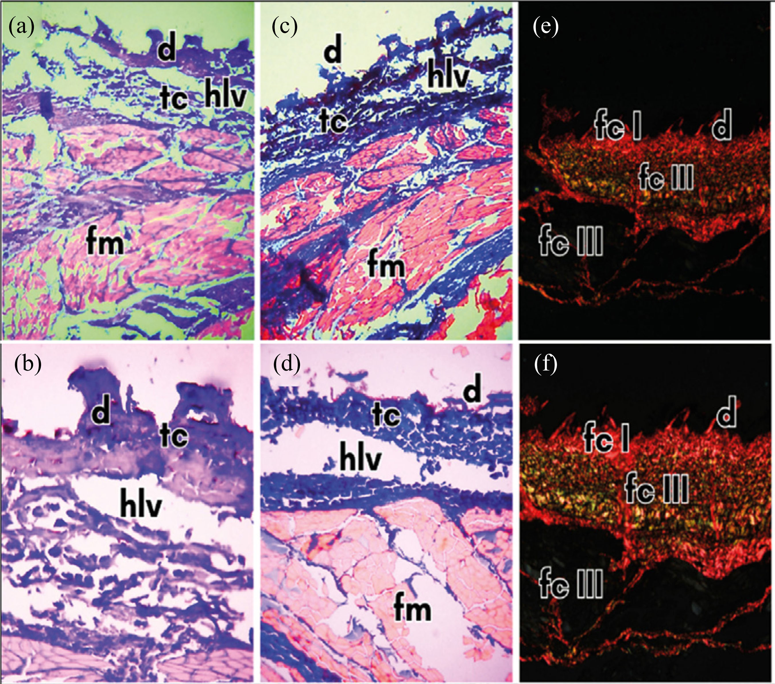
The species sharpnose shark R. lalandii showed hematoxylin-eosin staining 4x and 10x magnifications (Figs. 1a and 1b) of dermal denticles (d), with morphological variety and surrounded by epidermis composed of columnar cells, connective tissue (tc) in the dermis and epidermis, with hemolymphatic vessels (hlv) and muscle fiber (fm). In Masson’s trichrome staining 4x and 10x magnifications, respectively (Figs. 1c and 1d), the skin shows collagen fibers (fc), stained blue, present in the cells of the epidermis, dermis and musculature. This indicates that collagen is more uniform in all layers of the skin, with evidence of interspersed musculature in this region due to the traces of red coloring. It is also possible to see hemolymphatic conducting vessels between the epidermis and dermis. In Picrosirius red staining 4x and 10x magnifications, respectively (Figs. 1e and 1f), the skin sample showed type I collagen (fc I) and colored orange-red in the denticles, epidermis and dermis. In the dermis and muscles, it is possible to see the incidence of type III collagen, with a yellowish-green color (fc III).
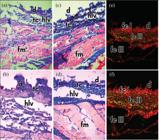
Photomicrograph of the histological section of the skin of the sharpnose shark Rhizoprionodon lalandii.
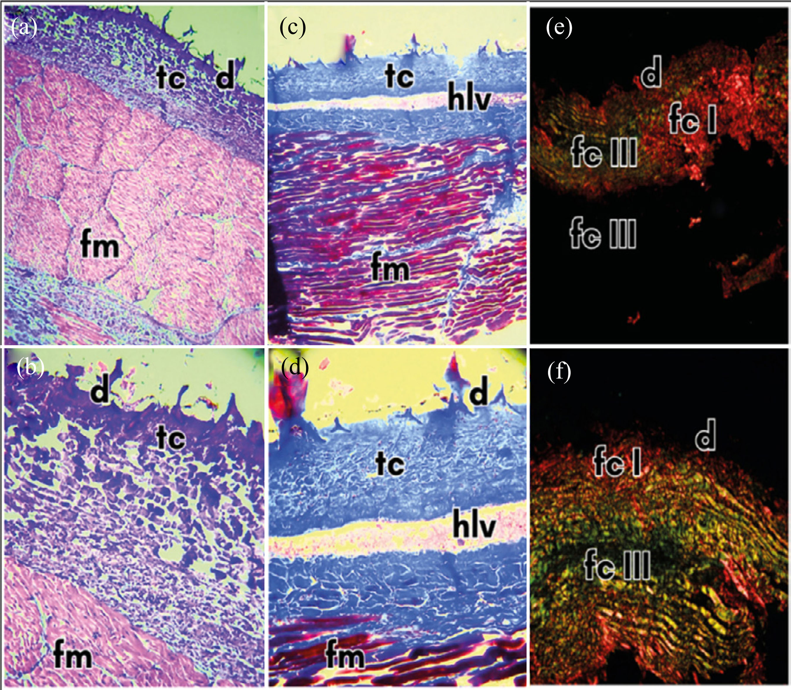
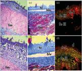
The dogfish S. albicaudus species showed a hematoxylin-eosin staining 4x and 10x magnifications, stain of dermal denticles (d), with morphological variety and surrounded by epidermis composed of columnar cells, connective tissue (tc) in the dermis and epidermis and muscle fibers (fm) (Figs. 2a and 2b). Masson’s trichrome staining 4x and 10x, respectively (Figs. 2c and 2d), showed collagen in the connective tissue (tc) in the epidermis, upper and lower dermis. At some points in the sample, the lower and upper dermis layers are separated by hemolymphatic vessels (hlv) and muscle tissue fiber (fm). In Picrosirius red staining 4x and 10x magnifications, respectively (Figs. 2e and 2f), the skin showed type I collagen (fc I) in the denticles, epidermis and dermis and type III collagen (fc III) in the dermis and musculature.
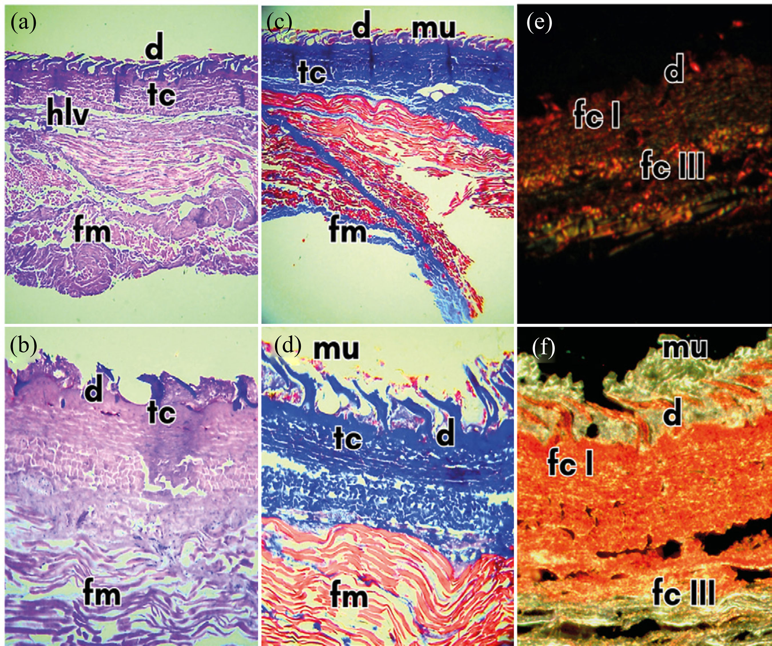
The hammerhead shark S. lewini species showed the same layered tissue as the other species, but more structured, including visualization of the S. laxum and S. compactum layers and transverse muscle fibers (fm) (Figs. 3a and 3b). In Masson’s trichrome staining 4x and 10x magnifications, respectively (Figs. 3c and 3d), the dermal denticles (d) are coloring red in their contours, which may indicate the presence of preserved supplemented mucus (mu) in the dwarf species Shortfin Mako Isurus oxyrinchus. The epidermis shows a dense condensed layer of connective tissue (tc), rich in collagen. It is possible to see the hemolymphatic conducting vessels and the transverse muscle fibers. In Picrosirius red staining 4x and 10x magnifications, respectively (Figs. 3e and 3f), the skin sample also showed type I collagen in the denticles, epidermis, and dermis. The denticle (d) showed a birefringence yellowish-green outline, which may indicate some protein component preserved in the supplemented mucus, also seen in Figs. 1e and 1f. In the dermis and musculature, it is possible to visualize the incidence of type III collagen.
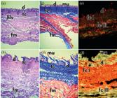
Photomicrograph of a histological section of the skin of a hammerhead shark Sphyrna lewini.
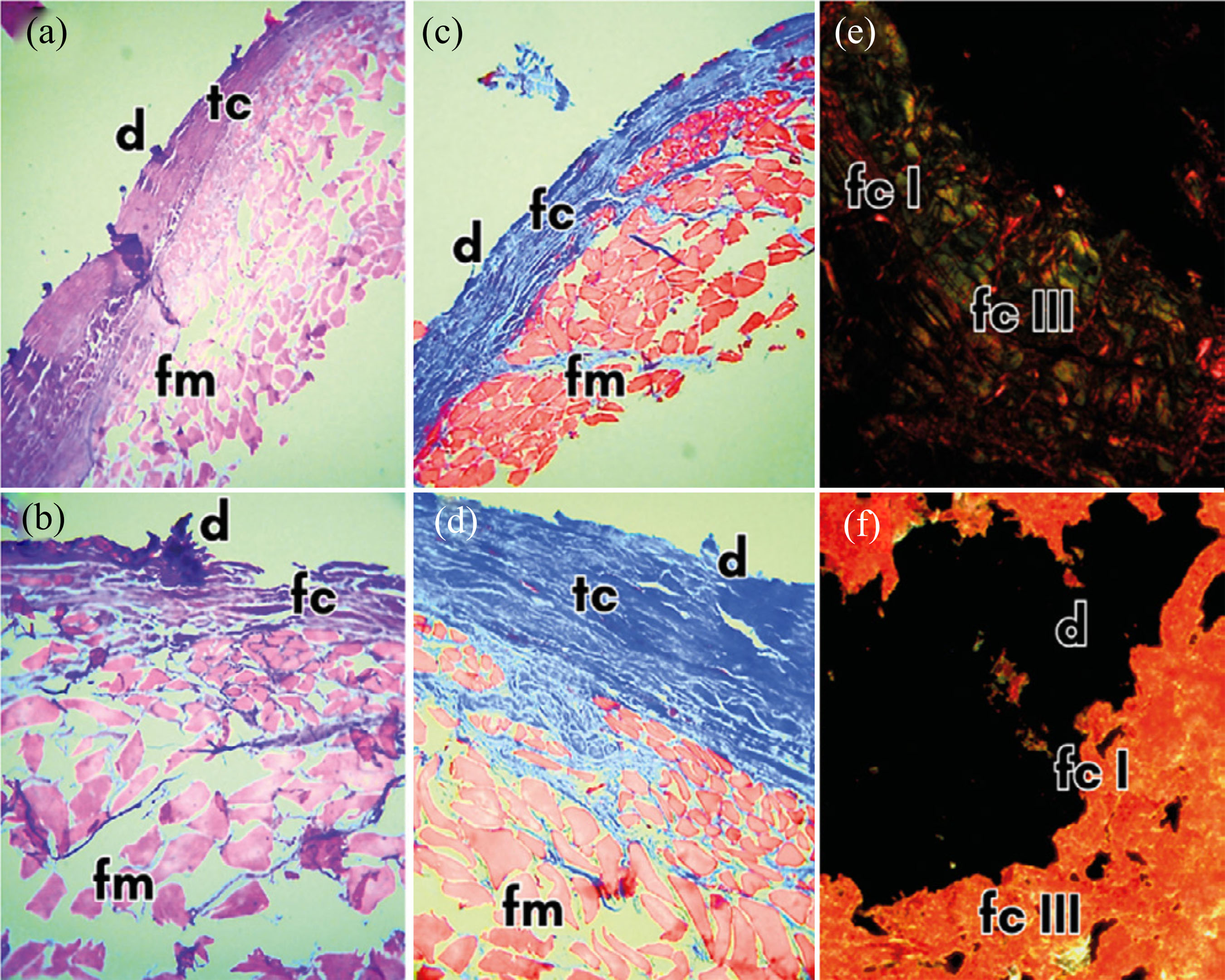
The skin of S. guggenheim showed greater rigidity in the layer of dermo-epidermal denticles and epidermis during the cutting of the samples. After the results of the analyses, it showed an arrangement of spaced dermo-epidermal denticles (d) (Figs. 4a and 4b), and it was possible to observe the epidermis and dermis with more condensed cells, but the musculature with freer and looser cells. Masson’s trichrome (Figs. 4c and 4d) staining 4x and 10x magnifications, respectively, showed a high concentration of condensed connective tissue (tc) and collagen in the epidermis and dermis layers, but little in the muscle fibers (mf). In the Picrosirius red staining 4x and 10x magnifications, respectively (Figs. 4e and 4f), the skin also showed type I collagen (fc I) in the dermo-epidermal denticles (d), in the epidermis and dermis. In these layers, there is also strongly intercalated type III collagen (FC III). In the dermis and musculature, it is possible to visualize the incidence of type III collagen.
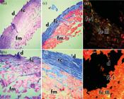
Photomicrograph of a histological section of the skin of the angular angelshark Squatina guggenheim.
DISCUSSION
The skins of the four shark species analyzed in this study were morphologically characterized by standard layers with dermal denticles, interspersed in the hypodermis, epidermis and dermis, and anchored to the basal plate. Their tip was hollow, open towards the pulp cavity, filled with fibroblasts and fibrocytes and with blood vessels and nerve fibers that connect them to the dermis system. This structure has the same type of collagen present in the dermis and epidermis of shark skin, as described by Hamlett (1999), Meyer and Seegers (2012), and Seegers and Meyer (2009).
The epidermis layer is made up of more condensed connective tissue cells and blood vessels that separate the dermis and epidermis from the musculature. In the dermis, there are looser muscle tissue cells, blood vessels, and type I collagen in both the epidermis and dermis, as described in other shark species such as the bamboo shark Chiloscyllium punctatum (Müller & Henle 1838), researched by Kittiphattanabawon et al. (2010), the blue shark, in the studies by Shen et al. (2017), and the mako shark I. oxyrinchus (Rafinesque 1810), which was studied by Lang et al. (2011) and Schuitema et al. (2025).
In hammerhead shark species, the presence of type I collagen was evidenced by Changfeng et al. (2013), with the extraction of the protein from the skin of S. lewini and evidenced by Vijayan et al. (2018) in studies with Sphyrna mokarran. In addition, Schuitema et al. (2025) found a total average collagen fiber cover of 71,6% in the head and from 50,8 to 79,6% in caudal coverage of S. mokarran. The study also highlighted the differences in fiber thickness between the regions. The presence of type III collagen in the contour of dermo-epidermal teeth of the hammerhead shark species S. lewini, analyzed via colorations Trichome of Masson and Picrosirius red, indicates that the supplemented mucus remained preserved even after handling, conservation, and processing. Lang et al. (2011), however, did not determined yet whether the collagen found in this structure was type I or type III and if there were such conservation in fact. This theory has remained in the current research and generates expectations for future and more in-depth studies.
The skin of S. guggenheim, whose epidermis and dermis have more condensed cells, and a high concentration of condensed connective tissue (tc) and collagen in the layers of the epidermis and dermis, is characteristic by harder skin, which is more difficult to cut, and is found in deep-sea animals, as shown in studies with Galeus or Scyliorhinus by Sotelo et al. (2016).
In all species studied, the lower region of the dermis has numerous horizontally arranged layers of strong, parallel collagen bundles, forming a tight texture. According to Schuitema et al. (2025) in a comparative study, the flank region of I. oxyrinchus had the highest value of elastic fibers, when compared to Ginglymostoma cirratum, which had thick skin, with the dermal denticle mirroring this trend. Similarly, this pattern was found in the present study: R. lalandii showed more uniform collagen distributed across all layers of the skin, while S. guggenheim exhibited greater rigidity in the layer of dermo-epidermal denticles and epidermis. This configuration found in I. oxyrinchus and R. lalandii can contribute to rapid swimming or increased flexibility, when hydrostatic pressure increases on the skin, the body remains rigid, allowing for the accumulation of substantial energy, as evidenced by Meyer and Seegers (2012). Despite of being a demersal species, R. lalandii exhibited collagen fiber structures resembling those of pelagic sharks, such as I. oxyrinchus, which may indicate functional convergence or reflect specific biomechanical demands.
The elastic fibers in the dermis of sharks found in this study are distributed and organized, creating a constant horizontal pattern of collagen-elastin-collagen-elastin, with thicker elastic fibers in the anterior parts of the body, as described in the Marracho swordfish shark species Scoliodon laticaudus (Müller & Henle 1838). This system helps to prevent hypertension of the stretching skin, especially during rapid swimming (Meyer & Seegers, 2012).
In all samples analyzed with Picrosirius red stain, the presence of type I and III collagen was observed in both the dermal and muscular layers. Su et al. (2022) evaluated the possible presence of type I and III collagen only in stages 4 and 5 of healing wounds on cetaceans’ skin, and it was associated with the formation of collagen bundles and restructuring of skin architecture. The detection of type III collagen in sharks is significant, particularly in whole tissues, as it may be related to rapid tissue regeneration and reflects the need for flexibility during swimming (Meyer & Seegers, 2012). Supporting this, Schuitema et al. (2025) observed that S. mokarran and I. oxyrinchus showed smaller sized collagen fibers, which may contribute to the species-specific ecomorphology demands.
Studies such as that by Seixas et al. (2020) have reported the presence of type I and type II collagen in collagenic extracts in Prionace glauca and rays, making the presence of type III collagen indicative of a feature still poorly described in this group.
CONCLUSION
The four species of shark showed all the structures present in shark skin, such as dermal denticles, with morphological variety and surrounded by an epidermis composed of columnar cells, connective tissue in the dermis and epidermis, with hemolymphatic vessels, muscle fibers and type I and type III collagen fibers, expressed using Masson’s Trichrome and Picrosirius red stains, which are unprecedented in the literature. Supplemented mucus in a possibly preserved state and with an abundance of collagen was only observed in the S. lewini species. Type I collagen fibers were found in high concentration in the dermal denticles, epidermis and dermis, while type III collagen fibers were present in the dermis layers and muscle fibers and found in mucus supplemented with S. lewini.
The points studied in this research can open a series of new themes and possibilities, supporting future research into both the study of collagen fibers in their entirety and their isolation for use as the basis of healing products.
ACKNOWLEDGMENTS
To the Postgraduate Program in Veterinary Hygiene and Technological Processing of Products of Animal Origin at the Veterinary School of the Universidade Federal Fluminense (UFF). To Professor Dr. Alberto Amorim, from the Instituto de Pesca de Santos. To Dr. Cherley Borda, from the Histology and Embryology Department of the Roberto Alcântara Gomes Biology Institute at the Universidade do Estado do Rio de Janeiro. To Professor Dr. Juliana da Silva Leite and Professor Dr. Angélica Consalter, from the Veterinary Pathology Laboratory of the UFF.
-
FUNDING
Conselho Nacional de Desenvolvimento Científico e TecnológicoGrant No.: 131598/2021-0.
-
DECLARATION OF USE OF ARTIFICIAL INTELLIGENCE TOOLS
Artificial intelligence tools were used only for minor language corrections in the manuscript translation. All scientific content remains the responsibility of the authors.
DATA AVAILABILITY STATEMENT
All dataset were generated or analyzed in the current study.
REFERENCES
-
Changfeng, C., Bin, W., Zhong-Rui, L., & Hong-Yu, L. (2013). Characterization of acid-soluble collagen from the skin of hammerhead shark (Sphyrna lewini). Journal of Food Biochemistry, 38(2). https://doi.org/10.1111/jfbc.12042
» https://doi.org/10.1111/jfbc.12042 -
Dillon, E. M., O’dea, A., & Norris, R. D. (2017). Dermal denticles as a tool to reconstruct shark communities. Marine Ecology Progress Series, 566, 117-134. https://doi.org/10.3354/meps12018
» https://doi.org/10.3354/meps12018 -
Ferraro, V., Anton, M., & Santé-Lhoutellier, V. (2016). The “sisters” α-helices of collagen, elastin and keratin recovered from animal by-products: Functionality, bioactivity and trends of Application. Trends in Food Science & Technology, 51, 65-75. https://doi.org/10.1016/j.tifs.2016.03.006
» https://doi.org/10.1016/j.tifs.2016.03.006 -
Gordon, M., & Hahn, R. (2010). Collagens. Cell and Tissue Research, 339, 247-257. https://doi.org/10.1007/s00441-009-0844-4
» https://doi.org/10.1007/s00441-009-0844-4 - Hamlett, W. C. (1999). Sharks, skates and rays: the biology of elasmobranch fishes Johns Hopkins University.
-
Kittiphattanabawon, P., Benjakul, S., Visessanguan, W., Kishimura, H., & Shahidi, F. (2010). Isolation and characterization of collagen from the skin of brown banded bamboo shark (Chiloscyllium punctatum). Food Chemistry, 119(4), 1519-1526. https://doi.org/10.1016/j.foodchem.2009.09.037
» https://doi.org/10.1016/j.foodchem.2009.09.037 -
Lang, A., Motta, P., Hueter, R., & Habegger, M. (2011). Shark skin separation control mechanisms. Marine Technology Society Journal, 45(4), 208-215. https://doi.org/10.4031/MTSJ.45.4.12
» https://doi.org/10.4031/MTSJ.45.4.12 - Lehninger, A. L. (1995). Princípios de bioquímica (2. ed.). Savier.
- Linden, G., & Lorient, D. (2000). New ingredients in food processing: biochemistry and agriculture CRC Press.
-
Luer, C., & Walsh, C. (2018). Potential human health applications from marine biomedical research with elasmobranch. Fishes, 3(4), 47. https://doi.org/10.3390/fishes3040047
» https://doi.org/10.3390/fishes3040047 -
Meyer, W., & Seegers, U. (2012). Basics of skin structure and function in elasmobranchs: a review. Journal of Fish Biology, 80(5), 1940-1967. https://doi.org/10.1111/j.1095-8649.2011.03207.x
» https://doi.org/10.1111/j.1095-8649.2011.03207.x - Moretti, B. R. (2009). Efeito da suplementação do leite com proteínas de diferentes fontes (soro de leite, soja e colágeno) e da composição da cultura lática em iogurtes [dissertation]. Universidade Estadual Paulista “Júlio de Mesquita Filho”, Instituto de Biociências, Letras e Ciências Exatas.
-
Motta, P., Habegger, M. L., Lang, A., Hueter, R., & Davis, J. (2012). Scale morphology and flexibility in the shortfin mako Isurus oxyrinchus and the blacktip shark Carcharhinus limbatus Journal of Morphology, 273(10), 1096-1110. https://doi.org/10.1002/jmor.20047
» https://doi.org/10.1002/jmor.20047 -
Oliveira, V. M., Assis, C. R. D., Herculano, P. N., Cavalcanti, M. T. H., Bezerra, R. S., & Porto, A. L. F. (2017a). Collagenase from smooth weakfish: extraction, partial purification, characterization and collagen. Boletim do Instituto de Pesca, 43(1), 52-64. https://doi.org/10.20950/1678-2305.2017v43n1p52
» https://doi.org/10.20950/1678-2305.2017v43n1p52 - Oliveira, V. M., Cunha, M. N. C., Assis, C. R. D., Nascimento, T. P., Herculano, P. N., Cavalcanti, M. T. H., & Porto, A. L. F. (2017b). Colagenases de pescado e suas aplicações industriais. Pubvet, 11(3), 243-255.
-
Oliveira, V. M., Cunha, M. N. C., Nascimento, T. P., Assis, C. R. D., Bezerra, R. S., & Porto, A. L. F. (2017c). Colágeno: características gerais e produção de peptídeos bioativos - uma revisão com ênfase nos subprodutos do pescado. Acta of Fisheries and Aquatic Resources, 5(2), 56-68. Available from https://periodicos.ufs.br/ActaFish/article/download/5833/5614/19660
» https://periodicos.ufs.br/ActaFish/article/download/5833/5614/19660 -
Ostrander, G. K., Cheng, K. C., Wolf, J. C., & Wolfe, M. J. (2004). Shark cartilage, cancer and the growing threat of pseudoscience. Cancer Research, 64(23), 8485-8491. https://doi.org/10.1158/0008-5472.CAN-04-2260
» https://doi.org/10.1158/0008-5472.CAN-04-2260 -
Santos, K. R. P., Aguiar Junior, F. C. A., Antonio, E. A., Silva, F. R., Silva, K. T., Marinho, K. S. N., & Lima Júnior, N. B. (2021). Manual de técnica histológica de rotina e de colorações Universidade Federal de Pernambuco. https://repositorio.ufpe.br/handle/123456789/40530
» https://repositorio.ufpe.br/handle/123456789/40530 -
Schuitema, O., Motta, P. J., Gelsleichter, J., Horton, M., & Habegger, M. L. (2025). Histological comparison of shark dermis across various ecomorphologies. The Anatomical Record, 308(5), 1463-1479. https://doi.org/10.1002/ar.25568
» https://doi.org/10.1002/ar.25568 - Seegers, U., & Meyer, W. (2009). Grundlegendes zur Struktur und Fuktion der Haut der Fische aus vergleichender Sicht. Kleintierpraxis, 54, 73-87.
-
Seixas, M. J., Martins, E., Reis, R. L., & Silva, T. H. (2020). Extraction and characterization of collagen from elasmobranch byproducts for potential biomaterial use. Marine Drugs, 18(12), 617. https://doi.org/10.3390/md18120617
» https://doi.org/10.3390/md18120617 -
Shen, X. R., Chen, X. L., Xie, H. X., He, Y., Chen, W., Luo, Q., Yuan, W. H., Tang, X., Hou, D. Y., Jiang, D. W., & Wang, Q. R. (2017). Beneficial effects of a novel shark-skin collagen dressing for the promotion of seawater immersion wound healing. Military Medical Research, 4(1), 33. https://doi.org/10.1186/s40779-017-0143-4
» https://doi.org/10.1186/s40779-017-0143-4 -
Sotelo, C. G., Comesaña, M. B., Ariza, P. R., & Pérez-Martín, R. I. (2016). Characterisation of collagen from different species of fish discarded off the west coast of the Iberian Peninsula. Journal of Aquatic Food Product Technology, 25(3), 388-399. https://doi.org/10.1080/10498850.2013.865283
» https://doi.org/10.1080/10498850.2013.865283 -
Su, W. C., Chang, Y. C., Chiang, W. C., Yang, C. W., Lin, Y. A., & Chen, H. C. (2022). Characterization of full-thickness skin wound healing in Fraser’s dolphins (Lagenodelphis hosei). Animals, 12(5), 537. https://doi.org/10.3390/ani12050537
» https://doi.org/10.3390/ani12050537 - Vijayan, D. K., Sreerekha, P. R., Elavarasan, K., Mathew, S., Ravi Shankar, C. N., & Anandan, R. (2018). Determination of electrophoretic subunit pattern and peptide mapping of collagen and collagen peptides extracted from skin of hammerhead shark (Sphyrnae mokkaran). SciFed Journal of Analitical Biochemistry, 1, 1-8.
- Wirth, U. (1999). Vergleichende Untersuchungen zur Struktur und Funktion der Hautschichten bei Fischen unter besonderer Berücksichtigung von Hautsekreten [thesis]. Hannover University.
- Wolf, K. L. (2007). Propriedades físico-químicas e mecânicas de biofilmes elaborados a partir de fibra e pó de colágeno [dissertation]. Universidade Estadual Paulista “Júlio de Mesquita Filho”, Instituto de Biociências, Letras e Ciências Exatas.
Edited by
-
Section editor:
Isabella Bordon https://orcid.org/0000-0001-9413-8227