 Comparative phytochemical and GC-MS analysis of leaves, stems, and roots of Euphorbia yaroslavii Poljakov.
Abstract
Euphorbia yaroslavii Poljakov. is a rare plant species endemic to Kazakhstan whose phytochemical composition has remained largely unexplored. This study presents the first organ-specific phytochemical and GC-MS characterization of the leaves, stems, and roots of this species. Quantitative analyses indicated pronounced metabolic differentiation among plant organs. Roots were characterized by high contents of total phenolics (58.2 ± 2.5 mg GAE/g DW), coumarins (2.48%), and mineral components (total ash 13.22%), while leaves exhibited the highest tannin content (2.35%). The maximum flavonoid content was observed in roots (0.27%), whereas stems contained the highest level of ascorbic acid (0.70 mg/g DW). GC-MS profiling tentatively identified 68 compounds in leaf extracts, 65 in stem extracts, and 39 in root extracts. Root extracts were dominated by fatty acids and triterpenoid-sterol constituents, including octadecanoic acid (18.9%), hexadecanoic acid (17.5%), β-amyrin (5.2%), and lanosterol (4.9%). Overall, the results indicate organ-specific metabolic specialization in Euphorbia yaroslavii Poljakov. and provide a reliable phytochemical baseline for future bioactivity-oriented and conservation-related studies.
Keywords:
Euphorbia yaroslavii Poljakov.; GC-MS profiling; phytochemistry; secondary metabolites; endemic plant
Resumo
Euphorbia yaroslavii Poljakov. é uma espécie vegetal rara e endêmica do Cazaquistão, cuja composição fitoquímica permanece pouco estudada. O presente estudo apresenta, pela primeira vez, uma caracterização fitoquímica organoespecífica e por GC-MS de folhas, caules e raízes dessa espécie. As análises quantitativas revelaram uma diferenciação metabólica acentuada entre os órgãos vegetais. As raízes apresentaram elevados teores de fenólicos totais (58,2 ± 2,5 mg EAG/g MS), cumarinas (2,48%) e componentes minerais (cinzas totais 13,22%), enquanto as folhas exibiram o maior conteúdo de taninos (2,35%). O maior teor de flavonoides foi observado nas raízes (0,27%), ao passo que os caules apresentaram a maior concentração de ácido ascórbico (0,70 mg/g MS). A análise por GC-MS permitiu a identificação preliminar de 68 compostos nos extratos foliares, 65 nos caulinares e 39 nos radiculares. Os extratos das raízes foram predominantemente compostos por ácidos graxos e constituintes triterpenoides-esterólicos, destacando-se o ácido octadecanoico (18,9%), o ácido hexadecanoico (17,5%), o β-amirina (5,2%) e o lanosterol (4,9%). De modo geral, os resultados evidenciam uma especialização metabólica dependente do órgão em Euphorbia yaroslavii Poljakov., fornecendo uma base fitoquímica confiável para futuras investigações orientadas à bioatividade e à conservação da espécie.
Palavras-chave:
Euphorbia yaroslavii Poljakov.; perfil por GC-MS; fitoquímica; metabólitos secundários; planta endêmica
1. Introduction
The genus Euphorbia L. (Euphorbiaceae) represents one of the most species-rich and morphologically diverse lineages of flowering plants, comprising more than 2,000 taxa distributed across a wide range of ecological conditions worldwide (Horn et al., 2012). Members of this genus are particularly well adapted to environments characterized by water deficit, high solar radiation, and pronounced seasonal variability, which has facilitated their successful radiation in arid and semi-arid regions (Xu et al., 2021). These adaptive traits are closely associated with the biosynthesis of a broad spectrum of secondary metabolites (Mamurova et al., 2025a; Tleuberlina et al., 2026), including terpenoids, phenolic compounds, sterols, and coumarins (Zhang et al., 2023) many of which exhibit pronounced biological activities such as antioxidant, anti-inflammatory, antimicrobial, and cytotoxic effects (Shi et al., 2008).
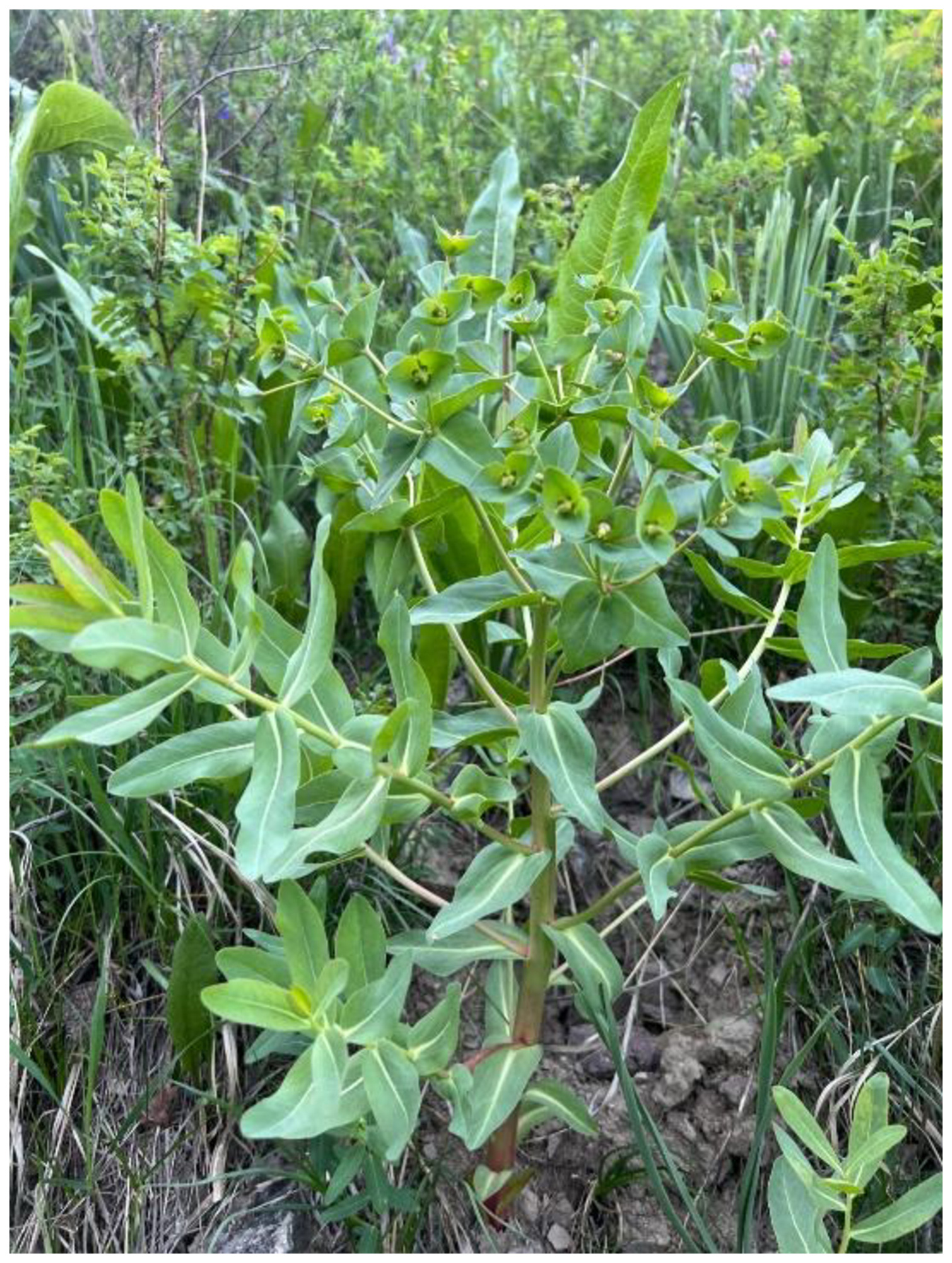
In Central Asia, several Euphorbia L. species exhibit narrow distribution ranges and a high degree of ecological specialization; however, they remain poorly studied compared with their Mediterranean and tropical counterparts (Sumbembayev et al., 2025). Euphorbia yaroslavii Poljakov. (Figure 1) is a rare herbaceous perennial geophyte endemic to southeastern Kazakhstan, where it occurs in a limited number of isolated localities in the foothills of the Zailiyskiy Alatau Range (Turgara et al., 2025). The species is listed in the Red Data Book of the Republic of Kazakhstan, reflecting its vulnerable conservation status, which is associated with restricted distribution, population fragmentation, and high sensitivity to anthropogenic disturbance, including land-use change and habitat degradation (Republic of Kazakhstan, 2014).
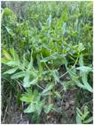
Natural growth of Euphorbia yaroslavii Poljakov. Zailiyskiy Alatau, Ushkonyr Gorge (Kazakhstan).
Ecological investigations have demonstrated that Euphorbia yaroslavii Poljakov. exhibits pronounced edaphic specificity, occurring predominantly on well-drained calcareous loamy soils with slightly alkaline reaction and high base saturation. Population abundance has been shown to correlate strongly with soil texture, humus content, and the availability of exchangeable cations, particularly magnesium, emphasizing the crucial role of soil chemistry in shaping the realized ecological niche of this species (Turgara et al., 2025). Plants inhabiting arid and semi-arid environments commonly accumulate secondary metabolites that enhance stress tolerance, protect against herbivory, and mitigate oxidative damage (Mamurova et al., 2025b). In the genus Euphorbia L., numerous studies have documented the presence of diterpenoids, triterpenes, flavonoids, phenolic acids, and related compounds with diverse biological activities (Figueiredo et al., 2008). However, despite recent advances in understanding the ecology and population structure of Euphorbia yaroslavii Poljakov., information on its phytochemical composition remains extremely limited. The integration of advanced analytical techniques, such as gas chromatography coupled with mass spectrometry (GC-MS), with classical pharmacognostic approaches provides a robust framework for characterizing the chemical diversity of rare endemic plants. Such comprehensive analyses are essential not only for evaluating pharmacological potential but also for elucidating the biochemical basis of ecological adaptation under environmental stress (Zhan et al., 2022). In the case of Euphorbia yaroslavii Poljakov., chemical profiling may offer novel insights into the mechanisms underlying drought tolerance and specialization to calcareous substrates.
Therefore, the present study aims to conduct a detailed investigation of the chemical composition of Euphorbia yaroslavii Poljakov., with particular emphasis on a comparative analysis of organ-specific metabolite profiles in leaves, stems, and roots. This approach enables assessment of the distribution of biologically active compounds among different plant organs, complements existing ecological data, and provides a scientific basis for future studies addressing the pharmacological potential and conservation-oriented utilization of this endemic species.
2. Experimental
2.1. Plant material
Euphorbia yaroslavii Poljakov. was collected in the Zailiyskiy Alatau, Ushkonyr Gorge, Kazakhstan (43.131590° N, 76.512721° E; GPS: Garmin Montana 710). Plant material was collected in the morning during the period of active vegetation in June 2025. The raw plant material was collected in ecologically clean areas, and the species was identified and authenticated by the staff of the IBP (Almaty, Kazakhstan). To confirm the identification, part of the collected material was preserved and transferred for storage to the herbarium of the Institute of Botany and Phytointroduction, Almaty, Kazakhstan (voucher no: 12/36). The samples were washed thoroughly in running tap water to remove soil particles and adhered debris and finally washed with sterile distilled water; dried in the dark at room temperature for at least ten days. The dried material was then processed into a uniform fine powder using an electric micro-grinding machine (110 V, 1400 rpm) to ensure consistency for further analysis. Grinding was performed separately for leaves, stems, and roots to prevent cross-contamination of plant organs.
2.2. Extraction procedure
Ultrasonic extraction was used to obtain plant extracts: powdered plant material was extracted three times with 70% aqueous ethanol (v/v) at a ratio of 1:8 (w/v) for 30 min each using a KQ5200B ultrasonic bath (35 kHz). The combined extracts were filtered and evaporated at 45 °C using an EYELA N-1300 rotary evaporator (Chemat et al., 2017). Fractionation of the crude extract was carried out using solvents of increasing polarity. The extract was dissolved in distilled water and sequentially partitioned with petroleum ether (1:1, three times), dichloromethane, ethyl acetate, and butanol. Each organic fraction was evaporated at 35-45 °C, and the remaining aqueous fraction at 50 °C, using an EYELA N-1300 rotary evaporator (Tokyo Rikakikai Co., Ltd., Japan). Qualitative profiling of the extracts and fractions was performed by thin-layer chromatography (Do et al., 2014).
2.3. Phytochemical and physicochemical analysis
Moisture content was determined by drying 1 g of powdered Euphorbia yaroslavii Poljakov. plant material to a constant mass at 100-105 °C. Total ash content was assessed by incinerating 2 g of material at 500-600 °C with subsequent correction of all quantitative results to absolutely dry weight (W) (Musinov and Tulagenova, 2016).
Total flavonoids were determined by extraction with 90% ethanol containing 1% HCl, hydrolysis, complexation with alcoholic AlCl3 solution, and quantification spectrophotometrically at 430 nm, with results expressed as quercetin equivalents and calculated with moisture correction, extraction repeated twice (Pękal and Pyrzynska, 2014).
Vitamin C (ascorbic acid) was quantified by aqueous extraction, followed by acidification and titration with 0.001 M 2,6-dichlorophenolindophenol (DCPIP) to a stable pink endpoint (1 mL ≡ 0.000088 g ascorbic acid), with results expressed on an absolutely dry basis (Almeida et al., 2011).
Polysaccharides were assessed after repeated hot-water extraction, combining extracts to 250 ml, precipitation of an aliquot with 95% ethanol, heating, centrifugation (5000 rpm, 30 min), filtration through a pre-weighed glass filter (POR 16), washing, and gravimetric determination after drying to constant mass at 100-105 °C (Kakar et al., 2022).
Total alkaloids were determined following alkaline extraction with chloroform or ethyl acetate, evaporation, addition of NaOH and HCl, filtration, and back-titration of excess acid with 0.01-0.1 M NaOH (methyl red indicator), with a parallel blank (control) experiment. Results expressed as thermopsine equivalents on dry basis (Harborne, 1998).
Coumarins were analyzed after repeated chloroform extraction, evaporation, dissolution in 96% ethanol, dilution, and UV spectrophotometry at 272 nm (specific absorbance 734 for coumarin standard) (Matos, 2009).
Total tannins were determined by repeated hot-water extraction, permanganatometric titration of the combined extract using indigosulfonic acid as indicator and 0.02 M KMnO4, with a control titration (equivalents: 0.004157 g hydrolysable or 0.00582 g condensed tannins per ml KMnO4) (European Pharmacopoeia Commission, 2019).
Free organic acids were quantified by hot-water extraction, filtration, and titration with 0.1 M NaOH using phenolphthalein (or mixed indicator) to pale pink endpoint, with results expressed as malic or valeric acid equivalents (AOAC, 2016).
Mineral composition (macro- and microelements) was determined after dry ashing of the plant material, optional treatment of the ash with concentrated nitric acid, subsequent calcination (at approximately 500 °C to constant mass, followed by additional treatment up to 600 °C when necessary), dissolution in HNO3 (1:1), transfer to 1 N HCl or HNO3, and dilution to 25 ml. A blank experiment was performed in parallel. Elemental quantification was carried out using atomic absorption spectrometry on the “ASSIN” instrument (Carl Zeiss, Germany), with additional arc emission spectroscopy (300 mg ash evaporated in DC arc, spectra photographed on DFS-13 spectrograph, reverse linear dispersion 1 Å/mm, range 2100-3600 Å). Sensitivity: 10−2-10−5 %. Accuracy controlled using standard copper slag samples TSO 2962-84 and 2964-84 (Welz and Sperling, 1999).
Thin-layer chromatography (TLC). Qualitative analysis of the obtained extracts and fractions was performed by thin-layer chromatography (TLC) on silica gel plates using a solvent system composed of ethyl acetate: toluene: formic acid in a ratio of 7:8:2 (v/v/v). After development, the plates were dried and examined under ultraviolet light at wavelengths of 254 and 365 nm (Barron, 1964). For additional visualization, the chromatograms were sprayed with 10% sulfuric acid in ethanol followed by heating, allowing visualization of separated components as characteristic-colored zones.
The total phenolic content (TPC) of the extracts was determined using the Folin-Ciocalteu method. The reaction mixture consisted of 2.5 μL of extract, 2.5 μL of Folin-Ciocalteu reagent, 50 μL of 7% Na2CO3 solution, and 195 μL of distilled water. After incubation for 30 min in the dark, absorbance was measured at 765 nm. Total phenolic content was calculated using a calibration curve constructed with gallic acid and expressed as mg gallic acid equivalents per gram of dry weight (mg GAE/g DW) (Mamurova et al., 2025b; Zhou et al. 2004).
2.4. Gas chromatography-mass spectrometry (GC-MS) analysis
GC-MS analysis was performed separately for leaf, stem, and root extracts. Chromatographic separation and compound identification were carried out using an Agilent 7890A gas chromatograph coupled with a 5975C mass-selective detector (Agilent Technologies, USA). Samples (0.5 μL) were injected in splitless mode at an injector temperature of 280 °C. Separation was achieved on a DB-17ms capillary column (30 m × 0.25 mm i.d., film thickness 0.25 μm) using helium as the carrier gas at a constant flow rate of 1.0 mL/min. The oven temperature program was set from 40 °C to 300 °C at a rate of 5 °C/min, followed by a final hold at 300 °C for 10 min, with a total analysis time of 67 min. Mass spectrometric detection was conducted in SCAN mode over m/z 34-750. Data acquisition and processing were performed using Agilent MSD ChemStation software (version 1701EA) (Mukanova et al., 2024). Compound identification was conducted by comparing the acquired mass spectra with the Wiley and NIST’02 mass spectral libraries (Wiley Science Solutions, 2023). Only compounds with acceptable spectral match probabilities (≥65-70%) were considered identified (Mamurova et al., 2025c). Relative abundances of detected compounds were calculated by peak area normalization and expressed as percentages of the total ion current. The GC-MS analysis was qualitative and semi-quantitative in nature and was performed without the use of external standards.
2.5. Statistical analysis.
Statistical analysis was performed using GraphPad Prism (GraphPad Software, 2020, San Diego, CA, USA). Quantitative data were obtained from three independently prepared samples, and the results are presented as mean ± standard deviation (SD). Differences among plant organs (leaves, stems and roots) were evaluated using one-way analysis of variance (ANOVA), followed by Tukey’s post hoc test for multiple comparisons. Differences were considered statistically significant at p < 0.05.
3. Results and Discussion
3.1. Phytochemical composition of leaves, stems, and roots
The analysis of plant material of Euphorbia yaroslavii Poljakov. (leaves, stems and roots), conducted using the described methodologies, revealed pronounced differences in the content of biologically active compounds and mineral elements. All quantitative results are expressed on an absolutely dry weight basis, with correction for moisture content (W); mineral elements are reported in mg/kg dry weight, while organic constituents are expressed as percentages or mg/g DW, as appropriate. The obtained data confirm the potential value of this species as a source of biologically active substances characteristic of the genus Euphorbia. Because the available literature contains very limited information specifically on Euphorbia yaroslavii Poljakov., a rare endemic species of Kazakhstan with scarce published data, comparative analysis was performed using closely related Euphorbia species (E.hirta, E.helioscopia, E.milii, E.granulata), in which similar patterns of compound accumulation in different plant organs have been reported (Ernst et al., 2015).
Moisture content varied slightly among plant organs and remained within a narrow range, amounting to 5.58 ± 0.12% in leaves, 5.05 ± 0.10% in stems, and 5.68 ± 0.15% in roots (Table 1). These values indicate well-prepared plant material suitable for storage and subsequent phytochemical analysis. Minor differences in moisture content between organs may be related to structural and functional characteristics of the tissues. Similar moisture ranges (5-10%) have been reported for other Euphorbia species adapted to arid and semi-arid environments (Kumar et al., 2010). Total ash content exhibited pronounced organ-specific variation, with the highest values observed in roots (13.22 ± 0.30%), followed by leaves (11.36 ± 0.20%), while stems contained the lowest ash content (6.48 ± 0.25%). This distribution suggests preferential accumulation of mineral elements in underground organs, which may be associated with reserve nutrition and osmoregulation. Comparable ash content ranges (5-15%) and similar organ-dependent trends have been reported for representatives of the genus Euphorbia, where roots frequently contain higher mineral fractions than aboveground parts (Saleem et al., 2014). In E.hirta, ash content ranges from 8-12%, which is lower than our root values, likely due to differences in soil conditions (tropical vs. mountainous) (Yang et al., 2021).
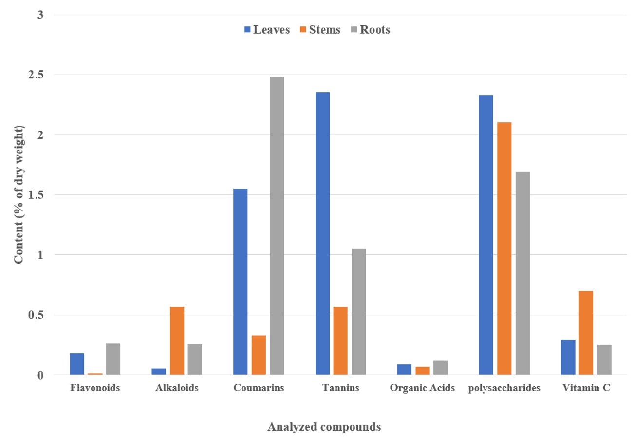
The sum of flavonoids was highest in roots (0.265 ± 0.008%), followed by leaves (0.18 ± 0.006%), while stems exhibited the lowest content (0.014 ± 0.002%). Such a distribution reflects the functional role of flavonoids as protective metabolites, particularly in plant organs exposed to environmental stress and involved in storage and defense mechanisms, although in this species their accumulation in roots appears to be species-specific. In representatives of the genus Euphorbia, flavonoids are often predominantly detected in leaves, reaching up to 2-3% in tropical species; however, the lower values observed in Euphorbia yaroslavii Poljakov. compared to E.hirta (up to 41 mg QE/g) and E.milii (19-20 mg QE/g) may be attributed to climatic conditions and differences in extraction and analytical approaches (Pollastri and Tattini, 2011; Shi et al., 2008).
The sum of alkaloids, recalculated as thermopsine, was highest in stems (0.565 ± 0.010%), followed by roots (0.258 ± 0.007%) and leaves (0.052 ± 0.003%) (Figure 2). This pattern is characteristic of the genus Euphorbia, where alkaloids function as protective toxic compounds and are often unevenly distributed among plant organs. The obtained values fall within the typical range of 0.05-1% reported for Euphorbia species, including E.hirta and E.guyoniana, where alkaloid accumulation contributes to defense against pathogens and herbivores (Moghaddam et al., 2012).
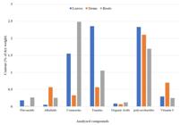
The sum of coumarins was highest in roots (2.483 ± 0.050%), followed by leaves (1.551 ± 0.035%) and stems (0.332 ± 0.020%). This distribution is consistent with the role of coumarins as antimicrobial and protective constituents associated with latex and underground tissues. The values obtained exceed the commonly reported range for the genus (0.1-1%), which may reflect species-specific metabolic features and adaptation of this endemic plant to local environmental stressors. Related species such as E.hirta and E.guyoniana have been reported to contain coumarins qualitatively, but at lower quantitative levels (Venugopala et al., 2013; Wiśniewski et al., 2016).
The highest tannin content was recorded in leaves (2.353 ± 0.040%), followed by roots (1.052 ± 0.030%) and stems (0.568 ± 0.025%). This distribution supports the role of tannins as astringent and antimicrobial compounds, particularly in photosynthetically active tissues. The obtained values are comparable to those reported for E.hirta (1-2%) and are consistent with the protective functions of tannins described for the genus Euphorbia (Kumar et al., 2010).
The content of free organic acids was relatively low across all organs (0.071-0.124%), with the highest value observed in roots, followed by leaves and stems. Such levels are typical for Euphorbia species and reflect their involvement in primary metabolic processes rather than storage functions. Similar qualitative patterns have been reported for E.greenwayi (Hammadi, 2023).
Polysaccharide content was highest in leaves (2.33 ± 0.060%), followed by stems (2.106 ± 0.045%), while roots contained the lowest amount (1.696 ± 0.05%). This distribution indicates preferential accumulation of polysaccharides in аaerial parts in Euphorbia yaroslavii Poljakov., which may be associated with active metabolic processes and structural carbohydrate deposition in photosynthetically active tissues. The observed values (1.7-2.3%) fall within the typical range reported for the genus Euphorbia (1-3%), although direct quantitative comparisons remain limited due to the predominance of qualitative reports in the literature (Gorshkova et al., 2018; Zhang et al., 2018).
Vitamin C (ascorbic acid) content reached its maximum in stems (0.698 ± 0.015 mg/g), with lower values detected in leaves (0.294 ± 0.010 mg/g) and roots (0.252 ± 0.008 mg/g). This pattern reflects the physiological role of ascorbic acid in actively metabolizing tissues, where it contributes to antioxidant defense and regulation of growth-related processes. The relatively high concentration in stems highlights their potential as a source of natural vitamin C, particularly under conditions of increased oxidative stress associated with high-altitude environments (Smirnoff, 2018).
The mineral composition is dominated by macroelements K (94.691-122.949 mg/kg) and Ca (45.135-179.601 mg/kg) (Table 2), with the highest concentrations observed mainly in leaves, which is important for metabolic activity, structural support, and osmoregulation. Microelements (Fe 0.2459-1.0335 mg/kg, Mn 0.1059-0.3756 mg/kg, etc.) are present at low levels and remain within safe limits; the contents of Pb and Cd do not exceed the maximum permissible values (WHO, 2007). This profile is typical for the genus Euphorbia, in which K and Ca are the predominant mineral elements contributing to adaptation under stressful environmental conditions (Hammadi, 2023). In E.hirta and E.milii, a similar dominance of K and Ca has been reported; however, the potassium content in Euphorbia yaroslavii Poljakov. (up to 122.9 mg/kg) is comparatively higher, possibly due to increased soil mineralization in the Zailiyskiy Alatau region (Kumar et al., 2010).
Thin-layer chromatography (TLC) profiling corroborated this distribution, revealing characteristic zones under UV light (254 and 365 nm) and after visualization with 10% sulfuric acid in ethanol followed by heating. The ethyl acetate fractions exhibited blue fluorescence at 365 nm, indicative of coumarins, as well as yellow-green zones attributable to flavonoids. Root fractions displayed more intense bands in the Rf range of 0.4-0.6, tentatively corresponding to terpenoid compounds, further supporting the organ-specific accumulation patterns observed in the quantitative analysis. The extraction yields obtained in this study (12.8-18.5%) are comparable to those reported for other Euphorbia species, such as Euphorbia larica Boiss. (10-20%), where similar solvent systems have been employed for the partitioning of latex-derived metabolites (Shah et al., 2023).
TPC showed clear organ-specific variation in Euphorbia yaroslavii Poljakov. Leaves exhibited a moderate TPC (25.4 ± 1.2 mg GAE/g DW), consistent with their flavonoid content (0.18% QE, DW) and the established role of aerial tissues in photoprotection and antioxidant defense. Stems displayed the lowest TPC (18.7 ± 0.9 mg GAE/g DW), reflecting lower accumulation of phenolic compounds in predominantly structural organs. In contrast, roots accumulated significantly higher levels of phenolics (58.2 ± 2.5 mg GAE/g DW), in agreement with the elevated concentrations of coumarins and tannins determined by quantitative assays, indicating dominance of non-flavonoid phenolics in underground tissues (Shahidi and Ambigaipalan, 2015). Similar organ-dependent phenolic allocation has been reported for medicinal plants, where roots function as reservoirs of defensive phenolic metabolites against soil-borne stressors (Cheynier et al., 2013).
Euphorbia yaroslavii Poljakov. demonstrates a characteristic Euphorbia phytochemical profile with pronounced accumulation of defensive and storage-related compounds in roots, while leaves preferentially accumulate photoprotective compounds such as flavonoids. The obtained data indicate potential applicability of root-derived extracts in mineral supplementation and antimicrobial formulations, whereas leaf and stem extracts may represent promising sources of antioxidant compounds. Further bioactivity-guided studies are required to substantiate these potential applications.
3.2. Comparative GC-MS Analysis of Leaf, Stem, and Root Extracts of Euphorbia yaroslavii Poljakov.
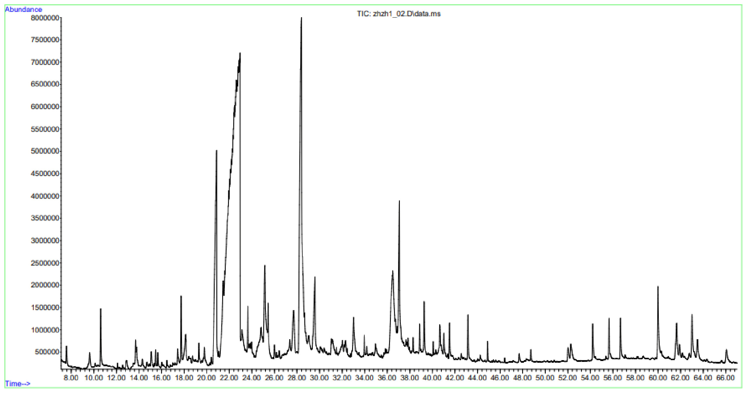
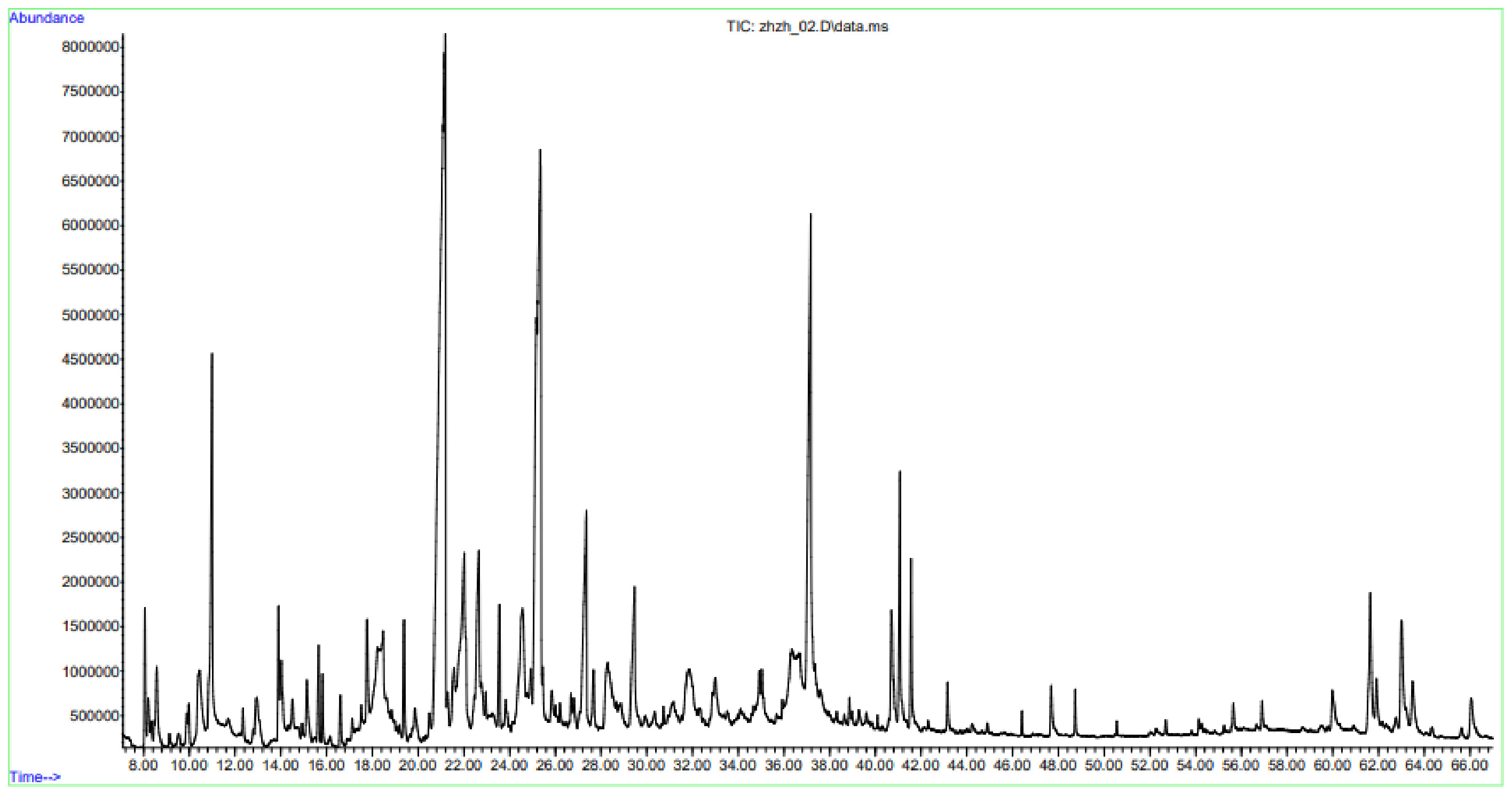
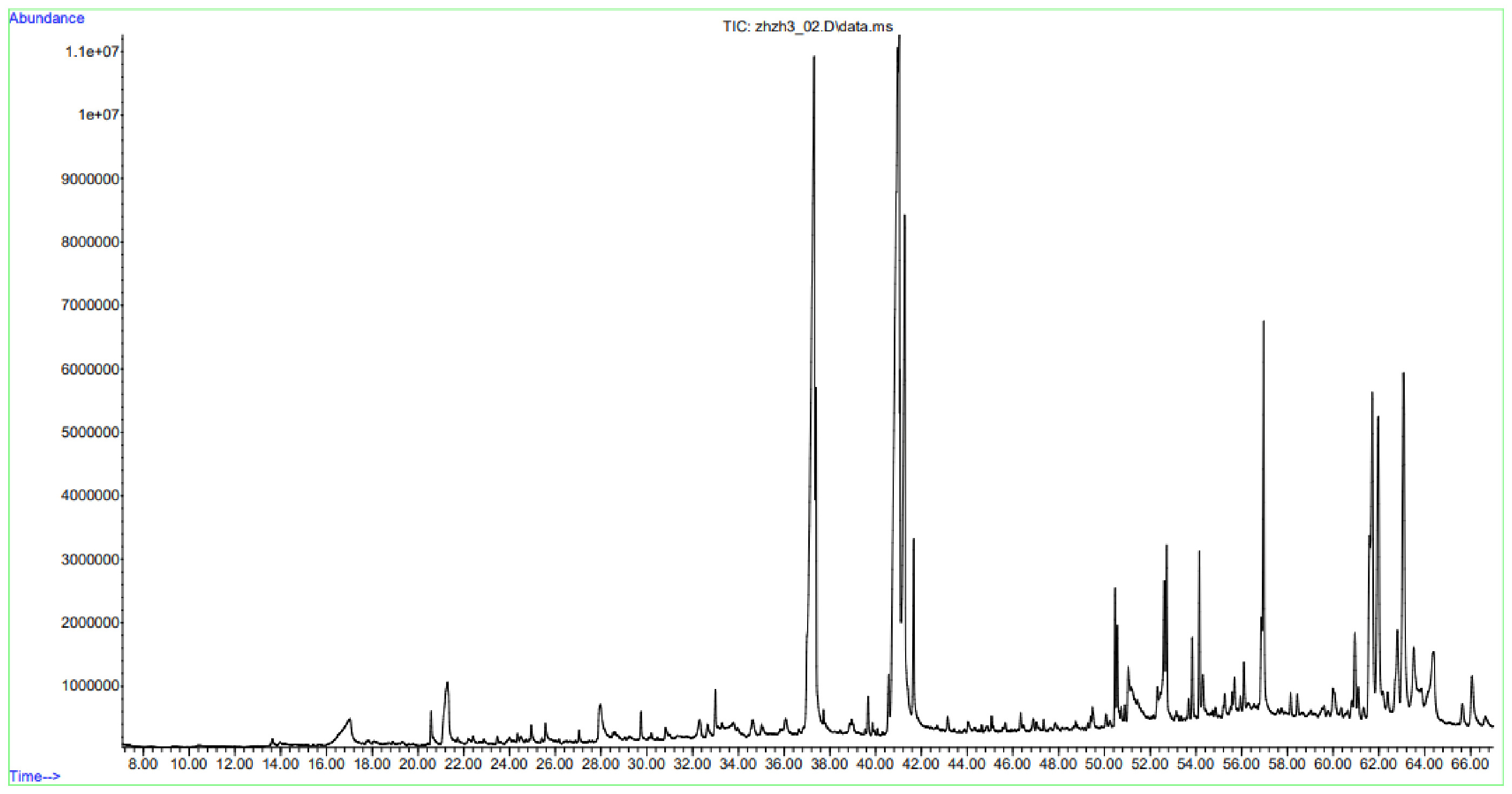
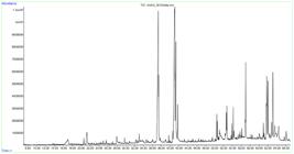
Gas chromatography-mass spectrometry (GC-MS) analysis of 70% aqueous-ethanol extracts obtained from the leaves, stems, and roots of Euphorbia yaroslavii Poljakov. revealed clear organ-specific differences in chemical composition. A total of 68 compounds were tentatively identified in leaf extracts (Figure 3), 65 compounds in stem extracts (Figure 4), and 39 compounds in root extracts (Figure 5), representing diverse chemical classes, including furan derivatives, phenolic compounds, fatty acids, terpenoids, sterols, and vitamins (Tables 3 to 5; Figures 3 to 5). Compound identification was based on retention times and mass spectral matching with the Wiley and NIST’02 mass spectral libraries. Only compounds with acceptable spectral match probabilities (≥65-70%) were considered. Relative abundances were calculated using peak area normalization and expressed as percentages of the total ion current.
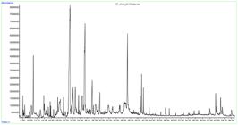
The leaf extract exhibited the highest chemical diversity among the analyzed organs. The predominant constituents were 2-propanamine, N-methyl-N-nitroso- (31.94%), 1-butanol, 3-methyl-, acetate (11.26%), and 1,2,3-benzenetriol (12.86%). In addition, minor amounts of phenolic compounds such as phenol (0.12%) and hydroquinone (0.30%) were detected. Fatty acids, including hexadecanoic acid (3.42%) and octadecanoic acid (0.87%), were present together with terpenoid-related metabolites such as phytol (0.58%) and lupeol (0.56%). Sterols, particularly γ-sitosterol (1.23%) and lanosterol (0.79%), were also detected (Table 3). The presence of tocopherols (α-, β-, γ-, and δ-forms), collectively accounting for approximately 1.5% of the chromatographic profile, indicates the presence of compounds commonly associated with antioxidant properties in leaf tissues. Similar enrichment of phenolics, tocopherols, and triterpenoids in aerial parts has been reported for species of the genus Euphorbia and is associated with protection against oxidative stress, ultraviolet radiation, and herbivory (Shi et al., 2008).
The stem extract was characterized by a predominance of oxygenated heterocyclic compounds tentatively associated with carbohydrate-derived constituents, with 4H-pyran-4-one, 2,3-dihydro-3,5-dihydroxy-6-methyl- (20.16%) and 5-hydroxymethylfurfural (14.04%) as major components (Table 4). In contrast to leaf extracts enriched in phenolic and antioxidant-related compounds and root extracts dominated by fatty acids and triterpenoid-sterol constituents, stems exhibited a profile typical of intermediate metabolic tissues (Xiao et al., 2025; Mamurova et al., 2025d).
Fatty acids, particularly hexadecanoic acid (8.35%) and 9,12-octadecadienoic acid (Z,Z)- (2.18%), were present at moderate levels but remained less abundant than in roots, indicating preferential lipid accumulation in underground organs. Phenolic compounds were detected in lower amounts than in leaves, while terpenoids and sterols occurred at intermediate concentrations, supporting the organ-specific distribution of metabolites and the functional role of stems between photosynthetically active leaves and storage-oriented roots (Kumar et al., 2010).
In contrast to aerial organs, the root extract was dominated by fatty acids and triterpenoid-sterol compounds. The principal constituents included octadecanoic acid (18.92%), hexadecanoic acid (17.46%), and 9,12-octadecadienoic acid (Z,Z)- (8.81%) (Table 5), indicating a markedly higher lipid fraction compared with leaves and stems. Terpenoids and sterols were particularly abundant, with β-amyrin (5.22%), lanosterol (4.85%), 9,19-cyclolanostan-3-ol, 24-methylene-, (3β)- (6.14%), and lupeol (2.63%) together accounting for more than 20% of the total chromatographic profile. Roots exhibited a profile dominated by storage- and structure-related metabolites. Such enrichment of triterpenoids and sterols in underground organs is commonly associated with protective and structural functions, including membrane stabilization and interactions with soil microorganisms, as plant sterols are integral components of membrane lipid bilayers regulating membrane integrity, while terpenoid compounds are widely involved in plant defense responses (Ninkuu et al., 2021). Similar dominance of β-amyrin and lupeol has been reported in roots of E.hirta and E.tirucalli, where these compounds are discussed in relation to antimicrobial and anti-inflammatory properties (Geetha and Varalakshmi, 2001).
The observed distribution of metabolites indicates organ-specific metabolic specialization in Euphorbia yaroslavii Poljakov. Leaf extracts were characterized by a higher relative abundance of phenolic and antioxidant-related constituents, stem extracts were enriched in carbohydrate-derived oxygenated metabolites, whereas root extracts were dominated by fatty acids and triterpenoid-sterol compounds, including octadecanoic acid, hexadecanoic acid, 9,12-octadecadienoic acid (Z,Z)-, β-amyrin, lanosterol, and lupeol. This pattern agrees with general phytochemical trends reported for the genus Euphorbia, where underground organs are enriched in defensive metabolites and aerial parts contain protective and regulatory compounds (Salehi et al., 2019). Quantitative analysis supported these results, showing higher levels of tannins and polysaccharides in leaves and increased coumarin content in roots, while stems displayed intermediate values. The detection of scopoletin (0.38-0.53%) in leaf and stem extracts confirms the presence of coumarin-type constituents consistent with reported antifungal and antimicrobial properties (Lemos et al., 2020). Unsaturated fatty acids, including 9,12,15-octadecatrienoic acid (Z,Z,Z)- (up to 2.05% in roots), represent a characteristic component of the root lipid fraction, similar to profiles reported for Euphorbia laricaBoiss. (Shah et al., 2023). ocopherols detected in leaf extracts further indicate the antioxidant role of aerial tissues, as reported for Euphorbia wallichii (Haq et al., 2012). Despite the informative nature of the GC-MS profiling, compound identification remains tentative because it is based on spectral library matching without confirmation using authentic standards.
4. Conclusion
This study presents the first comparative phytochemical and GC-MS analysis of leaves, stems, and roots of Euphorbia yaroslavii Poljakov., a species endemic to Kazakhstan. The results indicate pronounced organ-specific metabolic differentiation. Root tissues are characterized by high levels of triterpenoids, sterols, fatty acids, coumarins, and mineral components, whereas leaves exhibit elevated contents of phenolic compounds, including tannins. Stems display an intermediate metabolic profile, dominated mainly by carbohydrate-related oxygenated metabolites. By integrating quantitative pharmacognostic analyses with GC-MS profiling, this work provides a phytochemical baseline for a poorly studied endemic taxon. The findings contribute to a better understanding of organ-specific metabolite allocation in arid-adapted Euphorbia species and may serve as a framework for future studies focusing on compound isolation, bioactivity assessment, and conservation-oriented research.
Data Availability Statement
The entire data set that supports the results of this study was published in the article itself.
References
-
ALMEIDA, M.M.B., DE SOUSA, P.H.M., ARRIAGA, A.M.C., PRADO, G.M., MAGALHÃES, C.E.C., MAIA, G.A. and LEMOS, T.L.G., 2011. Bioactive compounds and antioxidant activity of fresh exotic fruits from northeastern Brazil. Food Research International, vol. 44, no. 7, pp. 2155-2159. https://doi.org/10.1016/j.foodres.2011.03.051
» https://doi.org/10.1016/j.foodres.2011.03.051 - ASSOCIATION OF OFFICIAL ANALYTICAL CHEMISTS – AOAC, 2016. Official methods of analysis of the Association of Official Analytical Chemists 20th ed. Washington, D.C.: AOAC.
-
BARRON, E.S., 1964. Thin-layer chromatography. Postgraduate Medicine, vol. 36, no. 3, pp. A44-A52. https://doi.org/10.1080/00325481.1964.11695265 PMid:14179265.
» https://doi.org/10.1080/00325481.1964.11695265 -
CHEMAT, F., ROMBAUT, N., SICAIRE, A.G., MEULLEMIESTRE, A., FABIANO-TIXIER, A.S. and ABERT-VIAN, M., 2017. Ultrasound assisted extraction of food and natural products: mechanisms, techniques, combinations, protocols and applications. Ultrasonics Sonochemistry, vol. 34, pp. 540-560. https://doi.org/10.1016/j.ultsonch.2016.06.035 PMid:27773280.
» https://doi.org/10.1016/j.ultsonch.2016.06.035 -
CHEYNIER, V., COMTE, G., DAVIES, K.M., LATTANZIO, V. and MARTENS, S., 2013. Plant phenolics: recent advances on their biosynthesis, genetics, and ecophysiology. Plant Physiology and Biochemistry : PPB, vol. 72, pp. 1-20. https://doi.org/10.1016/j.plaphy.2013.05.009 PMid:23774057.
» https://doi.org/10.1016/j.plaphy.2013.05.009 -
DO, Q.D., ANGKAWIJAYA, A.E., TRAN-NGUYEN, P.L., HUYNH, L.H., SOETAREDJO, F.E., ISMADJI, S. and JU, Y.-H., 2014. Effect of extraction solvent on total phenol content, total flavonoid content, and antioxidant activity of Limnophila aromatica Journal of Food and Drug Analysis, vol. 22, no. 3, pp. 296-302. https://doi.org/10.1016/j.jfda.2013.11.001 PMid:28911418.
» https://doi.org/10.1016/j.jfda.2013.11.001 -
ERNST, M., GRACE, O.M., SASLIS-LAGOUDÁKIS, C.H., NILSSON, N., SIMONSEN, H.T., and RØNSTED, N., 2015. Global medicinal uses of Euphorbia L. (Euphorbiaceae). Journal of Ethnopharmacology, vol. 176, pp. 90-101. https://doi.org/10.1016/j.jep.2015.10.025 PMid:26485050.
» https://doi.org/10.1016/j.jep.2015.10.025 - EUROPEAN PHARMACOPOEIA COMMISSION, 2019. European Pharmacopoeia 10th ed. Strasbourg: Council of Europe.
-
FIGUEIREDO, A.C., BARROSO, J.G., PEDRO, L.G. and SCHEFFER, J.J.C., 2008. Factors affecting secondary metabolite production in plants: volatile components and essential oils. Flavour and Fragrance Journal, vol. 23, no. 4, pp. 213-226. https://doi.org/10.1002/ffj.1875
» https://doi.org/10.1002/ffj.1875 -
GEETHA, T. and VARALAKSHMI, P., 2001. Anti-inflammatory activity of lupeol and lupeol linoleate in rats. Journal of Ethnopharmacology, vol. 76, no. 1, pp. 77-80. https://doi.org/10.1016/S0378-8741(01)00175-1 PMid:11378285.
» https://doi.org/10.1016/S0378-8741(01)00175-1 -
GORSHKOVA, T., CHERNOVA, T., MOKSHINA, N., AGEEVA, M. and MIKSHINA, P., 2018. Plant “muscles”: fibers with a tertiary cell wall. The New Phytologist, vol. 218, no. 1, pp. 66-72. https://doi.org/10.1111/nph.14997 PMid:29364532.
» https://doi.org/10.1111/nph.14997 -
GRAPHPAD SOFTWARE, 2020 [viewed 24 January 2025]. GraphPad Prism, version 9.0 for Windows [online]. San Diego: GraphPad Software. Available from: https://www.graphpad.com/
» https://www.graphpad.com/ -
HAMMADI, R., 2023. Chemical and pharmacological investigations of African Euphorbia species. Szeged: University of Szeged. Doctoral Dissertation in Pharmacognosy. https://doi.org/10.14232/phd.11515
» https://doi.org/10.14232/phd.11515 - HAQ, I.U., ULLAH, N., BIBI, G., KANWAL, S., MIRZA, B. and AHMAD, V.U., 2012. Antioxidant and cytotoxic activities of Euphorbia wallichii root extract. Iranian Journal of Pharmaceutical Research: IJPR, vol. 11, no. 1, pp. 241-249. PMid:24250446.
- HARBORNE, J.B., 1998. Phytochemical methods: a guide to modern techniques of plant analysis. 3rd ed. London: Chapman & Hall.
-
HORN, J.W., VAN EE, B.W., MORAWETZ, J.J., RIINA, R., STEINMANN, V.W., BERRY, P.E. and WURDACK, K.J., 2012. Phylogenetics and the evolution of growth forms in the giant genus Euphorbia L. (Euphorbiaceae). Molecular Phylogenetics and Evolution, vol. 63, no. 2, pp. 305-326. https://doi.org/10.1016/j.ympev.2011.12.022 PMid:22273597.
» https://doi.org/10.1016/j.ympev.2011.12.022 -
KAKAR, M.U., LI, J., MEHBOOB, M.Z., SAMI, R., BENAJIBA, N., AHMED, A., NAZIR, A., DENG, Y., LI, B. and DAI, R., 2022. Purification, characterization, and determination of biological activities of water-soluble polysaccharides from Mahonia bealei. Scientific Reports, vol. 12, no. 1, pp. 8160. https://doi.org/10.1038/s41598-022-11661-3 PMid:35581215.
» https://doi.org/10.1038/s41598-022-11661-3 -
KUMAR, S., MALHOTRA, R. and KUMAR, D., 2010. Euphorbia hirta: its chemistry, traditional and medicinal uses, and pharmacological activities. Pharmacognosy Reviews, vol. 4, no. 7, pp. 58-61. https://doi.org/10.4103/0973-7847.65327 PMid:22228942.
» https://doi.org/10.4103/0973-7847.65327 -
LEMOS, A.S.O., FLORÊNCIO, J.R., PINTO, N.C.C., CAMPOS, L.M., SILVA, T.P., GRAZUL, R.M., PINTO, P.F., TAVARES, G.D., SCIO, E., APOLÔNIO, A.C.M., MELO, R.C.N. and FABRI, R.L., 2020. Antifungal activity of the natural coumarin scopoletin against planktonic cells and biofilms from a multidrug-resistant Candida tropicalis strain. Frontiers in Microbiology, vol. 11, pp. 1525. https://doi.org/10.3389/fmicb.2020.01525 PMid:32733416.
» https://doi.org/10.3389/fmicb.2020.01525 -
MAMUROVA, A., KAIRANOVA, G., ZAPARINA, Y., MENGTAY, A., KUDAIBERGENOVA, A., YEDILOVA, A. and KALIYEV, B., 2025a. Soil morphological and physico-chemical characteristics of Zygophyllum fabago L. populations in the Ili-Balkhash Region of Kazakhstan. EQA, vol. 70, pp. 106-115. https://doi.org/10.6092/issn.2281-4485/22033
» https://doi.org/10.6092/issn.2281-4485/22033 -
MAMUROVA, A., KAIRANOVA, G., BEYATLI, A., SMAGULOVA, G., YEDILOVA, A., ZHALDYBAYEV, K., ZHUMALINA, K. and TOREGELDIYEVA, A., 2025b. Phytochemical analysis and antioxidant, antimicrobial, cytotoxic activities of different solvent extracts of Zygophyllum fabago L. Brazilian Journal of Biology = Revista Brasileira de Biologia, vol. 85, e293666. https://doi.org/10.1590/1519-6984.293666 PMid:40435090.
» https://doi.org/10.1590/1519-6984.293666 -
MAMUROVA, A., KAIRANOVA, G., AMERTAYEVA, G., AKHTAYEVA, N., BEYATLI, A., KIYEKBAYEVA, L., ISKAKOVA, Z. and ZHALDYBAEV, K., 2025c. Phytochemical profiling and antioxidant properties of Ajania fastigiata (C. Winkl.) Poljakov (Asteraceae). Farmacia, vol. 73, no. 4, pp. 1038-1046. https://doi.org/10.31925/farmacia.2025.4.23
» https://doi.org/10.31925/farmacia.2025.4.23 -
MAMUROVA, A., KAIRANOVA, G., AMERTAYEVA, G., YEDILOVA, A., RAKHYMZHAN, A., KAMALOVA, M., IGLIKOVA, A., KIYEKBAYEVA, L., SMAGULOVA, G., MUKANOVA, U., MUKHAMETZHAN, A., BEYATLI, A. and KALIYEV, B., 2025d. Phytochemical composition and antioxidant, antimicrobial, and cytotoxic activities of different solvent extracts of Ajania fastigiata (C. Winkl.) Poljakov. Brazilian Journal of Biology = Revista Brasileira de Biologia, vol. 85, e300899. https://doi.org/10.1590/1519-6984.300899
» https://doi.org/10.1590/1519-6984.300899 - MATOS, F.J.A., 2009. Introdução à fitoquímica experimental. 3. ed. Fortaleza: Edições UFC, 147 p.
-
MOGHADDAM, G., EBRAHIMI, S.A., RAHBAR-ROSHANDEL, N. and FOROUMADI, A., 2012. Antiproliferative activity of flavonoids: influence of the sequential methoxylation state of the flavonoid structure. Phytotherapy Research : PTR, vol. 26, no. 7, pp. 1023-1028. https://doi.org/10.1002/ptr.3678 PMid:22184071.
» https://doi.org/10.1002/ptr.3678 -
MUKANOVA, A., DATKHAYEV, U., ABDULLABEKOVA, R., ALIMZHANOVA, M., KHRUSTALEV, D., ISKAKOVA, Z., et al, 2024. Composition of Scabiosa ochroleuca L. extracts prepared by ultrasonic and microwave methods and their antiradical and antioxidant activity assessment. Farmacia, vol. 72, no. 4, pp. 867-874. https://doi.org/10.31925/farmacia.2024.4.15
» https://doi.org/10.31925/farmacia.2024.4.15 - MUSINOV, S.R. and TULAGENOVA, A.U., 2016. The State Pharmacopoeia: main standard of quality of medicines and medical devices in the Republic of Kazakhstan. The Bulletin of the Scientific Centre for Expert Evaluation of Medicinal Products, no. 2, pp. 26-30.
-
NINKUU, V., ZHANG, Z., YAN, J., FU, Z., YANG, T. and ZENG, H., 2021. Biochemistry of terpenes and recent advances in plant defense. International Journal of Molecular Sciences, vol. 22, no. 11, pp. 5710. https://doi.org/10.3390/ijms22115710 PMid:34071919.
» https://doi.org/10.3390/ijms22115710 -
PĘKAL, A. and PYRZYNSKA, K., 2014. Evaluation of aluminium complexation reaction for flavonoid content assay. Food Analytical Methods, vol. 7, no. 9, pp. 1776-1782. https://doi.org/10.1007/s12161-014-9814-x
» https://doi.org/10.1007/s12161-014-9814-x -
POLLASTRI, S. and TATTINI, M., 2011. Flavonols: old compounds for old roles. Annals of Botany, vol. 108, no. 7, pp. 1225-1233. https://doi.org/10.1093/aob/mcr234 PMid:21880658.
» https://doi.org/10.1093/aob/mcr234 -
REPUBLIC OF KAZAKHSTAN. Ministry of Ecology, Geology and Natural Resources, 2014 [viewed 24 January 2025]. Red data book of Kazakhstan: plants [online]. Almaty, vol. 2. Available from: https://bioreserve-almaty.kz/ru/o-zapovednike/krasnaya-kniga.html
» https://bioreserve-almaty.kz/ru/o-zapovednike/krasnaya-kniga.html - SALEEM, U., HUSSAIN, K., AHMAD, M., BUKHARI, N.I., MALIK, A. and AHMAD, B., 2014. Physicochemical and phytochemical analysis of Euphorbia helioscopia (L.). Pakistan Journal of Pharmaceutical Sciences, vol. 27, no. 3, pp. 577-585. PMid:24811820.
-
SALEHI, B., IRITI, M., VITALINI, S., ANTOLAK, H., PAWLIKOWSKA, E., KRĘGIEL, D., SHARIFI-RAD, J., OYELEYE, S.I., ADEMILUYI, A.O., CZOPEK, K., STANIAK, M., CUSTÓDIO, L., COY-BARRERA, E., SEGURA-CARRETERO, A., CÁDIZ-GURREA, M.L., CAPASSO, R., CHO, W.C. and SECA, A.M.L., 2019. Euphorbia-derived natural products with potential for use in health maintenance. Biomolecules, vol. 9, no. 8, pp. 337. https://doi.org/10.3390/biom9080337 PMid:31382529.
» https://doi.org/10.3390/biom9080337 -
SHAH, M., KHAN, F., ULLAH, S., MOHANTA, T.K., KHAN, A., ZAINAB, R., RAFIQ, N., ARA, H., ALAM, T., REHMAN, N.U. and AL-HARRASI, A., 2023. GC–MS profiling and biomedical applications of essential oil of Euphorbia larica Boiss.: a new report. Antioxidants, vol. 12, no. 3, pp. 662. https://doi.org/10.3390/antiox12030662 PMid:36978910.
» https://doi.org/10.3390/antiox12030662 -
SHAHIDI, F. and AMBIGAIPALAN, P., 2015. Phenolics and polyphenolics in foods, beverages and spices: antioxidant activity and health effects. Journal of Functional Foods, vol. 18, pp. 820-897. https://doi.org/10.1016/j.jff.2015.06.018
» https://doi.org/10.1016/j.jff.2015.06.018 -
SHI, Q.-W., SU, X.-H. and KIYOTA, H., 2008. Chemical and pharmacological research of the plants in genus Euphorbia. Chemical Reviews, vol. 108, no. 10, pp. 4295-4327. https://doi.org/10.1021/cr078350s PMid:18817355.
» https://doi.org/10.1021/cr078350s -
SMIRNOFF, N., 2018. Ascorbic acid metabolism and functions: a comparison of plants and mammals. Free Radical Biology & Medicine, vol. 122, pp. 116-129. https://doi.org/10.1016/j.freeradbiomed.2018.03.033 PMid:29567393.
» https://doi.org/10.1016/j.freeradbiomed.2018.03.033 -
SUMBEMBAYEV, A.A., KOTUKHOV, Y.A., DANILOVA, A.N. and AITZHAN, M., 2025. Endemic and endangered vascular flora of Kazakhstan’s Altai Mountains: a baseline for sustainable biodiversity conservation. Sustainability (Basel), vol. 17, no. 16, pp. 7283. https://doi.org/10.3390/su17167283
» https://doi.org/10.3390/su17167283 -
TLEUBERLINA, O., DAULBAYEVA, A., STAMKULOVA, K., KENESBAY, A., SATBAEVA, G., MAMUROVA, A., YEDILOVA, A., KAIRANOVA, G., KAIRANOVA, G., KENZHEBAYEVA, Z., OTEGENOVA, A., MUKASH, A., TOREGELDIYEVA, A., DEMZHANOVA, G., URNESH, N. and KAIRANOVA, A., 2026.. Soil morphological, chemical and salinity characteristics of Capparis herbacea Willd. populations in Southern Kazakhstan. EQA, vol. 72, pp. 15-27. https://doi.org/10.60923/issn.2281-4485/22936
» https://doi.org/10.60923/issn.2281-4485/22936 -
TURGARA, Z., AMETOV, A., KULYMBET, K., CHILDIBAYEVA, A., NAZARBEKOVA, S., RYSKALI, T., EREZHETOVA, U. and TASTANBEKOVA, A., 2025. Soil properties drive population abundance of the rare endemic Euphorbia yaroslavii in the Zailiyskiy Alatau, Kazakhstan. Biodiversitas (Surakarta), vol. 26, no. 11, pp. 5881-5890. https://doi.org/10.13057/biodiv/d261144
» https://doi.org/10.13057/biodiv/d261144 -
VENUGOPALA, K.N., RASHMI, V. and ODHAV, B., 2013. Review on natural coumarin lead compounds for their pharmacological activity. BioMed Research International, vol. 2013, pp. 963248. https://doi.org/10.1155/2013/963248 PMid:23586066.
» https://doi.org/10.1155/2013/963248 -
WELZ, B. and SPERLING, M., 1999. Atomic absorption spectrometry. 3rd ed. Weinheim: Wiley-VCH. https://doi.org/10.1002/9783527611690
» https://doi.org/10.1002/9783527611690 -
WILEY SCIENCE SOLUTIONS, 2023 [viewed 25 December 2025]. Wiley registry of mass spectral data 2023 [online]. Hoboken: Wiley. Available from: https://sciencesolutions.wiley.com/solutions/technique/gc-ms/wiley-registry-nist-mass-spectral-library/
» https://sciencesolutions.wiley.com/solutions/technique/gc-ms/wiley-registry-nist-mass-spectral-library/ - WIŚNIEWSKI, J., WESOŁOWSKA, O., ŚRODA-POMIANEK, K., PAPROCKA, M., BIELAWSKA-POHL, A., KRAWCZENKO, A., DUARTE, N., FERREIRA, M.J., DUŚ, D. and MICHALAK, K., 2016. Euphorbia species-derived diterpenes and coumarins as multidrug resistance modulators in human colon carcinoma cells. Anticancer Research, vol. 36, no. 5, pp. 2259-2264. PMid:27127131.
- WORLD HEALTH ORGANIZATION – WHO, 2007. WHO guidelines for assessing quality of herbal medicines with reference to contaminants and residues Geneva: WHO.
-
XIAO, C., ZHOU, G., HE, T. and LI, C., 2025. Transport of secondary metabolites in plants: mechanistic insights and transporter engineering for crop improvement. Plant Communications, vol. 6, no. 12, pp. 101536. https://doi.org/10.1016/j.xplc.2025.101536 PMid:41013897.
» https://doi.org/10.1016/j.xplc.2025.101536 -
XU, Y., TANG, P., ZHU, M., WANG, Y., SUN, D., LI, H. and CHEN, L., 2021. Diterpenoids from the genus Euphorbia: structure and biological activity (2013-2019). Phytochemistry, vol. 190, pp. 112846. https://doi.org/10.1016/j.phytochem.2021.112846 PMid:34229224.
» https://doi.org/10.1016/j.phytochem.2021.112846 -
YANG, Y., CHEN, X., LUAN, F., WANG, M., WANG, Z., WANG, J. and HE, X., 2021. Euphorbia helioscopia L.: a phytochemical and pharmacological overview. Phytochemistry, vol. 184, pp. 112649. https://doi.org/10.1016/j.phytochem.2020.112649 PMid:33440297.
» https://doi.org/10.1016/j.phytochem.2020.112649 -
ZHAN, Z.-J., LI, S., CHU, W. and YIN, S., 2022. Euphorbia diterpenoids: isolation, structure, bioactivity, biosynthesis, and synthesis (2013-2021). Natural Product Reports, vol. 39, no. 11, pp. 2132-2174. https://doi.org/10.1039/D2NP00047D PMid:36111621.
» https://doi.org/10.1039/D2NP00047D -
ZHANG, Q.-W., LIN, L.-G. and YE, W.-C., 2018. Techniques for extraction and isolation of natural products: a comprehensive review. Chinese Medicine, vol. 13, no. 1, pp. 20. https://doi.org/10.1186/s13020-018-0177-x PMid:29692864.
» https://doi.org/10.1186/s13020-018-0177-x -
ZHANG, X., ZHENG, M., FU, A., LI, Q., CHEN, C., ZHU, H. and ZHANG, Y., 2023. Natural sesquiterpenoids, diterpenoids, sesterterpenoids, and triterpenoids with intriguing structures from 2017 to 2022. Chinese Journal of Chemistry, vol. 41, no. 12, pp. 1441-1476. https://doi.org/10.1002/cjoc.202300275
» https://doi.org/10.1002/cjoc.202300275 -
ZHOU, K., SU, L. and YU, L.L., 2004. Phytochemicals and antioxidant properties in wheat bran. Journal of Agricultural and Food Chemistry, vol. 52, no. 20, pp. 6108-6114. https://doi.org/10.1021/jf049214g PMid:15453674.
» https://doi.org/10.1021/jf049214g
Edited by
-
Editor:
Takako Matsumura Tundisi