 Diversity and distribution of Staphylococcus spp. in humans, animals, and healthcare settings: A One Health study in Brazil
Abstract
Species of the genus Staphylococcus are commensals and opportunistic pathogens of relevance to One Health, occurring in humans, animals, and healthcare-associated environments. This study aimed to characterize the diversity and distribution of Staphylococcus spp. in primary healthcare units in Recife, Brazil, using samples from humans, companion animals living in the surrounding area, and environmental surfaces and healthcare equipment within the units. Nasal and hand swabs were collected from humans (n = 83), oropharyngeal swabs from dogs (n = 27) and cats (n = 19), and swabs from environmental surfaces and healthcare equipment (n = 28) between July 2022 and January 2023. Isolates were obtained by culture and identified by MALDI-TOF MS. In humans, S. epidermidis was the most frequent species (33.7%), with a higher proportion among healthcare workers. In animals, S. felis (26.1%) and S. pseudintermedius (13.0%) predominated. In environmental surface and healthcare equipment samples, S. aureus was the most frequent species (23.8%). Species occurrence was associated with sample groups (χ2 = 46.05; p < 0.0001), indicating distinct distribution patterns and overlap in species occurrence across human, animal, and environmental sources. MALDI-TOF MS enabled rapid identification of diverse species, including less commonly reported taxa, highlighting its potential for microbiological surveillance in primary healthcare from a One Health perspective.
Keywords:
MALDI-TOF MS; healthcare workers; environmental surfaces; companion animals
Resumo
Espécies do gênero Staphylococcus são comensais e patógenos oportunistas de relevância para a Saúde Única, presentes em humanos, animais e ambientes assistenciais em saúde. Este estudo teve como objetivo caracterizar a diversidade e a distribuição de Staphylococcus spp. em unidades de atenção primária à saúde de Recife, Brasil, incluindo amostras humanas, animais de companhia residentes no entorno e superfícies ambientais/equipamentos das unidades. Foram coletados swabs nasais e de mãos de humanos (n = 83), swabs de orofaringe de cães (n = 27) e gatos (n = 19), além de swabs de superfícies ambientais e equipamentos (n = 28), entre julho de 2022 e janeiro de 2023. Os isolados foram obtidos por cultivo e identificados por MALDI-TOF MS. Em humanos, S. epidermidis foi a espécie mais frequente (33,7%), com maior proporção entre os profissionais de saúde. Nos animais, as espécies predominantes foram S. felis (26,1%) e S. pseudointermedius (13,0%). Em amostras de superfícies ambientais e equipamentos, S. aureus foi a espécie mais frequente (23,8%). Houve associação entre espécies e grupos amostrais (χ2 = 46,05; p < 0,0001), evidenciando padrões distintos de distribuição e sobreposição na ocorrência de espécies entre fontes humanas, animais e ambientais. MALDI-TOF MS permitiu a identificação rápida de espécies diversas, incluindo táxons menos comuns, reforçando seu potencial para a vigilância microbiológica na atenção primária sob a perspectiva da Saúde Única.
Palavras-chave:
MALDI-TOF MS; profissionais de saúde; superfícies ambientais; animais de companhia
1. Introduction
Species of the genus Staphylococcus are widely distributed bacteria that colonize humans and animals as commensals and may act as opportunistic pathogens. In humans, they are implicated in a broad clinical spectrum, ranging from skin and soft tissue infections to invasive diseases such as pneumonia, endocarditis, and sepsis (Tong et al., 2015). In veterinary medicine, Staphylococcus spp. are also frequent etiologic agents of dermatological, otic, and systemic infections, reinforcing their relevance within the One Health framework (Prescott et al., 2013).
Staphylococcus spp. contribute to the burden of infection and are associated with increased length of hospital stay, morbidity, mortality, and healthcare costs (Otto, 2012). This impact is amplified by antimicrobial resistance (AMR), particularly among methicillin-resistant strains, which complicate therapy and facilitate persistence and dissemination in clinical environments (DeLeo et al., 2010). Although methicillin-resistant Staphylococcus aureus (MRSA) remains a major concern, other staphylococcal species, especially coagulase-negative staphylococci such as S. epidermidis and S. lugdunensis, are also clinically relevant, notably in device-associated infections. Their capacity for biofilm formation and frequent multidrug resistance underscore the importance of surveillance that extends beyond a single species (Becker et al., 2014).
From a One Health perspective, describing the diversity and distribution of Staphylococcus spp. across humans, animals, and healthcare-associated environments, as well as understanding potential interfaces for colonization and transmission, is essential. Therefore, this study aimed to characterize the diversity and distribution of Staphylococcus spp. in (i) environmental surface samples collected within Primary Health Care Units (PHCUs), (ii) samples from healthcare workers and patients/companions present at the PHCUs during the sampling visits, and (iii) samples from domiciled companion animals (dogs and cats) living in households in the surrounding area of the PHCUs. As secondary objectives, we compared species composition among these sources and identified the most frequent species in each group.
2. Material and Methods
2.1. Study design and sample
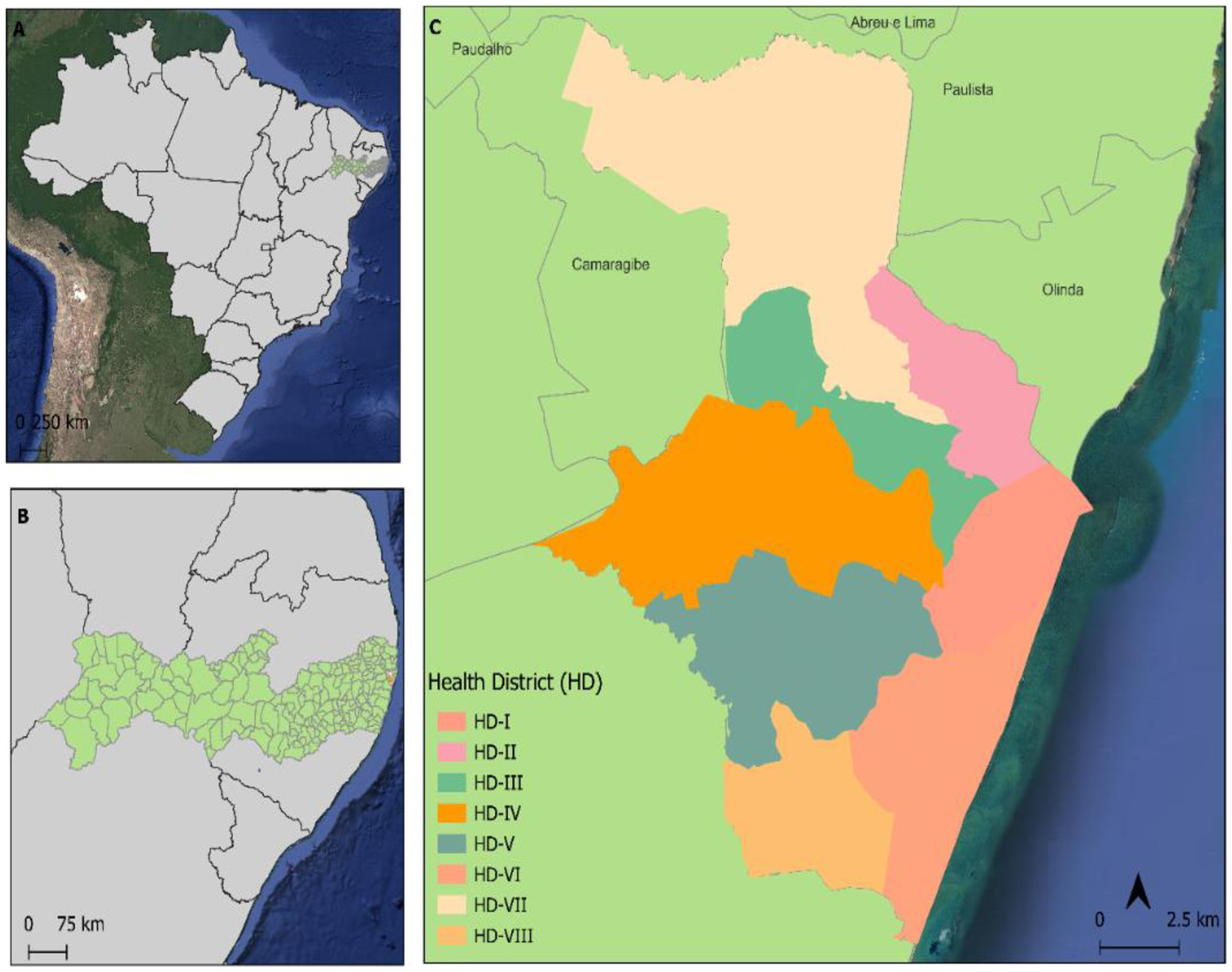
This cross-sectional study was approved by the Human Research Ethics Committee (protocol no. 50861320.7.0000.9547) and by the Ethics Committee on the Use of Animals (protocol no. 6600270921). Sampling was conducted between July 2022 and January 2023 in two Primary Health Care Units (PHCUs) located in Sanitary District III, Recife, northeastern Brazil (Figure 1).
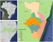
Map of the Sanitary Districts Division of Recife-PE. (A) Map of Brazil highlighting the state of Pernambuco; (B) Map of Pernambuco highlighting the Recife municipality (study area); (C) Detailed map of Recife showing the Health Districts (HD I–VIII).
2.2. Sampling and microbiological analysis
Samples were collected from three compartments linked to Primary Health Care Units (PHCUs): (i) people present in the PHCUs (healthcare professionals and patients/companions), (ii) surfaces and environmental equipment associated with healthcare within the PHCUs, and (iii) pets (dogs and cats) living in households within the PHCUs’ catchment area.
Samples were obtained from healthcare professionals and patients/companions who were present in the PHCUs during sample collection visits and agreed to participate. A total of 83 human swabs were collected from 60 individuals. Of these, 46 swabs corresponded to 23 participants from whom paired nasal and hand samples were obtained, while the remaining 37 swabs were collected from a single anatomical site.
For descriptive analyses, human isolates were summarized both at the swab level, according to sampling site (nasal or hand swabs), and at the participant level, according to participant category (healthcare workers or patients/companions).
Environmental and equipment samples were collected from frequently handled surfaces and objects within the PHCUs, including examination tables, procedure chairs, restrooms, computers, and break rooms. Environmental samples (n = 7) were collected from bathrooms, kitchens, consulting rooms, and treatment rooms. Equipment samples were collected from cell phones, tablets, computers, stethoscopes, and blood pressure monitors.
In addition, oropharyngeal samples were collected from dogs (n = 27) and cats (n = 19) that lived in the catchment area of the Primary Health Care Units (PHCUs) (i.e., neighboring residences/neighborhoods), and not within the PHCUs.
Samples were collected using sterile swabs applied directly to the selected sampling sites according to sample type. In humans, swabs were obtained from the nasal cavity and hands; in dogs and cats, oropharyngeal swabs were collected; and in environmental and equipment samples, swabs were applied to selected surfaces (Table 1). After collection, swabs were placed into labeled Falcon tubes containing 1% peptone water and transported to the Laboratory for the Diagnosis of Infectious Diseases (LDIC), Federal Rural University of Pernambuco (UFRPE), within 2 hours for microbiological processing.
2.3. Staphylococcus spp. isolation
Swabs were inoculated onto Mannitol Salt Agar (Difco Laboratories Inc., Detroit, USA) and incubated at 37 °C (±1 °C) for 24–48 h (Murray and Baron, 2003; Leite et al., 2023). Plates were inspected for growth, and one presumptive staphylococcal colony per sample was selected for Gram staining and catalase testing (Carter et al., 1989; Fan et al., 1995). Selected colonies were subcultured on Mannitol Salt Agar to obtain pure growth for downstream procedures. In parallel, isolates were inoculated in Brain Heart Infusion (BHI) broth (Difco Laboratories Inc., Detroit, USA) and incubated at 37 °C for 24 h, then cryopreserved at −80 °C in BHI with 20% glycerol (Winn and Koneman, 2006).
2.4. Identification of Staphylococcus species by MALDI-TOF MS
All presumptive Staphylococcus isolates were subjected to identification by MALDI-TOF MS following the laboratory’s established protocol, using direct deposition onto the MALDI target plate followed by application of the matrix solution. Protein spectra were acquired and compared against the reference database for species assignment (Seng et al., 2009; Croxatto et al., 2012). Isolates that did not yield a reliable species-level identification were recorded as not identified.
2.5. Statistical analysis
The dataset was organized in Microsoft Excel and analyzed in Python using pandas, SciPy, and statsmodels libraries. Descriptive analyses were initially performed to summarize the absolute frequencies of Staphylococcus species according to sample type. For human samples, species frequencies were further described according to anatomical sampling site (nasal and hand swabs) and according to participant category (healthcare workers and patients/companions). Species richness was defined as the number of Staphylococcus species identified per sample. Species richness was first described according to the six sample types. For inferential comparisons, these sample types were subsequently grouped into three major compartments: human, animal, and environmental. Mean richness values were calculated for each sample type, including human nasal swabs, human hand swabs, healthcare equipment samples, environmental surface samples, canine oropharyngeal swabs, and feline oropharyngeal swabs. Differences in mean species richness among sample types were assessed using one-way analysis of variance (ANOVA), adopting a significance level of 0.05. When the overall ANOVA was significant, post hoc pairwise comparisons were performed with adjustment for multiple testing (Fisher and Flowerdew, 1995; Thrusfield, 2007).
For broader ecological comparisons, samples were also grouped into three major compartments: human, animal, and environment. The human compartment included nasal and hand swabs collected from healthcare workers and patients/companions; the animal compartment included oropharyngeal swabs from dogs and cats; and the environmental compartment included swabs obtained from healthcare-associated surfaces and equipment within the PHCUs. Associations between Staphylococcus species and these sample groups were evaluated using contingency tables and the chi-square test; Fisher’s exact test was considered when expected cell counts were lower than 5 (Thrusfield, 2007).
Alpha diversity within each major compartment (human, animal, and environmental) was summarized using the Shannon and Simpson indices based on species frequencies. In addition, Multiple Correspondence Analysis (MCA) was performed using a presence/absence matrix of species by sample group to explore patterns of association among species and compartments. MCA results were displayed in two-dimensional plots. A p-value < 0.05 was considered statistically significant (Kim, 2017).
3. Results
In 23 individuals, paired samples were collected from the nares (nasal site) and from the hands, totaling 46 samples and enabling within-individual comparison of colonization sites. Staphylococcus epidermidis was isolated from both sites. In the remaining paired cases, different species were recovered between sites, with variable combinations.
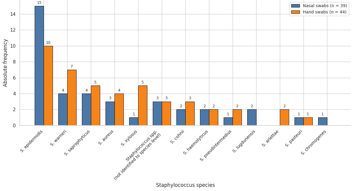
Figure 2 summarizes the Staphylococcus species identified according to sampling site, including 39 nasal swabs and 44 hand swabs. S. epidermidis was the most frequently detected species at both sites, being identified in 15/39 nasal samples and 10/44 hand samples. In nasal samples, other frequently identified species included S. warneri (4/39), S. saprophyticus (4/39), S. aureus (3/39), and Staphylococcus spp. not identified to species level (3/39). Additional species detected at lower frequencies included S. cohnii (2/39), S. haemolyticus (2/39), S. lugdunensis (2/39), S. pseudintermedius (1/39), S. pasteuri (1/39), and S. chromogenes (1/39).
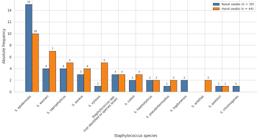
Hand samples showed higher frequencies of S. warneri (7/44), S. saprophyticus (5/44), S. xylosus (5/44), and S. aureus (4/44), in addition to Staphylococcus spp. not identified to species level (3/44), S. cohnii (3/44), S. haemolyticus (2/44), S. pseudintermedius (2/44), S. alfettae (2/44), and S. pasteuri (1/44). S. lugdunensis and S. chromogenes were detected only in nasal samples, whereas S. xylosus and S. alfettae were detected only in hand samples (Figure 2).
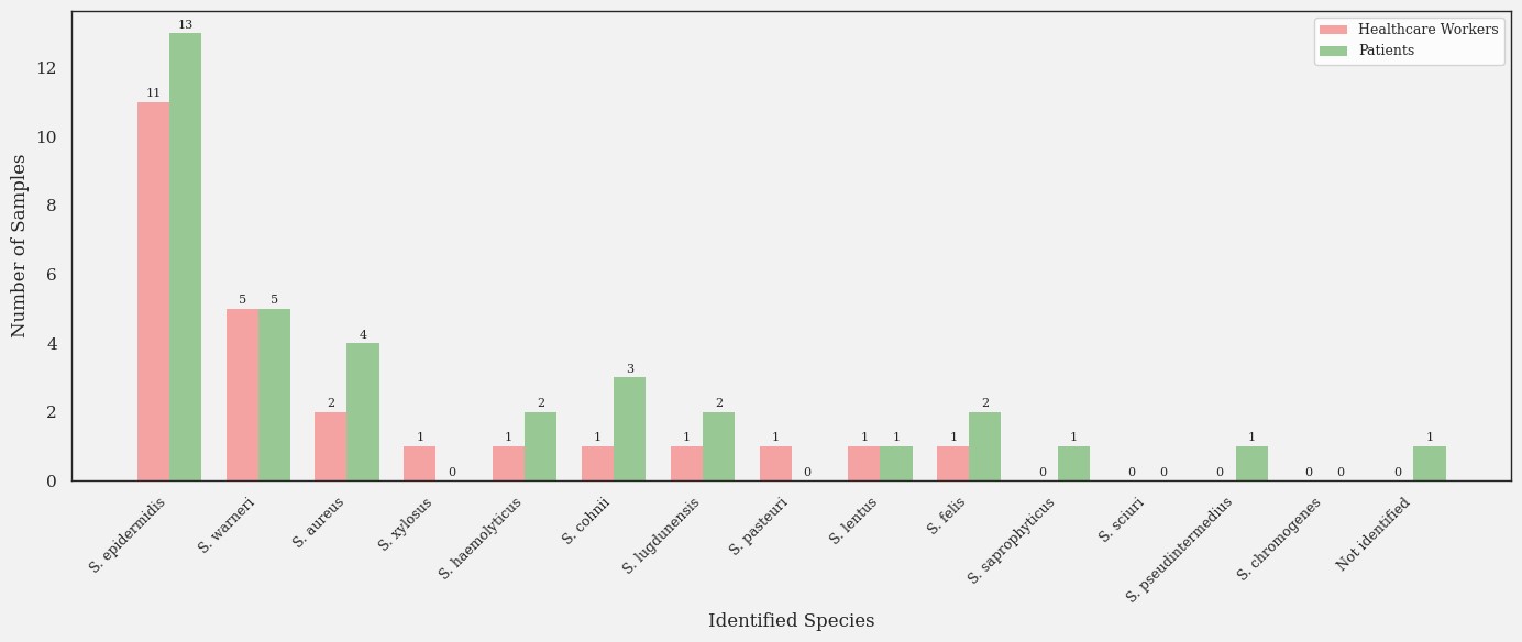
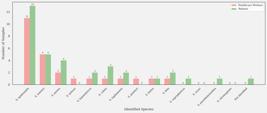
Among the human participants shown in Figure 3, 25 were healthcare workers and 35 were patients/companions, with distinct species distributions among groups. In healthcare workers, S. epidermidis was the most frequent species (11/25), followed by S. warneri (5/25) and S. aureus (2/25). Other species occurred sporadically (1/25 each), including S. xylosus, S. haemolyticus, S. cohnii, S. lugdunensis, S. pasteuri, S. lentus, and S. felis (Figure 3). Among patients/companions, S. epidermidis was also the most frequent species (13/35), followed by S. warneri (5/35), S. aureus (4/35), S. cohnii (3/35), and S. haemolyticus, S. lugdunensis, and S. felis (2/35 each). Less frequent findings included S. lentus, S. saprophyticus, and S. pseudintermedius (1/35 each), as well as one result not identified to species level (1/35) (Figure 3).
Oropharyngeal swabs collected from 19 cats yielded the following species: S. cohnii, S. epidermidis, Staphylococcus felis, Staphylococcus gallinarum, Staphylococcus pseudintermedius, S. sciuri, and Staphylococcus xylosus. Among the 27 oropharyngeal swabs obtained from dogs, the identified species were S. aureus, S. cohnii, S. epidermidis, S. felis, S. gallinarum, S. haemolyticus, Staphylococcus intermedius, S. pseudintermedius, S. saprophyticus, S. sciuri, Staphylococcus simulans, and S. xylosus.
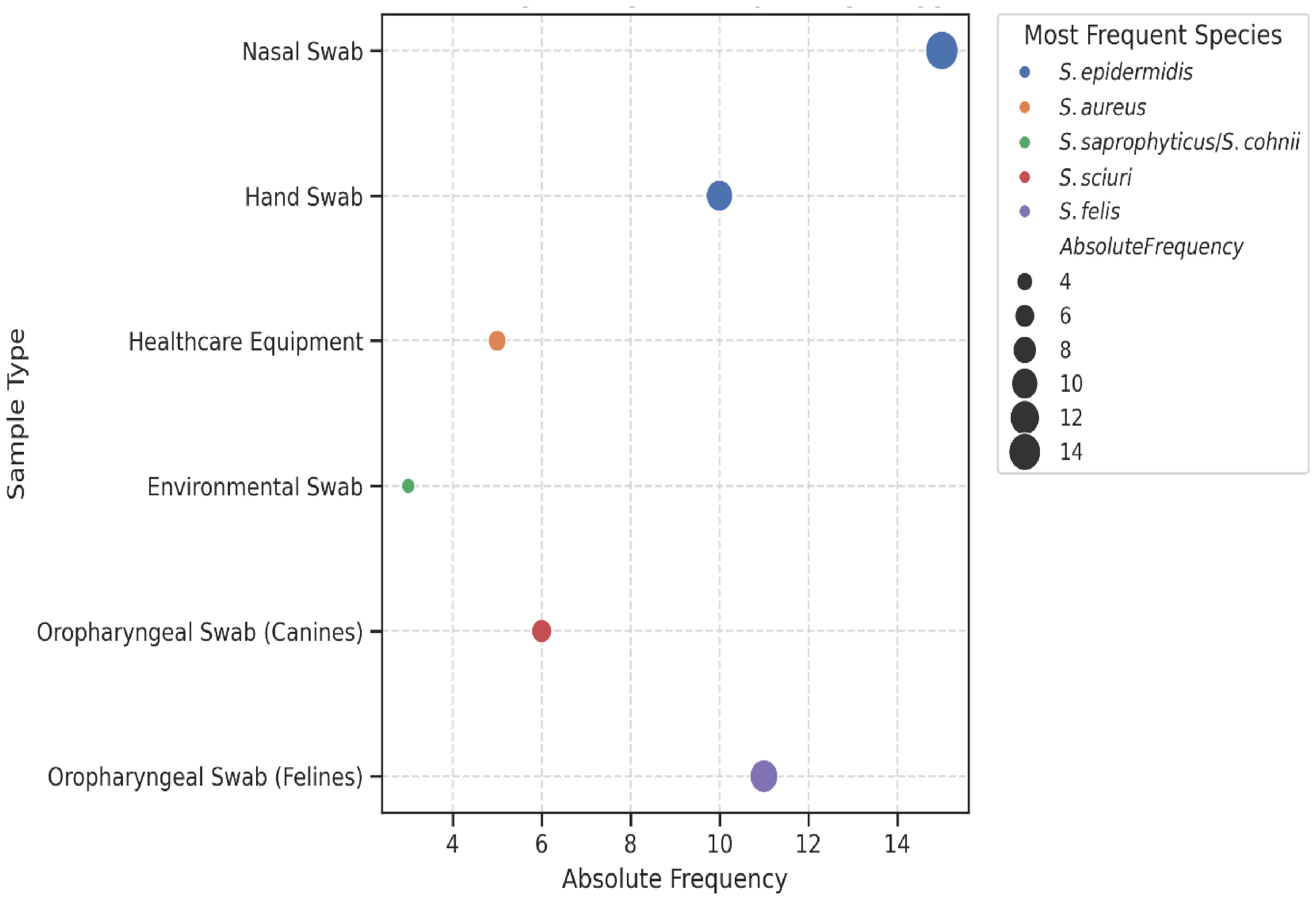
The absolute frequencies of the most prevalent Staphylococcus species varied according to sample type. In human samples, S. epidermidis predominated in both nasal and hand swabs. In healthcare equipment samples, S. aureus was the most frequently detected species. Among environmental swabs, S. saprophyticus and S. cohnii were the most prevalent species. In animal samples, S. sciuri was more frequently detected in dogs, whereas S. felis predominated in cats (Figure 4).
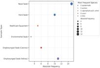
Among healthcare equipment samples collected in the primary healthcare units (n = 21), S. aureus was the most frequently detected species (5/21; 23.8%). S. saprophyticus and S. cohnii were each identified in 3/21 samples (14.3%). Among environmental swabs collected from PHCU areas (n = 7), the detected species included S. aureus, S. cohnii, and S. saprophyticus. In addition, three isolates from the environmental compartment, comprising healthcare equipment and environmental swabs, could not be conclusively identified by MALDI-TOF MS.
In human samples, comprising nasal and hand swabs, S. epidermidis was the most frequently detected species, followed by S. saprophyticus and S. aureus. When human isolates were stratified by anatomical site, S. epidermidis remained the predominant species in both nasal and hand swabs (Figure 2). When stratified by participant category, this species also predominated among both healthcare workers and patients/companions (Figure 3).
Among animal-derived samples (n = 46), corresponding to oropharyngeal swabs from dogs and cats, Staphylococcus felis was the most frequently isolated species and was predominantly detected in feline samples. This was followed by Staphylococcus sciuri (8/46; 17.4%) and Staphylococcus pseudointermedius (6/46; 13.0%). S. felis was more commonly found in cats, whereas S. sciuri was detected in both dogs and cats.
At the compartment level, S. sciuri was detected in both animal and environmental compartments, whereas S. pseudointermedius was identified exclusively in the animal compartment. These findings indicate overlap in species occurrence across compartments; however, transmission pathways and directionality cannot be inferred from the present data without genomic typing.
Species richness, defined as the number of Staphylococcus species identified per sample, varied among sample types. Nasal swabs showed a mean richness of 1.75 (±0.83) species, whereas hand swabs showed a mean richness of 1.45 (±0.69). Among environmental sample types, healthcare equipment samples exhibited the highest mean richness (2.10 ± 1.12), followed by environmental swabs (1.80 ± 0.78). Among animal samples, dogs showed greater richness (2.04 ± 0.89) than cats (1.63 ± 0.74). One-way ANOVA revealed significant differences in species richness among sample types (F = 5.94; p = 0.0161). Post hoc analysis indicated a significant difference between animal and environmental samples (p = 0.0148), whereas no significant differences were observed between human and animal samples (p = 0.0823) or between human and environmental samples (p = 0.6088).
When samples were grouped into the three major compartments (human, animal, and environmental), the chi-square test showed a significant association between Staphylococcus species distribution and sample group (χ2 = 46.05; df = 12; p < 0.0001). S. epidermidis was most frequently observed in human samples, S. pseudointermedius occurred exclusively in animal samples, and S. saprophyticus was more frequent in environmental samples.
Alpha diversity analysis showed lower diversity in the human compartment, with Shannon and Simpson indices of 0.875 and 0.444, respectively. Higher diversity values were observed in the animal compartment (Shannon = 1.245; Simpson = 0.674) and in the environmental compartment (Shannon = 1.200; Simpson = 0.643). Multiple correspondence analysis (Figure 5) produced an ordination pattern broadly consistent with these findings, with S. epidermidis more closely associated with the human compartment and S. pseudointermedius associated with the animal compartment, whereas the remaining species showed weaker or intermediate associations across compartments.

Multiple correspondence analysis of Staphylococcus species distribution across sample groups.
4. Discussion
This study characterizes the diversity and distribution of Staphylococcus spp. across a primary healthcare context, integrating human samples (healthcare workers and patients), companion animals living within the PHCU catchment area, and healthcare-associated environmental surfaces and equipment. Using MALDI-TOF MS for species-level identification, we observed a structured pattern across compartments. S. epidermidis predominated in human samples, S. aureus was most frequent among equipment-associated isolates, and animal-associated species such as S. felis, S. sciuri, and S. pseudointermedius were concentrated in companion animals. Together, these findings support the One Health rationale for considering humans, animals, and built environments as interconnected compartments in surveillance frameworks.
In humans, the predominance of S. epidermidis is consistent with its role as a common skin commensal and frequent cause of opportunistic, healthcare-associated infections (Otto, 2013; Becker et al., 2014). The higher frequency observed among healthcare workers compared with patients/companions may reflect repeated contact with the healthcare environment and high-touch surfaces, although the study design does not allow attribution of directionality or source. From a clinical standpoint, the relevance of S. epidermidis extends beyond colonization because strains adapted to healthcare settings are often associated with biofilm formation and device-related infections (Becker et al., 2014; Sabaté Brescó et al., 2017).
Beyond S. epidermidis, the detection of S. warneri, S. saprophyticus, S. aureus, S. lugdunensis, and S. pseudintermedius illustrates the heterogeneity of Staphylococcus spp. in primary healthcare interfaces. S. lugdunensis, despite being coagulase-negative, has been associated with clinical presentations that resemble those caused by S. aureus (Argemi et al., 2017). The identification of S. pseudintermedius, a species commonly linked to dogs and increasingly recognized in human infections, is relevant in One Health contexts because close human-animal contact may facilitate opportunities for exposure (Somayaji et al., 2016). However, the present dataset supports only the co-occurrence of species across compartments and does not demonstrate interspecies transmission.
Healthcare-associated surfaces and equipment yielded multiple Staphylococcus species, including S. aureus, S. epidermidis, and S. cohnii. This pattern is consistent with the role of fomites as potential contributors to microbial persistence and indirect exposure in clinical settings, particularly for organisms with environmental resilience and frequent hand-contact interfaces (Russotto et al., 2015; Peters et al., 2017). The presence of species more often described in animal hosts, such as S. cohnii, on equipment may suggest possible cross-compartment overlap, although this cannot be confirmed without strain-level typing, but confirmation would require strain-level typing.
In companion animals, the predominance of S. felis in cats and the detection of S. pseudintermedius and S. sciuri in dogs are aligned with known host associations. The concurrent detection of species commonly reported in humans, including S. epidermidis and S. aureus, in animal oropharyngeal samples highlights overlapping species pools at the human-animal interface (Vincze et al., 2014; Gómez-Sanz et al., 2019). This overlap is relevant for integrated surveillance, especially in settings where animals share households and daily contact with people, but the direction and frequency of transmission cannot be inferred without longitudinal sampling and genomic comparisons.
MALDI-TOF MS supported rapid species-level identification and enabled the recognition of less frequently reported taxa (e.g., S. alfettae and S. pasteuri), reinforcing its utility in routine surveillance workflows (Seng et al., 2009; Croxatto et al., 2012; Hou et al., 2019). Nonetheless, the occurrence of isolates that could not be conclusively identified underscores a known limitation related to database coverage and spectral representation, particularly for uncommon or recently described taxa (Kostrzewa et al., 2013; Patel, 2015). Continuous expansion and curation of reference libraries, alongside complementary confirmatory methods when needed, remain important to maximize diagnostic accuracy.
The One Health relevance of these findings becomes more apparent when considered alongside antimicrobial resistance (AMR). The presence of clinically relevant species across humans, animals, and healthcare-associated environments highlights interfaces where resistant strains could persist and be encountered by different hosts. Although this study was not designed to determine the origin of resistance determinants or transmission routes, its results provide baseline information to support future work that integrates phenotypic resistance, genomic typing, and contextual exposure data. In this regard, whole-genome sequencing (WGS) and comparative genomics would allow assessment of relatedness among isolates across compartments and would clarify epidemiological links not accessible through species-level identification alone (Tacconelli et al., 2018; Becker et al., 2017). Advances in MALDI-TOF MS applications for resistance screening and strain-level approaches may also complement routine surveillance when appropriately validated (Croxatto et al., 2012; Becker et al., 2017).
Finally, some limitations should be considered when interpreting these findings. This cross-sectional design provides a snapshot and does not capture temporal dynamics. Sampling was restricted to two PHCUs and to defined specimen types, and detailed information on exposures and contact networks was not available for all participants. In addition, species overlap across compartments should not be interpreted as evidence of transmission without genomic confirmation. Despite these constraints, the study documents a diverse Staphylococcus community spanning humans, companion animals, and healthcare-associated environments in a primary healthcare context, supporting the value of integrated surveillance under a One Health perspective.
5. Conclusion
This study documents the diversity and distribution of Staphylococcus spp. across human, companion animal, and healthcare-associated environmental samples in a primary healthcare context. S. epidermidis predominated in human samples, whereas S. aureus was frequently detected in healthcare equipment, and animal-associated species such as S. pseudointermedius and S. felis were concentrated in companion animals. The occurrence of overlapping species across compartments highlights relevant interfaces for integrated surveillance under a One Health perspective. However, species overlap should not be interpreted as evidence of transmission in the absence of genomic confirmation. Further studies combining longitudinal sampling and strain-level typing, including genomic approaches, are warranted to clarify epidemiological links and to better assess antimicrobial resistance dynamics across these compartments.
Data Availability Statement
All data generated or analyzed during this study are included in this published article.
References
- ARGEMI, X., HANSMANN, Y., PROLA, K. and PRÉVOST, G., 2017. Coagulase-negative Staphylococci pathogenomics. International Journal of Molecular Sciences, vol. 18, no. 8, pp. 1713.
-
BECKER, K., HEILMANN, C. and PETERS, G., 2014. Coagulase-negative Staphylococci. Clinical Microbiology Reviews, vol. 27, no. 4, pp. 870-926. https://doi.org/10.1128/CMR.00109-13 PMid:25278577.
» https://doi.org/10.1128/CMR.00109-13 - BECKER, K., SCHAUMBURG, F., FEGELER, C., FRIEDRICH, A.W. and KÖCK, R., 2017. Staphylococcus aureus from the German general population is highly diverse. International Journal of Medical Microbiology: IJMM, vol. 307, no. 1, pp. 21-27. PMid:28017539.
- CARTER, G.R., CLAUS, G.W. and RIKIHISA, Y., 1989. Fundamentos de bacteriología y micología veterinaria. Zaragoza: Acribia, 750 p.
-
CROXATTO, A., PROD'HOM, G. and GREUB, G., 2012. Applications of MALDI-TOF mass spectrometry in clinical diagnostic microbiology. FEMS Microbiology Reviews, vol. 36, no. 2, pp. 380-407. https://doi.org/10.1111/j.1574-6976.2011.00298.x PMid:22092265.
» https://doi.org/10.1111/j.1574-6976.2011.00298.x -
DELEO, F.R., OTTO, M., KREISWIRTH, B.N. and CHAMBERS, H.F., 2010. Community-associated meticillin-resistant Staphylococcus aureus. Lancet, vol. 375, no. 9725, pp. 1557-1568. https://doi.org/10.1016/S0140-6736(09)61999-1 PMid:20206987.
» https://doi.org/10.1016/S0140-6736(09)61999-1 - FAN, H.H., KLEVEN, S.H. and JACKWOOD, M.W., 1995. Application of polymerase chain reaction with arbitrary primers to strain identification of Mycoplasma gallisepticum. Avian Diseases, vol. 39, pp. 729-735.
- FISHER, M.M. and FLOWERDEW, A.D.J., 1995. An introduction to biostatistics 2nd ed. New York: Oxford University Press, 270 p.
-
GÓMEZ-SANZ, E., CEBALLOS, S., RUIZ-RIPA, L., ZARAZAGA, M. and TORRES, C., 2019. Clonally diverse methicillin and multidrug resistant coagulase negative Staphylococci are ubiquitous and pose transfer ability between pets and their owners. Frontiers in Microbiology, vol. 10, pp. 485. https://doi.org/10.3389/fmicb.2019.00485 PMid:30972035.
» https://doi.org/10.3389/fmicb.2019.00485 - HOU, T.Y., CHIANG-NI, C. and TENG, S.H., 2019. Current status of MALDI-TOF mass spectrometry in clinical microbiology. Yao Wu Shi Pin Fen Xi, vol. 27, no. 2, pp. 404-414. PMid:30987712.
- KIM, J., 2017. Exploratory data analysis using multiple correspondence analysis (MCA) and principal components analysis (PCA). Annals of Translational Medicine, vol. 5, no. 5, pp. 103.
-
KOSTRZEWA, M., SPARBIER, K., MAIER, T. and SCHUBERT, S., 2013. MALDI-TOF MS: an upcoming tool for rapid detection of antibiotic resistance in microorganisms. Proteomics. Clinical Applications, vol. 7, no. 11-12, pp. 767-778. https://doi.org/10.1002/prca.201300042 PMid:24123965.
» https://doi.org/10.1002/prca.201300042 -
LEITE, D.P.S.B.M., BARBOSA, I.C., SILVA, R.A., FERNANDES, P.R., ABAD, A.C.A., SILVA, J.G., MOTA, R.A. and PORTO, T.S., 2023. Occurrence of antimicrobial-resistant Staphylococcus aureus in a Brazilian veterinary hospital environment. Brazilian Journal of Microbiology, vol. 54, no. 3, pp. 2393-2401. https://doi.org/10.1007/s42770-023-01035-w PMid:37407882.
» https://doi.org/10.1007/s42770-023-01035-w - MURRAY, P.R. and BARON, E.J., 2003. Manual of clinical microbiology. Washington, D.C.: ASM Press.
-
OTTO, M., 2012. MRSA virulence and spread. Cellular Microbiology, vol. 14, no. 10, pp. 1513-1521. https://doi.org/10.1111/j.1462-5822.2012.01832.x PMid:22747834.
» https://doi.org/10.1111/j.1462-5822.2012.01832.x -
OTTO, M., 2013. Coagulase-negative Staphylococci as reservoirs of genes facilitating MRSA infection. BioEssays: News and Reviews in Molecular, Cellular and Developmental Biology, vol. 35, no. 1, pp. 4-11. https://doi.org/10.1002/bies.201200112 PMid:23165978.
» https://doi.org/10.1002/bies.201200112 -
PATEL, R., 2015. MALDI-TOF MS for the diagnosis of infectious diseases. Clinical Chemistry, vol. 61, no. 1, pp. 100-111. https://doi.org/10.1373/clinchem.2014.221770 PMid:25278500.
» https://doi.org/10.1373/clinchem.2014.221770 - PETERS, G., BECKER, K. and HEILMANN, C., 2017. The changing face of Staphylococcus aureus in healthcare settings. The Journal of Hospital Infection, vol. 95, no. 4, pp. 385-391.
- PRESCOTT, R.F., BAGGOT, J.D. and WALKER, R.D., 2013. Antimicrobial therapy in veterinary medicine. 5th ed. Ames: Wiley-Blackwell, 704 p.
-
RUSSOTTO, V., CORTEGIANI, A., RAINERI, S.M. and GIARRATANO, A., 2015. Bacterial contamination of inanimate surfaces and equipment in the intensive care unit. Journal of Intensive Care, vol. 3, no. 1, pp. 54. https://doi.org/10.1186/s40560-015-0120-5 PMid:26693023.
» https://doi.org/10.1186/s40560-015-0120-5 -
SABATÉ BRESCÓ, M., HARRIS, L.G., THOMPSON, K., STANIC, B., MORGENSTERN, M., O’MAHONY, L., RICHARDS, R.G. and MORIARTY, T.F., 2017. Pathogenic mechanisms and host interactions in Staphylococcus epidermidis device-related infection. Frontiers in Microbiology, vol. 8, pp. 1401. https://doi.org/10.3389/fmicb.2017.01401 PMid:28824556.
» https://doi.org/10.3389/fmicb.2017.01401 -
SENG, P., DRANCOURT, M., GOURIET, F., LA SCOLA, B., FOURNIER, P.-E., ROLAIN, J.M. and RAOULT, D., 2009. Ongoing revolution in bacteriology: routine identification of bacteria by MALDI-TOF MS. Clinical Infectious Diseases, vol. 49, no. 4, pp. 543-551. https://doi.org/10.1086/600885 PMid:19583519.
» https://doi.org/10.1086/600885 -
SOMAYAJI, R., PRIYANTHA, M.A., RUBIN, J.E. and CHURCH, D., 2016. Human infections due to Staphylococcus pseudintermedius, an emerging zoonosis of canine origin: report of 24 cases. Diagnostic Microbiology and Infectious Disease, vol. 85, no. 4, pp. 471-476. https://doi.org/10.1016/j.diagmicrobio.2016.05.008 PMid:27241371.
» https://doi.org/10.1016/j.diagmicrobio.2016.05.008 -
TACCONELLI, E., CARRARA, E., SAVOLDI, A., HARBARTH, S., MENDELSON, M., MONNET, D.L., PULCINI, C., KAHLMETER, G., KLUYTMANS, J., CARMELI, Y., OUELLETTE, M., OUTTERSON, K., PATEL, J., CAVALERI, M., COX, E.M., HOUCHENS, C.R., GRAYSON, M.L., HANSEN, P., SINGH, N., THEURETZBACHER, U., MAGRINI, N., ABODERIN, A.O., AL-ABRI, S.S., AWANG JALIL, N., BENZONANA, N., BHATTACHARYA, S., BRINK, A.J., BURKERT, F.R., CARS, O., CORNAGLIA, G., DYAR, O.J., FRIEDRICH, A.W., GALES, A.C., GANDRA, S., GISKE, C.G., GOFF, D.A., GOOSSENS, H., GOTTLIEB, T., GUZMAN BLANCO, M., HRYNIEWICZ, W., KATTULA, D., JINKS, T., KANJ, S.S., KERR, L., KIENY, M.-P., KIM, Y.S., KOZLOV, R.S., LABARCA, J., LAXMINARAYAN, R., LEDER, K., LEIBOVICI, L., LEVY-HARA, G., LITTMAN, J., MALHOTRA-KUMAR, S., MANCHANDA, V., MOJA, L., NDOYE, B., PAN, A., PATERSON, D.L., PAUL, M., QIU, H., RAMON-PARDO, P., RODRÍGUEZ-BAÑO, J., SANGUINETTI, M., SENGUPTA, S., SHARLAND, M., SI-MEHAND, M., SILVER, L.L., SONG, W., STEINBAKK, M., THOMSEN, J., THWAITES, G.E., VAN DER MEER, J.W.M., VAN KINH, N., VEGA, S., VILLEGAS, M.V., WECHSLER-FÖRDÖS, A., WERTHEIM, H.F.L., WESANGULA, E., WOODFORD, N., YILMAZ, F.O. and ZORZET, A., 2018. WHO priority list of antibiotic-resistant bacteria and tuberculosis. The Lancet. Infectious Diseases, vol. 18, no. 3, pp. 318-327. https://doi.org/10.1016/S1473-3099(17)30753-3 PMid:29276051.
» https://doi.org/10.1016/S1473-3099(17)30753-3 - THRUSFIELD, M., 2007. Veterinary epidemiology. 3rd ed. Oxford: Blackwell Science, 610 p.
-
TONG, S.Y., DAVIS, J.S., EICHENBERGER, E., HOLLAND, T.L. and FOWLER JUNIOR, V.G., 2015. Staphylococcus aureus infections: epidemiology, pathophysiology, clinical manifestations, and management. Clinical Microbiology Reviews, vol. 28, no. 3, pp. 603-661. https://doi.org/10.1128/CMR.00134-14 PMid:26016486.
» https://doi.org/10.1128/CMR.00134-14 -
VINCZE, S., STAMM, I., KOPP, P.A., HERMES, J., ADLHOCH, C., SEMMLER, T., WIELER, L.H., LÜBKE-BECKER, A. and WALTHER, B., 2014. Alarming proportions of MRSA in wound samples from companion animals, Germany 2010-2012. PLoS One, vol. 9, no. 1, e85656. https://doi.org/10.1371/journal.pone.0085656 PMid:24465637.
» https://doi.org/10.1371/journal.pone.0085656 - WINN, W.C. and KONEMAN, E.W., 2006. Atlas and textbook of diagnostic microbiology Philadelphia: Lippincott, 1550 p.
Edited by
-
Editor:
Marcelo A.M. Esquisatto