 In vitro and in vivo biocontrol potential of Pseudomonas aeruginosa Bac4 against Fusarium equiseti causing leaf spot disease in Saudi mangroves
Abstract
Mangrove ecosystems provide essential ecological services, including coastal protection, carbon sequestration, and biodiversity conservation, yet they are increasingly threatened by emerging plant diseases. During a disease survey conducted in February 2024 in the Sihat mangrove ecosystem (Eastern Saudi Arabia), Avicennia marina leaves exhibiting necrotic leaf spot symptoms were collected and examined. Based on morphological characteristics and ITS rDNA sequence analysis, the causal agent was identified as Fusarium equiseti, representing the first report of this pathogen causing leaf spot disease in Saudi mangroves. Pathogenicity was confirmed by fulfilling Koch’s postulates on detached healthy leaves. To explore environmentally sustainable disease management strategies, a soil-derived bacterial strain (Bac4) was evaluated for antifungal activity. Molecular identification using 16S rRNA gene sequencing revealed 100% similarity with Pseudomonas aeruginosa. In vitro assays demonstrated strong antagonistic activity of Bac4 against F. equiseti. The cell-free supernatant (CFS) significantly inhibited mycelial growth, biomass production, and spore germination in a dose-dependent manner, achieving up to 65.74% radial growth inhibition at a 50% CFS concentration. Microscopic observations revealed severe hyphal deformation, swelling, and cytoplasmic disintegration in treated fungi. Volatile organic compounds (VOCs) emitted by Bac4 also suppressed fungal growth and pigmentation, and this effect was partially reversed by activated charcoal, confirming VOC-mediated antifungal activity. Gas chromatography–mass spectrometry (GC–MS) analysis of the CFS identified several bioactive metabolites, including alcohols, diketopiperazines, and actinomycin derivatives. In vivo assays using detached mangrove leaves demonstrated a disease control efficiency of 70.33%, exceeding that of the commercial fungicide Hymexazol. Overall, the results indicate that P. aeruginosa Bac4 produces both diffusible and volatile antifungal metabolites and represents a promising biocontrol agent for the sustainable management of leaf spot disease in mangrove ecosystems.
Keywords:
Avicennia marina; Fusarium equiseti; Pseudomonas aeruginosa; biocontrol; antifungal metabolites; volatile organic compounds; mangrove disease
Resumo
Os ecossistemas de manguezais desempenham um papel essencial na proteção costeira, no sequestro de carbono e na conservação da biodiversidade, porém estão cada vez mais ameaçados por doenças fúngicas. Durante um levantamento fitossanitário realizado em fevereiro de 2024 no ecossistema de manguezais de Saihat, no leste da Arábia Saudita, folhas de Avicennia marina com sintomas de mancha necrótica foram coletadas e analisadas. As análises morfológicas e moleculares identificaram Fusarium equiseti como agente causal, constituindo o primeiro registro desse patógeno causando doença foliar em manguezais da Arábia Saudita. A patogenicidade foi confirmada pelo cumprimento dos postulados de Koch em folhas saudáveis destacadas. Com o objetivo de avaliar estratégias sustentáveis de manejo da doença, uma linhagem bacteriana (Bac4), isolada de solo agrícola, foi testada quanto à atividade antifúngica. A identificação molecular baseada no sequenciamento do gene 16S rRNA revelou 100% de similaridade com Pseudomonas aeruginosa. Ensaios in vitro demonstraram forte antagonismo contra F. equiseti. O sobrenadante livre de células (CFS) inibiu significativamente o crescimento radial, a biomassa e a germinação de esporos do fungo de forma dependente da concentração, alcançando até 65,74% de inibição a 50% de CFS. Observações microscópicas revelaram deformações hifais severas, inchaço e desintegração citoplasmática. Os compostos orgânicos voláteis (VOCs) produzidos pela Bac4 também reduziram o crescimento e a pigmentação do fungo, efeitos parcialmente revertidos pelo carvão ativado, confirmando a ação antifúngica mediada por VOCs. A análise por cromatografia gasosa acoplada à espectrometria de massas (GC–MS) identificou diversos metabólitos bioativos no CFS, incluindo álcoois, dicetopiperazinas e derivados de actinomicina. Ensaios in vivo em folhas destacadas de mangue demonstraram eficiência de controle da doença de 70,33%, superior ao fungicida comercial Hymexazol. Os resultados indicam que P. aeruginosa Bac4 possui elevado potencial como agente de biocontrole sustentável contra F. equiseti em manguezais.
Palavras-chave:
Avicennia marina; Fusarium equiseti; Pseudomonas aeruginosa; biocontrole; metabólitos antifúngicos; compostos orgânicos voláteis; doenças de manguezais
1. Introduction
Mangrove forests are among the most productive coastal ecosystems, providing essential ecological services such as shoreline stabilization, carbon sequestration, nutrient cycling, and habitat provision for diverse biological communities. Despite their ecological and economic importance, mangrove ecosystems are increasingly exposed to anthropogenic pressures, including coastal development, pollution, and climate-driven stressors, which can compromise plant health and ecosystem resilience (Alongi, 2022). In this context, emerging plant diseases represent an additional and often underappreciated threat to mangrove sustainability.
In Saudi Arabia, mangrove vegetation is dominated by Avicennia marina, which forms narrow but ecologically significant stands along the Arabian Gulf coastline. Although large-scale mangrove loss in the region has been relatively limited, increasing evidence suggests that local environmental stressors may predispose mangrove trees to biotic agents, particularly fungal pathogens. Foliar diseases such as leaf spot are of particular concern, as they directly affect photosynthetic tissues, accelerate leaf senescence, and may reduce overall plant vigor. However, information on the diversity, pathogenicity, and ecological relevance of mangrove-associated fungal pathogens in the Arabian Gulf region remains scarce. Members of the genus Fusarium are widely distributed phytopathogens known to infect a broad range of hosts, causing diseases such as wilts, rots, and leaf spots. Fusarium equiseti has been reported as a pathogen of several agricultural and wild plant species, yet its association with mangrove leaf spot disease has not been previously documented in Saudi Arabia. Accurate identification of fungal pathogens and confirmation of their pathogenicity are therefore critical for understanding disease etiology and for developing effective management strategies. Conventional disease control in plant systems often relies on chemical fungicides; however, their use in sensitive coastal environments raises concerns regarding environmental contamination, non-target effects, and the development of resistant pathogen populations. Consequently, increasing attention has been directed toward biologically based control approaches that are environmentally compatible and sustainable. Among these, soil- and plant-associated bacteria have attracted considerable interest due to their ability to suppress fungal pathogens through the production of diffusible secondary metabolites and volatile organic compounds (VOCs). Species of the genus Pseudomonas are well recognized for their antagonistic activity against phytopathogenic fungi, mediated by a diverse array of antimicrobial compounds, including antibiotics, siderophores, lytic enzymes, and VOCs. These metabolites can inhibit fungal growth, interfere with spore germination, and induce structural damage to hyphae. Importantly, bacterial metabolites may provide disease suppression while minimizing ecological disturbance, making them attractive candidates for biocontrol applications in natural ecosystems.
The present study aimed to (i) isolate and identify the fungal pathogen associated with leaf spot disease on Avicennia marina in the Sihat mangrove ecosystem of Eastern Saudi Arabia, (ii) confirm its pathogenicity through Koch’s postulates, and (iii) evaluate the antifungal potential of metabolites produced by a soil-derived bacterial strain identified as Pseudomonas aeruginosa Bac4. An integrated approach combining in vitro assays, volatile-mediated inhibition, chemical profiling of metabolites, and in vivo evaluation on detached mangrove leaves was employed. By providing the first detailed documentation of F. equiseti as a causal agent of mangrove leaf spot disease in Saudi Arabia and demonstrating the biocontrol potential of bacterial metabolites, this study contributes to mangrove disease ecology and supports the development of sustainable management strategies for coastal forest conservatio
2. Materials and Methods
2.1. Sample collection and disease survey
Field sampling was conducted on 6 February 2024 in the Sihat mangrove ecosystem (26°30′13.8″ N, 50°02′36.9″ E), Eastern Saudi Arabia. The study area was stratified into three ecological zones (shoreline, mid-zone, and inland) to account for spatial variability in disease occurrence. From each zone, 50 Avicennia marina leaves showing varying degrees of disease severity were randomly collected. Disease severity was assessed using a 0–5 rating scale as described by Rath and Meher (2019). Collected samples were placed in sterile bags, transported on ice, and processed within 24 h.
2.2. Isolation, purification, and preservation of fungal pathogen
Leaf segments (approximately 2 × 2 cm) excised from the margins of symptomatic tissue were surface-sterilized by immersion in 70% ethanol for 30 s, followed by two rinses with sterile distilled water. The disinfected tissues were placed on potato dextrose agar (PDA; Sigma-Aldrich) supplemented with lactic acid (1 mL L−1) to suppress bacterial growth. Plates were incubated at 29 °C for 7 days (Thilagam et al., 2018). Emerging fungal colonies with distinct morphological features were subcultured repeatedly on fresh PDA to obtain pure isolates. Pure cultures were preserved in 15% (v/v) sterile glycerol and stored at −80 °C for long-term maintenance (Ozerskaya et al., 2022).
2.3. Pathogenicity test
Pathogenicity was evaluated using detached healthy A. marina leaves following Koch’s postulates. Leaves were surface-sterilized and slightly wounded with a sterile needle. A conidial suspension (1 × 10⁶ spores mL−1) prepared from 7-day-old PDA cultures was applied to the wound sites. Control leaves received sterile distilled water. Each treatment consisted of six replicates incubated in sealed humid chambers at 20 °C in the dark for 7 days. Symptom development was monitored daily, and lesion formation was recorded to confirm pathogenicity (Brauna-Morževska et al., 2023).
2.4. Morphological characterization
Colony morphology was assessed on PDA after incubation at 29 °C, with observations including colony color, texture, and growth pattern. Microscopic features were examined using the slide culture technique (Prakash and Bhargava, 2016). Agar blocks inoculated with the fungal isolate were incubated in a moist chamber for 7 days, stained with methyl blue, and examined under a light microscope at 10×, 40×, and 100× magnifications to observe hyphal and conidial structures (Mustafa et al., 2023).
2.5. Molecular identification of the fungal isolate
Genomic DNA was extracted from actively growing mycelium using a modified Dellaporta method (Fazhan et al., 2016).The internal transcribed spacer (ITS) region of rDNA was amplified using primers ITS5 and ITS4. PCR products were purified and sequenced commercially. Obtained sequences were compared with reference sequences in the NCBI GenBank database using BLASTn. Phylogenetic analysis was performed using MEGA X software to confirm species-level identification (Al-Nadabi et al., 2020).
2.6. Bacterial isolate and molecular identification
The bacterial strain Bac4 was isolated from agricultural soil and screened for antifungal activity. Molecular identification was conducted by amplification and sequencing of the 16S rRNA gene. Sequence similarity was determined using BLASTn, and phylogenetic relationships were inferred using maximum likelihood analysis in MEGAX.
2.7. In vitro antifungal assays
2.7.1. Dual culture assay
Antagonistic activity of Bac4 against Fusarium equiseti was evaluated using a dual culture assay on PDA. A 5-mm fungal mycelial plug was placed at the center of the plate, while the bacterial isolate was streaked near the periphery. Plates were incubated at 29 °C for 7 days, and inhibition of fungal growth was recorded (Sabry, 2016).
2.7.2. Growth kinetics and cell-free supernatant preparation
Pseudomonas aeruginosa Bac4 was cultivated in nutrient broth at 28 °C with shaking at 200 rpm. Bacterial growth was monitored by measuring optical density at 600 nm (OD600) at 24-h intervals. At selected time points, cultures were centrifuged, and the supernatant was sterilized by sequential filtration through 0.45 µm and 0.2 µm membrane filters to obtain cell-free supernatant (CFS) (Dullah et al., 2021).
2.7.3. Radial growth inhibition assay
The antifungal activity of CFS was assessed by incorporating CFS into PDA at final concentrations of 15%, 25%, 35%, and 50% (v/v). Plates were inoculated with 7-day-old fungal plugs and incubated at 29 °C. Colony diameters were measured along two perpendicular axes once control plates reached full growth. To distinguish fungistatic from fungicidal effects, inhibited fungal plugs were transferred to fresh PDA and monitored for regrowth (Lee et al., 2019).
2.7.4. Biomass reduction assay
The effect of CFS on fungal biomass was evaluated by measuring wet and dry weights. Flasks containing nutrient broth supplemented with CFS (15–50%, v/v) were inoculated with a fungal spore suspension (1 × 10⁷ spores mL−1) and incubated under optimal conditions. Biomass was harvested by filtration, weighed fresh, then dried at 60–70 °C for 24–48 h before final weighing (Slowik et al., 2023).
2.7.5. Spore germination inhibition
Fungal spores were incubated with CFS, and germination was assessed microscopically after 24 h. The percentage inhibition of spore germination was calculated relative to untreated controls (Ji et al., 2018).
2.7.6. Volatile Organic Compound (VOC) assay
The antifungal activity of volatile organic compounds (VOCs) produced by Bac4 was evaluated using the sealed plate (sandwich) method. PDA plates inoculated with the fungal pathogen were paired with nutrient agar plates containing the bacterial culture. Plates were sealed to allow VOC diffusion without physical contact. Activated charcoal was included in selected treatments to adsorb VOCs. Plates were incubated at 29 °C until control colonies reached full growth (Yang et al., 2017).
2.8. GC–MS analysis of bacterial metabolites
Bioactive metabolites in the CFS were extracted using ethyl acetate and analyzed by gas chromatography–mass spectrometry (GC–MS). Compounds were identified by comparison with mass spectral libraries (Pringgenies et al., 2021).
2.9. In vivo evaluation on detached mangrove leaves
Detached A. marina leaves were surface-disinfected, wounded, and treated with either 50% CFS or a commercial fungicide. Preventive and curative treatments were applied, and inoculated and uninoculated controls were included. Leaves were incubated at 29 °C under high humidity, and disease severity was scored using a 0–5 scale. Disease control efficacy was calculated relative to untreated controls (Wang et al., 2011).
2.10. Statistical analysis
All experiments were conducted in triplicate. Data were analyzed using IBM SPSS Statistics 26 and expressed as mean ± standard deviation. Treatment effects were evaluated for statistical significance at p < 0.05. Phylogenetic analyses were performed using MEGA softwar
3. Result
3.1. Study area and sample collection
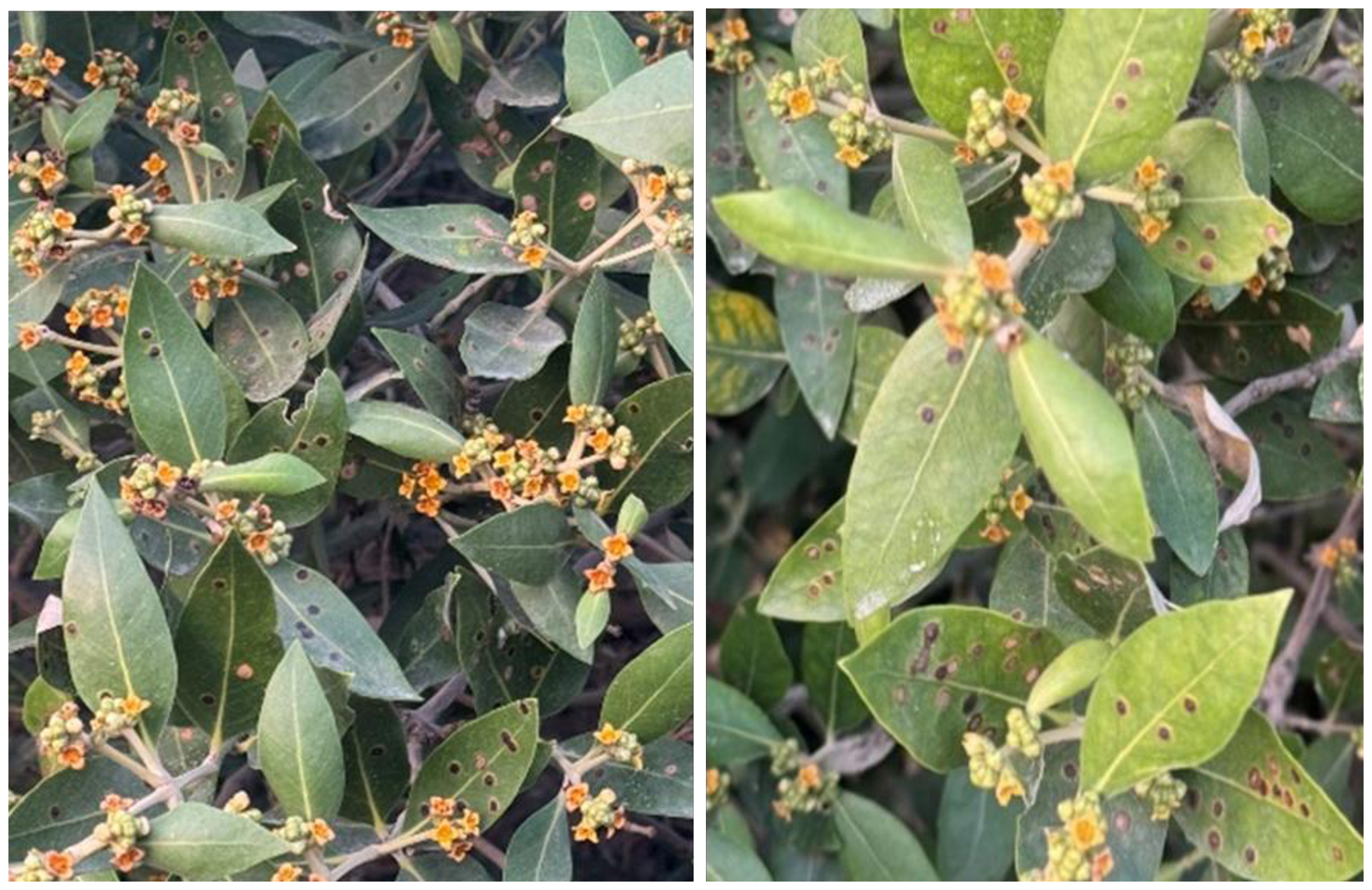
Field surveys conducted in the Sihat mangrove ecosystem revealed clear symptoms of leaf spot disease on Avicennia marina. Symptoms progressed from small, circular whitish lesions to enlarged, irregular necrotic spots surrounded by chlorotic tissue. Advanced stages were characterized by blackened necrotic areas, indicating active disease development. Leaves representing different stages of symptom severity were collected for laboratory analyses (Figure 1).

Representative Leaf Spot Symptoms on Mangrove Leaves. Mangrove leaves exhibit leaf spot disease, Lesions start as small white spots often representing the early stage of infection,and turn to black irregular necrotic spots as the disease development and tissue degradation occurs and this indicate fungus active sporulation.
3.2. Pathogenicity assessment
Following artificial inoculation of detached healthy mangrove leaves, symptoms identical to those observed in the field developed within seven days. Inoculated leaves exhibited expanding necrotic lesions, whereas control leaves remained asymptomatic. Re-isolation of the fungus from symptomatic tissues fulfilled Koch’s postulates, confirming the pathogenicity of the isolate (Figure 2).

Pathogenicity tests of fungal isolates on mangrove leaves seven days after inoculation. (c) Healthy control leaves remained asymptomatic, whereas inoculation with isolated fungi resulted in the formation of characteristic black spot lesions, confirming their pathogenic potential.
3.3. Morphological characterization of the fungal isolate
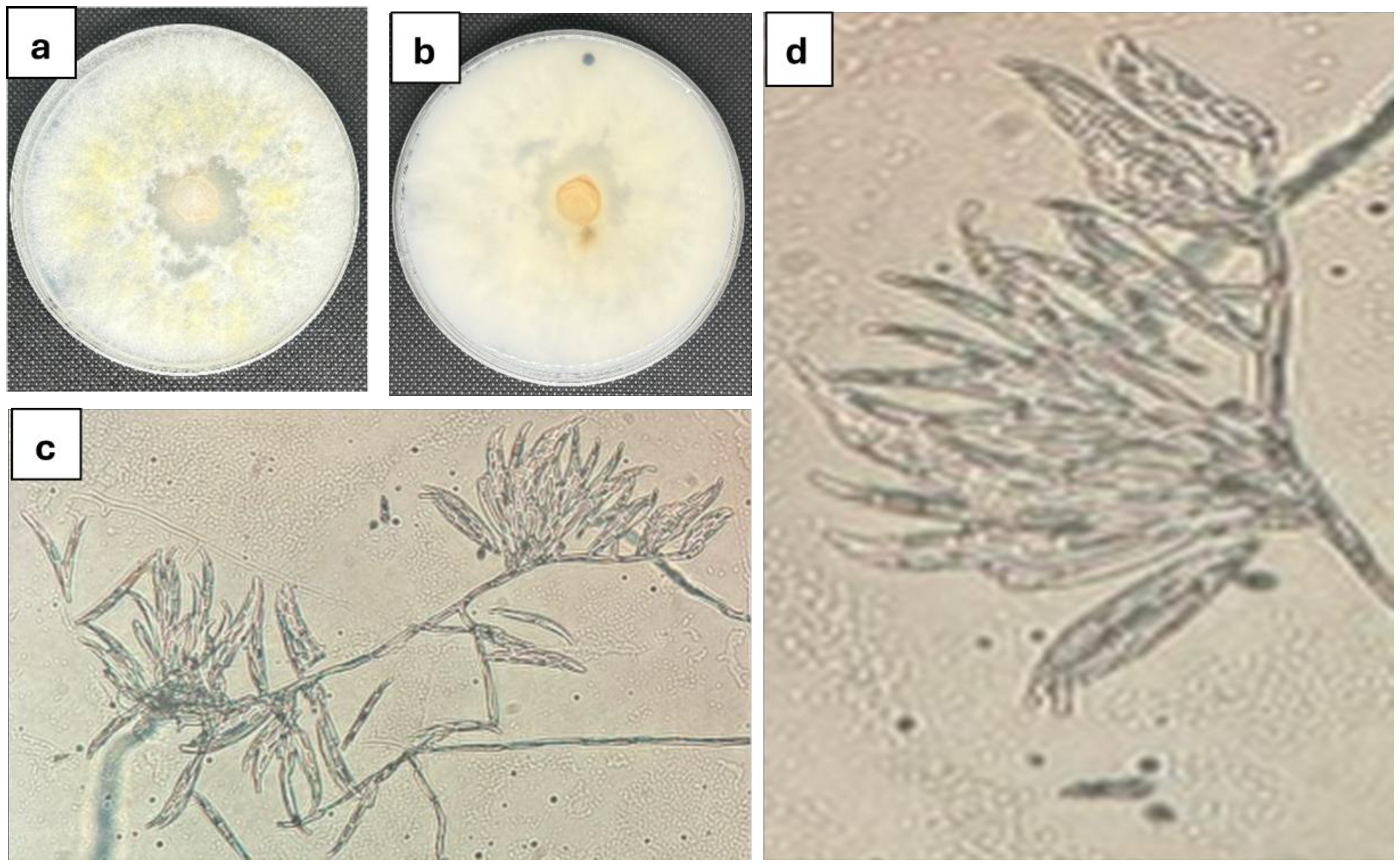
On potato dextrose agar, the fungal isolate produced rapidly growing colonies with distinct pigmentation and surface texture. Microscopic examination revealed septate hyphae and characteristic multicellular conidia consistent with Fusarium species. These morphological features supported the preliminary identification of the isolate as Fusarium equiseti (Figure 3).

Cultural morphology of fungal isolate on potato dextrose agar after 5 days’ incubation at 29◦C (a) front view and (b) back view. (c))Microscopic examination of the fungal isolateunder a light microscope (400×) revealed Multicellular conidia were formed in branched acropetal.
3.4. Molecular identification of the fungal pathogen
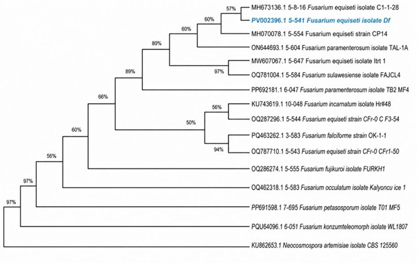
Amplification and sequencing of the ITS rDNA region confirmed the identity of the fungal isolate as Fusarium equiseti. The obtained sequence showed 100% similarity with reference sequences deposited in the NCBI GenBank database (accession OQ629143). Phylogenetic analysis clustered the isolate within a well-supported F. equiseti clade, confirming species-level identification (Figure 4).

Phylogenetic Analysis of Fusarium equiseti (Isolate PV022350.1) Based on ITS Sequences Using Maximum Likelihood Method.
3.5. Antagonistic activity of bacterial isolate Bac4
Dual culture assays demonstrated strong antagonistic activity of Bac4 against F. equiseti. Fungal radial growth was markedly restricted, and a distinct inhibition zone developed toward the bacterial streak after seven days of incubation at 29 °C. Molecular identification based on 16S rRNA gene sequencing revealed 100% similarity of Bac4 to Pseudomonas aeruginosa (GenBank accession KU937381), confirming its taxonomic identity (Figure 5).
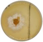
Dual-culture assay showing the antagonistic activity of Bac4 against F. equiseti on PDA after 7 days at 29 °C. Fungal growth was inhibited with a clear zone of inhibition toward Bac4, indicating strong antifungal activiy.
3.6. Phylogenetic analysis of bacterial isolate
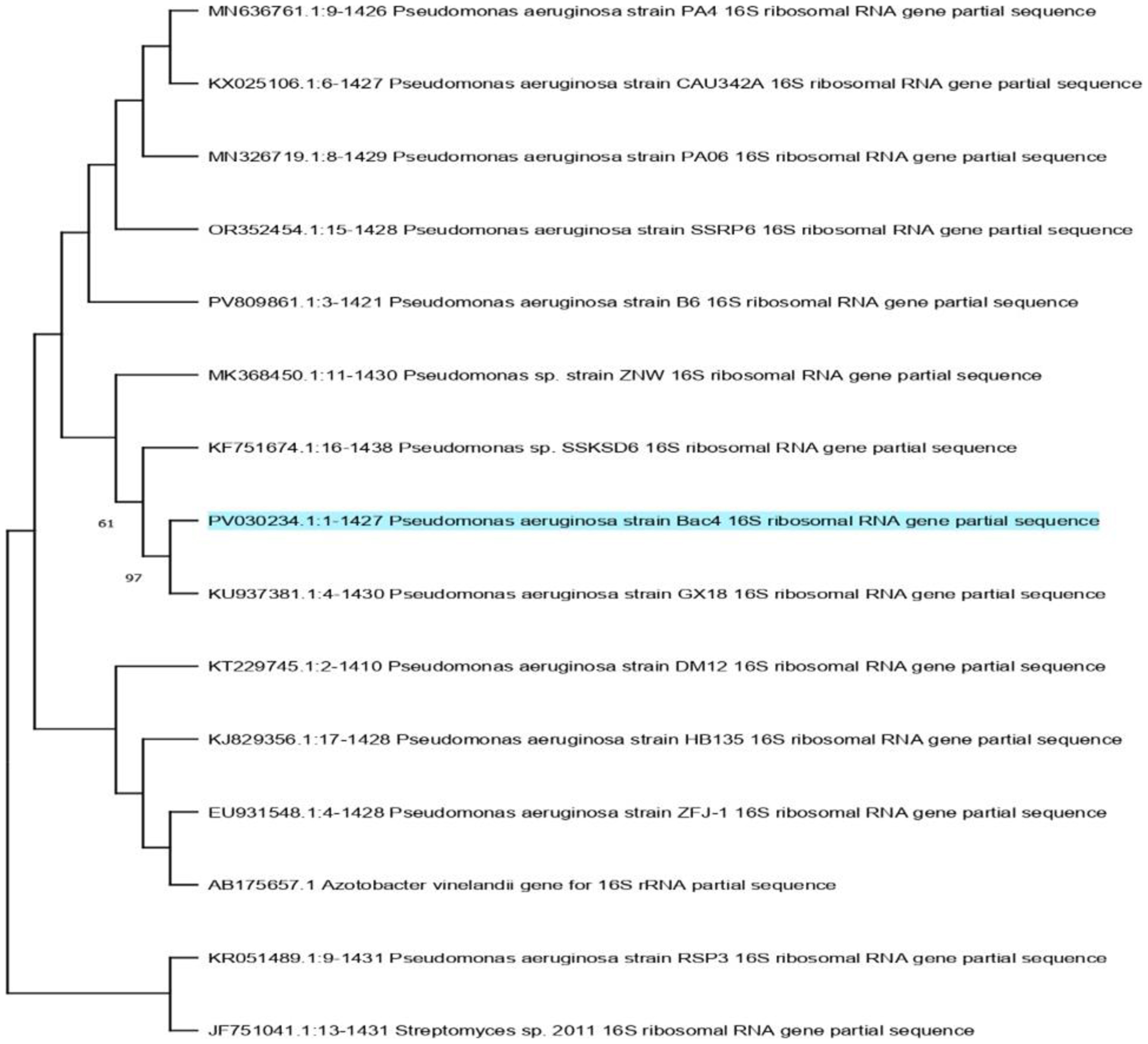
Phylogenetic analysis of the 16S rRNA gene sequence placed Bac4 within a well-supported clade of Pseudomonas aeruginosa strains. The isolate clustered closely with reference strains retrieved from GenBank, further corroborating its molecular identification (Figure 6).

Maximum likelihood tree obtained through heuristic searches of the 16S rRNA gene region sequences of 15 strains. Values of Bootstrap (BS) (1000 replicates) are provided at the nodes.Azotobacter vinelandii (AB175657.1) is treated as an outgroup. The sequences obtained in the current study are highlighted in blue.
3.7. Bacterial growth kinetics and antifungal activity
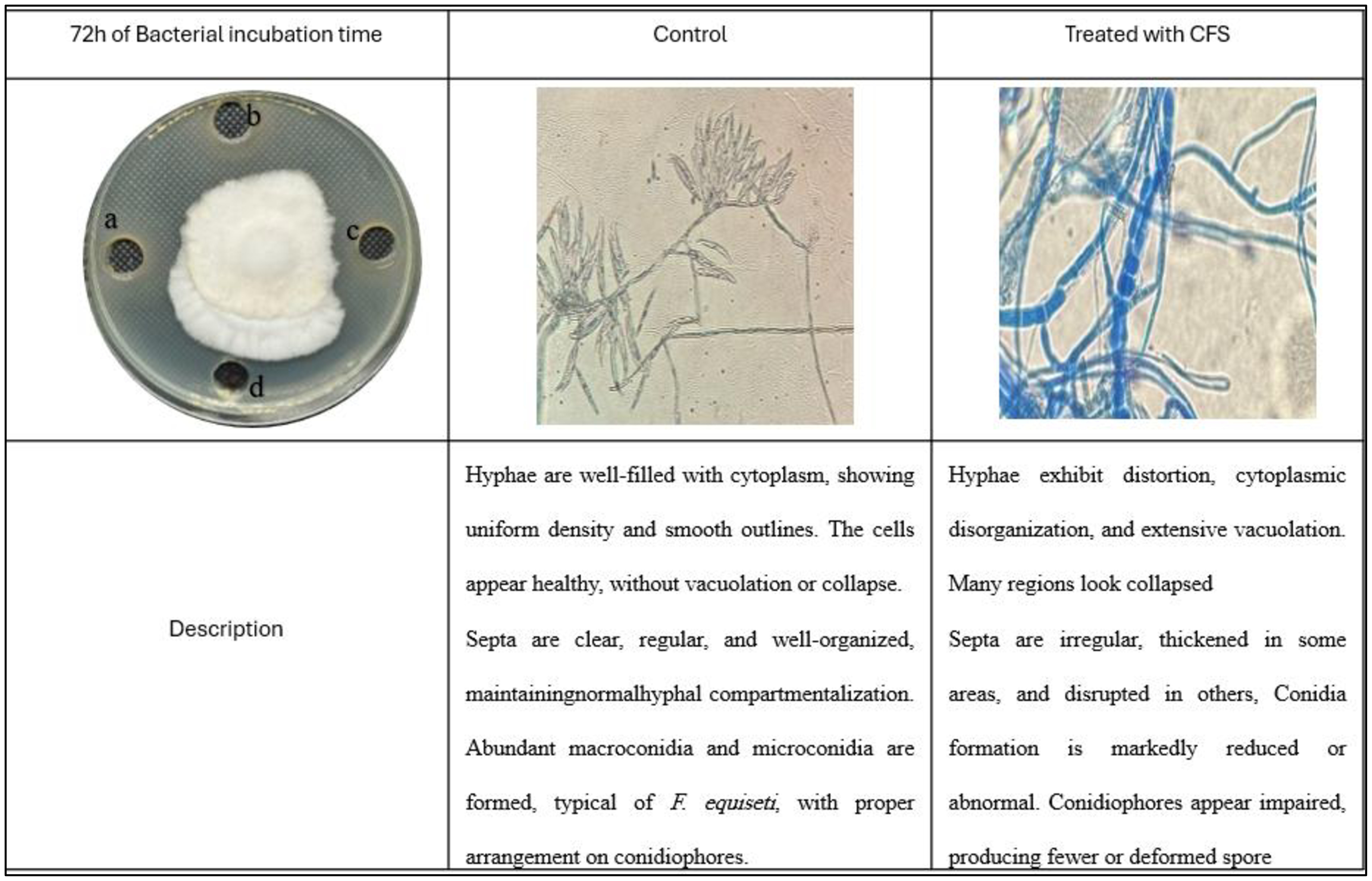
The growth curve of P. aeruginosa Bac4 showed a typical pattern, reaching the exponential phase within 48 h and peaking at 72 h. Maximum antifungal activity of the cell-free supernatant (CFS) against F. equiseti coincided with this peak growth phase. At 72 h, CFS inhibited fungal growth by 46.07% in the well diffusion assay. Microscopic observations revealed vacuolation, hyphal fragmentation, and structural abnormalities in treated fungi compared with untreated controls (Figures 7 and 8).
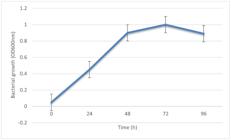
Growth curve of P.s aeruginosa Bac4 showing mean OD600 values measured at 0, 24, 48, 72, and 96 h. Error bars indicate ± SE. Antifungal activity F. equiseti was monitored at 24-hour intervals, showing peak inhibition at 72 h.
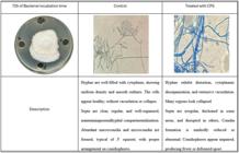
Antifungal activity of 72-h bacterial cell-free supernatant (CFS) against Fusarium equiseti assessed by well diffusion and microscopic observation. (a–c) Inhibition zones and associated microscopic alterations in fungal hyphae following CFS treatment; (d) nutrient broth control showing normal fungal growth.
3.8. Radial growth inhibition by cell-free supernatant
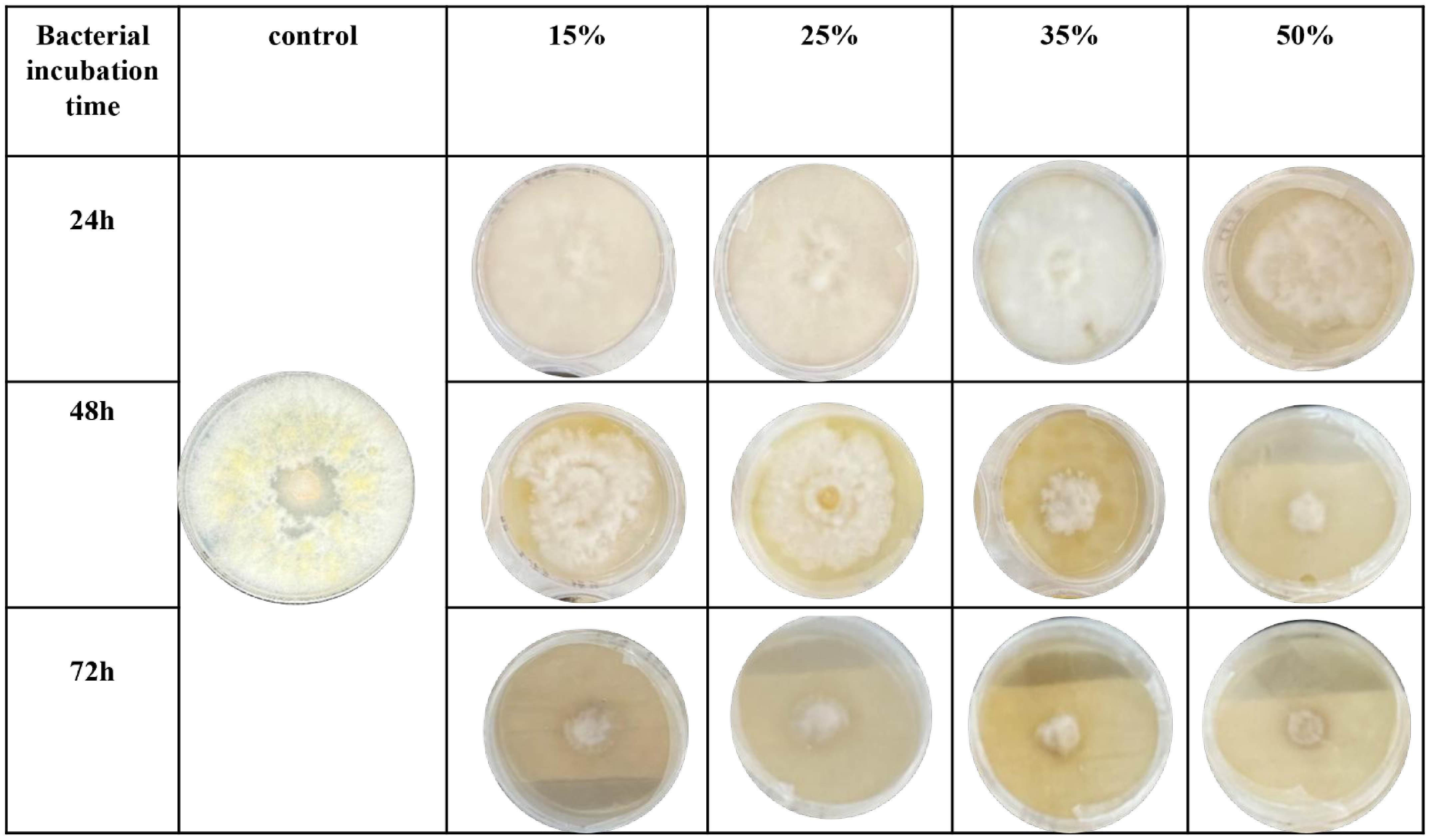
The radial growth of F. equiseti was significantly inhibited by Bac4 CFS in a concentration- and time-dependent manner. Growth inhibition was first observed at 50% CFS after 24 h and increased with prolonged incubation. Higher CFS concentrations resulted in reduced colony diameter, sparse mycelial growth, and altered pigmentation relative to controls (Figure 9).

3.9. Effect of CFS on fungal biomass
Exposure of F. equiseti to Bac4 CFS resulted in a significant reduction in both wet and dry biomass compared with untreated controls. The inhibitory effect increased with CFS concentration, with maximum biomass reduction observed at 50% CFS. Statistical analysis confirmed significant differences among treatments (p < 0.05), indicating high sensitivity of the pathogen to Bac4 metabolites (Figure 10).
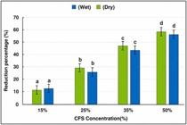
Effect of P.s aeruginosa Bac4 cell-free supernatant (CFS) on wet and dry biomass of F. equiseti at different concentrations. Error bars represent ± SE. Different letters indicate significant differences among treatments (p < 0.05, one-way ANOVA with post-hoc test).
3.10. Inhibition of spore germination
The cell-free supernatant of P. aeruginosa Bac4 significantly suppressed spore germination of F. equiseti. Approximately 47% of spores failed to germinate after exposure to CFS, representing a significant reduction relative to the control treatment (p < 0.05).
3.11. Antifungal activity of volatile organic compounds
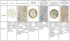
Volatile organic compounds (VOCs) produced by Bac4 significantly inhibited the growth, pigmentation, and sporulation of F. equiseti in sealed plate assays. Microscopic examination revealed hyphal deformation and cytoplasmic disruption in VOC-exposed fungi. The inclusion of activated charcoal partially restored fungal growth, confirming that inhibition was mediated by VOCs (Figure 11).
3.12. GC–MS profiling of antifungal metabolites
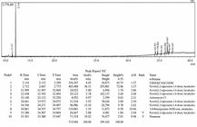
GC–MS analysis of the ethyl acetate extract of Bac4 CFS identified multiple metabolites associated with antifungal activity. Major compounds included alcohols and diketopiperazine derivatives, along with minor constituents such as actinomycin C2 and nonacosane. These compounds collectively support the bioactive potential of Bac4 metabolites (Figure 12).
3.13. In vivo evaluation of disease control
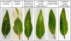
In vivo assays on detached A. marina leaves demonstrated that Bac4 CFS effectively reduced leaf spot disease severity. Inoculated control leaves exhibited extensive necrotic lesions, whereas preventive and curative CFS treatments significantly limited symptom development. Curative treatment with CFS achieved the highest disease control efficacy, outperforming the commercial fungicide Hymexazol. Disease severity indices and control efficacy values confirmed the superior performance of Bac4 CFS under experimental conditions(Figure13)
4. Discussion
Mangrove ecosystems are increasingly recognized as vulnerable to emerging plant diseases; however, information on the identity of causal agents and effective management strategies remains limited in many regions, including the Arabian Gulf. The present study provides the first confirmed report of Fusarium equiseti causing leaf spot disease on Avicennia marina in Saudi Arabia, thereby expanding the known host range and geographic distribution of this pathogen. The development of characteristic necrotic lesions following artificial inoculation and the successful re-isolation of the fungus fulfilled Koch’s postulates and unequivocally established its pathogenic role.
Species of Fusarium are well known for their adaptability to diverse environmental conditions and their capacity to cause foliar diseases under plant stress. The occurrence of F. equiseti in the Sihat mangrove ecosystem suggests that local environmental pressures may favor pathogen establishment. Mangrove habitats are subject to fluctuating salinity, temperature, and sediment chemistry, which can influence host susceptibility and pathogen aggressiveness. Although the present study did not experimentally assess environmental drivers, the observed disease incidence underscores the need for continued monitoring of mangrove health in the region.
The isolation of Pseudomonas aeruginosa Bac4 and its pronounced antagonistic activity against F. equiseti highlight the potential of soil-associated bacteria as biological control agents. In vitro assays demonstrated that Bac4 metabolites effectively suppressed fungal radial growth, biomass accumulation, and spore germination in a concentration-dependent manner. Microscopic observations revealed severe hyphal deformation, vacuolation, and cytoplasmic disintegration, indicating that bacterial metabolites interfere with essential structural and physiological processes of the pathogen. These findings are consistent with previous reports describing antifungal mechanisms of Pseudomonas spp., including disruption of cell wall integrity and inhibition of fungal development (Dullah et al., 2021).
In addition to diffusible metabolites present in the cell-free supernatant, volatile organic compounds (VOCs) emitted by Bac4 contributed significantly to fungal growth inhibition. The partial restoration of fungal growth following the addition of activated charcoal confirmed the involvement of VOC-mediated antagonism. Volatile compounds have been increasingly recognized as important components of microbial interactions, particularly in soil and plant-associated environments, where they can act over distances without direct contact. The combined activity of diffusible and volatile metabolites suggests a multifaceted mode of action by Bac4, enhancing its effectiveness against F. equiseti.
Chemical profiling by GC–MS identified several classes of compounds with known antimicrobial properties, including alcohols, diketopiperazine derivatives, and actinomycin-related compounds. While the present analysis does not establish direct causality between individual metabolites and antifungal activity, the presence of these compounds supports the observed biological effects and suggests potential synergistic interactions. Further studies involving purification and functional characterization of individual metabolites would be required to elucidate their specific roles.
Importantly, in vivo assays using detached mangrove leaves demonstrated that Bac4 cell-free supernatant provided effective disease suppression under controlled conditions, surpassing the performance of the commercial fungicide Hymexazol. This finding underscores the practical relevance of bacterial metabolites as alternative disease management tools. However, it should be noted that P. aeruginosa is an opportunistic human pathogen, and its direct application in natural environments requires careful biosafety consideration. The results presented here emphasize the potential use of bacterial metabolites rather than live bacterial inoculants, which may mitigate associated risks while retaining antifungal efficacy.
Overall, the integrated in vitro and in vivo evidence indicates that metabolites produced by Pseudomonas aeruginosa Bac4 effectively suppress Fusarium equiseti, offering a promising and environmentally compatible approach for managing mangrove leaf spot disease. This study contributes to the understanding of mangrove disease ecology in the Arabian Gulf and provides a foundation for future research aimed at developing safe, metabolite-based biocontrol strategies to support the conservation of these ecologically valuable coastal ecosystem
Acknowledgements
The authors thank the Department of Biology, College of Science, Imam Abdulrahman bin Faisal University, Kingdom of Saudi Arabia, for support. This study was approved by the Institutional Review Board of Imam Abdulrahman Bin Faisal University (IRB No. IRB-PGS-2024-10-486).
Data Availability Statement
Research data is available in the body of the article.
Reference
-
AL-NADABI, H., MAHARACHCHIKUMBURA, S.S.N., AL-GAHAFFI, Z.S., AL-HASANI, A.S., VELAZHAHAN, R. and AL-SADI, A.M., 2020. Molecular identification of fungal pathogens associated with leaf spot disease of date palms (Phoenix dactylifera). All Life, vol. 13, no. 1, pp. 587-597. https://doi.org/10.1080/26895293.2020.1835740
» https://doi.org/10.1080/26895293.2020.1835740 -
ALONGI, D.M., 2022. Climate change and mangroves. In: S. C. DAS, P. PULLAIAH, and E. C. ASHTON, eds. Mangroves: biodiversity, livelihoods and conservation Singapore: Springer Nature Singapore, pp. 175-198. https://doi.org/10.1007/978-981-19-0519-3_8
» https://doi.org/10.1007/978-981-19-0519-3_8 -
BRAUNA-MORŽEVSKA, E., STODDARD, F.L., BANKINA, B., KAŅEPS, J., BIMŠTEINE, G., PETROVA, I., NEUSA-LUCA, I., ROGA, A. and FRIDMANIS, D., 2023. Evaluation of pathogenicity of Botrytis species isolated from different legumes. Frontiers in Plant Science, vol. 14, pp. 1069126. https://doi.org/10.3389/fpls.2023.1069126 PMid:37051088.
» https://doi.org/10.3389/fpls.2023.1069126 -
DULLAH, S., HAZARIKA, D.J., PARVEEN, A., KAKOTI, M., BORGOHAIN, T., GAUTOM, T., BHATTACHARYYA, A., BAROOAH, M. and BORO, R.C., 2021. Fungal interactions induce changes in hyphal morphology and enzyme production. Mycology, vol. 12, no. 4, pp. 279-295. https://doi.org/10.1080/21501203.2021.1932627 PMid:34900382.
» https://doi.org/10.1080/21501203.2021.1932627 -
FAZHAN, H., WAIHO, K. and SHAHREZA, M., 2016. A simple and efficient total genomic DNA extraction method for individual zooplankton. SpringerPlus, vol. 5, no. 1, pp. 2049. https://doi.org/10.1186/s40064-016-3724-x PMid:27995026.
» https://doi.org/10.1186/s40064-016-3724-x -
JI, D., CHEN, T., MA, D., LIU, J., XU, Y. and TIAN, S., 2018. Inhibitory effects of methyl thujate on mycelial growth of Botrytis cinerea and possible mechanisms. Postharvest Biology and Technology, vol. 142, pp. 46-54. https://doi.org/10.1016/j.postharvbio.2018.04.003
» https://doi.org/10.1016/j.postharvbio.2018.04.003 -
LEE, N.L.Y., HUANG, D., QUEK, Z.B.R., LEE, J.N. and WAINWRIGHT, B.J., 2019. Mangrove-associated fungal communities are differentiated by geographic location and host structure. Frontiers in Microbiology, vol. 10, pp. 2456. https://doi.org/10.3389/fmicb.2019.02456 PMid:31736902.
» https://doi.org/10.3389/fmicb.2019.02456 -
MUSTAFA, H.K., ANWER, S.S. and ZRARY, T.J., 2023. Influence of pH, agitation speed, and temperature on growth of fungi isolated from Koya, Iraq. Kuwait Journal of Science, vol. 50, no. 4, pp. 657-664. https://doi.org/10.1016/j.kjs.2023.02.036
» https://doi.org/10.1016/j.kjs.2023.02.036 -
OZERSKAYA, S.M., IVANUSHKINA, N.E., KOCHKINA, G.A., DANILOGORSKAYA, A.A., PINCHUK, I.P. and VASILENKO, A.N., 2022. Various methods of long-term preservation of fungal cultures in all-russian collection of microorganisms (VKM). In: V. K. GUPTA and M. TUOHY, eds. Laboratory protocols in fungal biology Cham: Springer International Publishing, pp. 1-67. https://doi.org/10.1007/978-3-030-83749-5_1
» https://doi.org/10.1007/978-3-030-83749-5_1 -
PRAKASH, P.Y. and BHARGAVA, K., 2016. A modified micro chamber agar spot slide culture technique for microscopic examination of filamentous fungi. Journal of Microbiological Methods, vol. 123, pp. 126-129. https://doi.org/10.1016/j.mimet.2016.02.015 PMid:26924158.
» https://doi.org/10.1016/j.mimet.2016.02.015 -
PRINGGENIES, D., SANTOSA, G.W., DJUNAEDI, A. and SUSANTO, A.B., 2021. Potential of bioactive compounds of Holothuria atra- Associated bacteria as a raw material in bioindustry. In: S. BOREK, ed. New visions in biological science Vol. 4 Bhanjipur: Book Publisher International, pp. 66-75. https://doi.org/10.9734/bpi/nvbs/v4/4436F
» https://doi.org/10.9734/bpi/nvbs/v4/4436F -
RATH, A.K. and MEHER, J.K., 2019. Disease detection in infected plant leaf by computational method. Archiv für Phytopathologie und Pflanzenschutz, vol. 52, no. 19-20, pp. 1348-1358. https://doi.org/10.1080/03235408.2019.1708546
» https://doi.org/10.1080/03235408.2019.1708546 -
SABRY, S., 2016. Studies on cabbage alternaria leaf spot disease Zagazig, Egypt: Zagazig University. Doctoral dissertation. https://doi.org/10.13140/RG.2.2.28084.81285
» https://doi.org/10.13140/RG.2.2.28084.81285 -
SLOWIK, A.R., HESKETH, H., SAIT, S.M. and DE FINE LICHT, H.H., 2023. A rapid method for measuring in vitro growth in entomopathogenic fungi. Insects, vol. 14, no. 8, pp. 703. https://doi.org/10.3390/insects14080703 PMid:37623413.
» https://doi.org/10.3390/insects14080703 -
THILAGAM, R., KALAIVANI, G. and HEMALATHA, N., 2018. Isolation and identification of phytopathogenic fungi from infected plant parts. International Journal of Current Pharmaceutical Research, vol. 10, no. 1, pp. 26. https://doi.org/10.22159/ijcpr.2018v10i1.24404
» https://doi.org/10.22159/ijcpr.2018v10i1.24404 -
WANG, G.-W., HU, W.-T., HUANG, B.-K. and QIN, L.-P., 2011. Illicium verum: A review on its botany, traditional use, chemistry and pharmacology. Journal of Ethnopharmacology, vol. 136, no. 1, pp. 10-20. https://doi.org/10.1016/j.jep.2011.04.051 PMid:21549817.
» https://doi.org/10.1016/j.jep.2011.04.051 -
YANG, J., HSIANG, T., BHADAURIA, V., CHEN, X.-L. and LI, G., 2017. Plant fungal pathogenesis. BioMed Research International, vol. 1-2, pp. 9724283. https://doi.org/10.1155/2017/9724283 PMid:28194426.
» https://doi.org/10.1155/2017/9724283
Edited by
-
Editor:
Takako Matsumura Tundisi