 Restorative effects of Thai Mucuna pruriens seed extract on reproductive, hormonal, and fertility impairments in letrozole-induced PCOS rats
Abstract
Polycystic ovary syndrome (PCOS) is a complex endocrine disorder, leading cause of female infertility, disrupted estrous cycles, hormonal dysregulation, and abnormal ovarian morphology. This study aimed to evaluate the restorative effects of Thai Mucuna pruriens (TMP) seed extract on reproductive, hormonal, and fertility impairments in a PCOS rat model induced by letrozole. Following PCOS induction, female rats received TMP extract at 150, 300, and 600 mg/kg BW for 30 consecutive days. Untreated PCOS rats displayed persistent diestrus, hormonal dysregulation, and polycystic ovaries, and no pregnancies were observed. After TMP treatment, especially at 300 mg/kg BW, significantly reversed these impairments: estrous cyclicity normalized, hormonal profiles improved (increasing FSH and estradiol, decreasing LH and testosterone levels), and ovarian histology showed active folliculogenesis and reappearance of multiple corpus lutea. In addition, TMP could reduce the body weight and downregulate the ovarian androgen receptor (AR), estrogen receptor alpha (ERα), and tumor necrosis factor-alpha (TNF-α) protein expression. Moreover, TMP restored the fertility index and promoted embryo implantation, comparable to control animals. In conclusion, Thai Mucuna pruriens seed extract, particularly at 300 mg/kg, effectively reversed the reproductive, endocrine, and fertility impairments in letrozole-induced PCOS animal model, suggesting its promising potential as a phytotherapeutic agent for managing PCOS-associated reproductive dysfunction.
Keywords:
Thai Mucuna pruriens; PCOS; fertility; hormonal balance; ovary
Resumo
A síndrome dos ovários policísticos (SOP) é um distúrbio endócrino complexo caracterizado por anovulação, desequilíbrio hormonal e alterações morfológicas ovarianas. Este estudo avaliou os efeitos restauradores do extrato de sementes de Thai Mucuna pruriens (TMP) sobre os prejuízos reprodutivos, hormonais e de fertilidade em um modelo de SOP induzida por letrozol em ratas. Após a indução da SOP, as ratas receberam TMP nas doses de 150, 300 e 600 mg/kg por 30 dias consecutivos. As ratas com SOP não tratadas apresentaram diestro persistente, alterações hormonais e ausência de gravidez. O tratamento com TMP, especialmente na dose de 300 mg/kg, reverteu significativamente essas alterações, normalizando o ciclo estral, melhorando os perfis hormonais e restaurando a histologia ovariana com reaparecimento de folículos ativos e corpos lúteos. Além disso, TMP reduziu o peso corporal, diminuiu a expressão ovariana de AR, ERα e TNF-α, e restaurou a fertilidade, com presença de sítios de implantação embrionária. Conclui-se que o extrato de sementes de TMP, particularmente na dose de 300 mg/kg, exerce potente efeito fitoterapêutico na restauração das disfunções reprodutivas associadas à SOP.
Palavras-chave:
Thai Mucuna pruriens; SOP; fertilidade; hormônios; ovário
1. Introduction
Polycystic ovary syndrome (PCOS) is one of the most prevalent endocrine disorders among women of reproductive age, with global prevalence estimates ranging from 6% to 13% (WHO, 2025), and a reported prevalence of 5.29% among Thai adolescent girls (Kaewnin et al., 2017). Among women experiencing infertility due to anovulation, approximately 80% of cases are attributable to PCOS (Balen et al., 2016). The PCOS is clinically defined by features such as hyperandrogenism, ovulatory dysfunction, hormonal imbalance, disrupted steroidogenic signaling, and molecular alterations linked to inflammation (Rosenfield and Ehrmann, 2016; Dey et al., 2023; Su et al., 2025). Standard pharmacological interventions to induce ovulation in PCOS typically include the use of clomiphene citrate, an estrogen receptor modulator, and metformin, an insulin-sensitizing agent, each targeting specific symptoms like anovulation and insulin resistance (Palomba et al., 2006; Legro et al., 2014). Nevertheless, such medications have been reported to be associated with adverse effects, including gastrointestinal disturbances frequently observed with metformin, and vision changes or mood alterations related to clomiphene (Lord et al., 2003; Duleba and Dokras, 2012; Murri et al., 2013). Furthermore, the conventional drugs do not completely address the diverse biological and molecular mechanisms underlying PCOS, such as chronic inflammation, oxidative stress, and persistent hormonal imbalance (Rudnicka et al., 2021).
In light of these limitations, there is a growing need to reveal the medicinal plants for their potential to restore reproductive and metabolic homeostasis in PCOS. Recently, the research interest in medicinal plants has increased due to the presence of bioactive compounds and their generally favorable safety profiles. Previously, the powder extracts from plants such as Phyllanthus muellerianus (Mvondo et al., 2020), Curcuma longa (Akter et al., 2023), and Terminalia chebula (Kalimuthu et al., 2025) have demonstrated efficacy in improving hormonal profiles, enhancing insulin sensitivity, and promoting ovarian function recovery in the PCOS rat models. These findings suggest that plants with anti-androgenic and homeostatic regulatory properties may represent promising alternatives to conventional PCOS therapies. Thai Mucuna pruriens (TMP), a traditional herbal remedy, has been shown to protect against testicular damage in animal studies, primarily attributed to its high L-DOPA content and resulting improvements in testosterone levels, sperm quality, and sexual performance in models exposed to stress or alcohol (Lapyuneyong et al., 2022; Tangsrisakda et al., 2022; Choowongin et al., 2022). Interestingly, TMP has been demonstrated to exhibit strong antioxidant activity, did not induce reproductive toxicity in female rats, and increased estrogen levels, effects primarily ascribed to its L-DOPA, flavonoid, and phenolic constituents (Iamsaard et al., 2020). Although the evidence for Mucuna pruriens in female reproductive models is limited, prior studies using Indian and Nigerian seed varieties have shown improvements in hormonal status and ovarian histology (Ojo et al., 2014; Sogbesan et al., 2022). However, there are no studies investigating TMP seed extract in the PCOS rat model. Therefore, the present study aimed to investigate the therapeutic potential of TMP seed extract using a letrozole-induced PCOS rat model.
2. Materials and Methods
2.1. Ethical approval
All animal experimental procedures in this study were approved by the Institutional Animal Care and Use Committee of Khon Kaen University (IACUC-KKU-54/65) under the guidelines on the Ethics of Animal Experimentation of the National Research Council of Thailand.
2.2. Preparation of Thai Mucuna pruriens (TMP) seed extract
The aqueous Thai Mucuna pruriens seed extract used in this study was prepared in our laboratory, yielding approximately 16.29% of stock powder from the initial seed materials. The phytochemical composition of TMP, including L-DOPA (approximately 10% of total dried weight), has been previously characterized (Choowong-In et al., 2022), and the extract was prepared using the same standardized protocol to ensure batch-to-batch consistency. The TMP extract’s antioxidant activity and capacity, as well as the absence of reproductive toxicity, have been previously validated in animal models (Iamsaard et al., 2020; Tangsrisakda et al., 2022). The selected doses of TMP (150, 300, and 600 mg/kg BW) were based on previous toxicological and reproductive studies demonstrating biological activity without reproductive toxicity (Iamsaard et al., 2020; Choowong-In et al., 2022).
2.3. Animals and experimental designs
Sixty female Sprague-Dawley rats (180–200 g) were purchased from the National Laboratory Animal Center, Mahidol University (Salaya, Nakhon Pathom, Thailand). Animals were housed in standard plastic cages under controlled environmental conditions (12-hour dark/light cycle, temperature 22 ± 2 °C, humidity 30–60%, noise level <85 dB, and light intensity 350–400 lux), with free access to a standard pellet diet and water ad libitum. Animals were randomly divided into two groups: the control group (received 0.1% carboxymethylcellulose [CMC]) and the PCOS group (received letrozole 1 mg/kgBW in 0.1% CMC) once daily for 21 consecutive days to induce PCOS as previously described (Mvondo et al., 2020). Vaginal lavage was collected from all animals between days 15 and 21 to determine their estrous cyclicity. Representative estrous cycles of control and PCOS rats with the diestrus period are shown in Supplementary material available in Bunsueb and Iamsaard (2025). The animals were randomly divided into five experimental groups (12 rats per group): control, PCOS, PCOS+TMP150, PCOS+TMP300, and PCOS+TMP600. The PCOS group continued to receive letrozole alone, whereas the treatment groups received letrozole co-administered with TMP seed extract at doses of 150, 300, or 600 mg/kg, respectively, for 30 consecutive days. The 30-day treatment duration was selected to encompass multiple estrous cycles in rats, enabling reliable evaluation of estrous cyclicity, hormonal profiles, ovarian morphology, and fertility-related outcomes following TMP administration. Vaginal lavage samples were collected again from days 45 to 51, before ending the experiment, to assess alterations in the estrous cycle. At the end of the treatment period, six rats from each group were randomly selected for collection of blood, ovary, and uterus to perform hormonal assays, histological examination, and western blot analysis, respectively, while the remaining six animals per group were retained for evaluation of fertility outcomes.
2.4. Vaginal lavage collection and estrous cycle assessment
Vaginal lavage was gently performed and collected in the morning (between 08:00 and 10:00 h) at the same time each day by flushing 100 µl of PBS (pH 7.4) into the vaginal canal using a sterile micropipette tip. Then, the lavage (10 µl) was dropped onto a glass slide and air-dried before staining with 0.1% crystal violet. The vaginal cells stained were examined under a light microscope to determine the status of rat estrous cycle stage as previously described (McLean et al., 2012). Briefly, the proestrus was defined by the predominance of nucleated epithelial cells, whereas the estrus was characterized by that of cornified epithelial cells. Metestrus phase showed the mixture of neutrophils, nucleated epithelial cells, and cornified cells. Abundant neutrophils in the lavage were classified to indicate the diestrus period.
2.5. Blood collection and hormonal analyses
At the end of the experiment (day 52), six rats from each group were randomly selected for blood collection to determine sex hormone levels in serum. Each animal was anesthetized with thiopental sodium (80 mg/kgBW, i.p.) and euthanized by cervical dislocation. Then, the blood sample was collected immediately by cardiac puncture and centrifuged at 13,000 rpm for 10 min at 4 °C to separate serum from blood cells. The serum levels of testosterone (ng/ml), estradiol (pg/ml), luteinizing hormone (LH; mIU/ml), and follicle-stimulating hormone (FSH; mIU/ml) were determined using electrochemiluminescence immunoassay (ECLIA; cobas e analyzer, Roche Diagnostics, Mannheim, Germany) according to the manufacturer’s protocol. All assays were performed at the Clinical Immunology Unit, Srinagarind Hospital, Faculty of Medicine, Khon Kaen University, Thailand.
2.6. Relative weight and ovarian histological analyses
The weights of rat body, ovaries, and uteri were recorded and calculated for their relative organ weights by using a formula of: 100 × absolute organ weight / final body weight. Subsequently, the tissues were captured and fixed in 10% formalin fixative before histological processing. Each paraffin block of left ovary was sectioned at 5 μm thickness and stained with hematoxylin and eosin (H&E). Then, ovarian sections of all groups were observed under a light microscope (ZEISS Axio Imager.A2, Germany). The representative images were captured using an AxioCam ICc 5 digital camera (ZEISS, Germany).
2.7. Western blot analysis
The right ovary of each animal was homogenized with 1X RIPA lysis buffer (Merk Millipore, Cat. No. 20-188) containing protease inhibitor cocktail tablet (Sigma, Cat. No. S8830) and centrifuged at 13,000 rpm for 10 min at 4 °C to separate the supernatant from tissue pellets. Total protein concentration in the supernatant was determined using a NanoDrop ND-1000 (NanoDrop Technologies, Inc., USA) to read the absorbance of protein at a wavelength of 280 nm. Subsequently, total proteins with equal amounts (100 μg) from each sample were separated into 10% SDS–PAGE gel and electrotransferred onto nitrocellulose membranes. Non-specific protein binding was blocked with 5% skim milk in TBST (TBS containing 0.1% Tween-20, pH 7.4). Then, each membrane was separately incubated with primary antibody against androgen receptor (AR, 1:1000; Sigma-Aldrich, cat. 06-680), estrogen receptor α (ERα, 1:1000; Sigma-Aldrich, cat. 06-935), or tumor necrosis factor-α (TNF-α, 1:1000; MyBioSource, cat. MBS9700322), overnight at 4 °C. After washing unbound primary antibodies, membrane was incubated with HRP-conjugated secondary antibody against its primary antibody. The specific antibody–antigen complexes of protein expressions were detected using the enhanced chemiluminescence (ECL) substrate kit (GE Healthcare Life Science, USA) and visualized under the Gel Documentation 4 system (ImageQuant 600, GE Healthcare, USA). Immunoblots were performed in triplicate to analyze the intensities of AR, ERα, and TNF-α expressions by using ImageJ software (version 1.53a) (ImageJ, 2025). β-actin, an internal control protein, was used for normalization, and the relative intensities of the targeted protein expression were expressed as one relative unit.
2.8. Fertility assessment
The remaining six animals per group were mated with males (2:1 female:male ratio) for five consecutive days. Daily vaginal lavage was performed to detect sperm presence, with detection day designated as gestational day 1 (GD1). Sperm-positive females were individually housed until GD13, then they were euthanized to observe the number of implantation sites in the uteri for estimation of female fertility as previously explained (Awounfack et al., 2018). The fertility index (%) was calculated as follows: (number of pregnant females/ number of sperm-positive females) × 100 (Baek et al., 2022).
2.9. Statistical analysis
All data were analyzed using GraphPad Prism software version 10.3.1 (GraphPad Software, San Diego, CA, USA). Results are expressed as mean ± standard error of mean (SEM). To compare differences among groups, including the control group, PCOS group, and TMP-treated PCOS groups (150, 300, and 600 mg/kg), a one-way analysis of variance (ANOVA) was applied, followed by Tukey’s multiple comparisons post hoc test to determine pairwise differences between groups. Statistical differences were considered significant when the p-value was less than 0.05.
3. Results
3.1. TMP seed extract improved the normal estrous cyclicity of PCOS rats
Representative estrous cycle patterns of control, PCOS, and TMP treated groups are shown in Supplementary 2 (Zenodo, DOI: https://doi.org/10.5281/zenodo.17698668). The results demonstrated that untreated PCOS rats remained in persistent diestrus, whereas TMP extract improved estrous cyclicity, with the PCOS+TMP300 group showing the most consistent recovery. PCOS+TMP150 and PCOS+TMP600 groups showed the delayed progression of estrous cyclicity as shown in Supplementary 2.
3.2. TMP seed extract changed sex hormone levels in PCOS rats
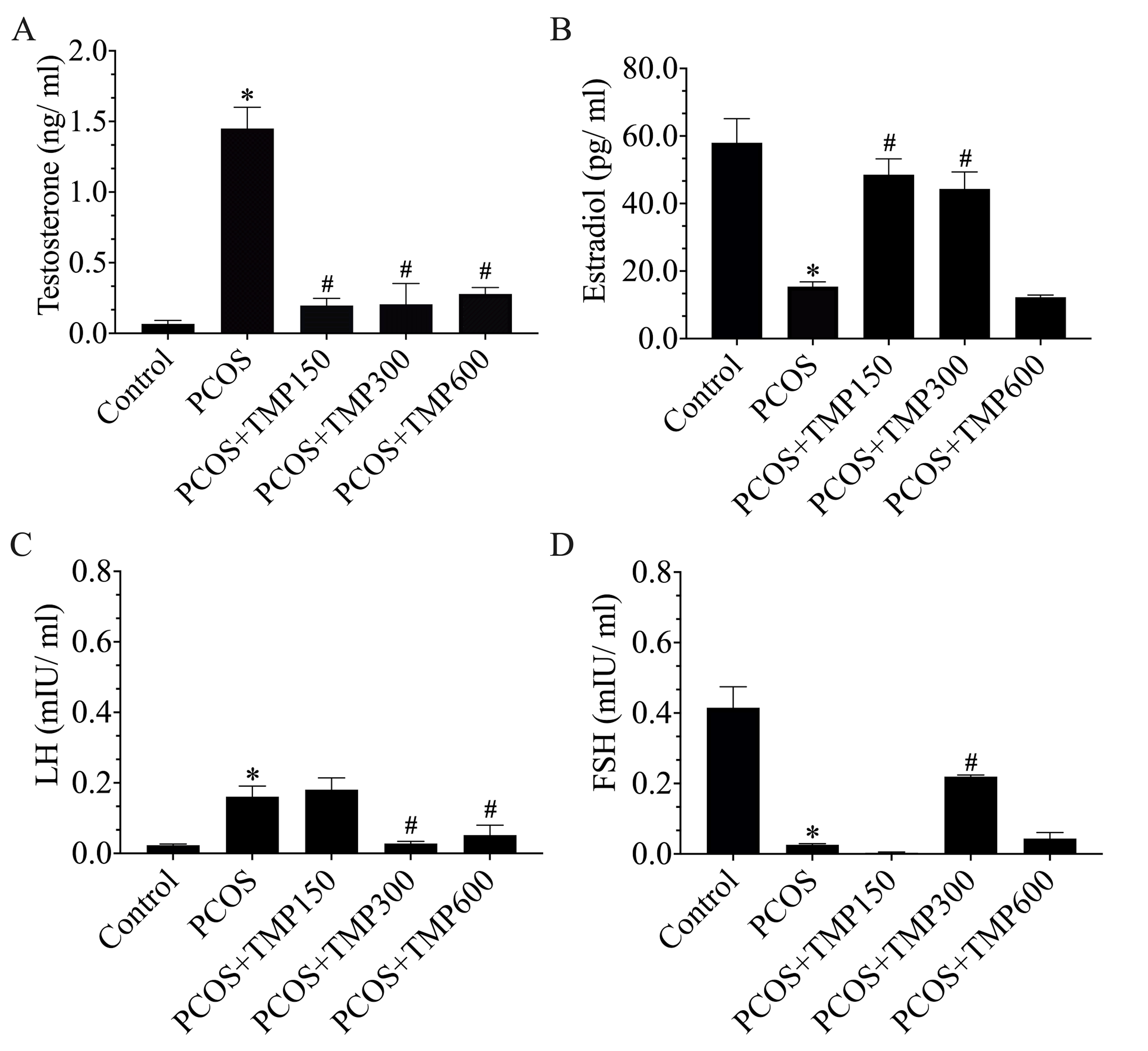
As compared to control, PCOS rats showed the significant increase of testosterone and LH levels but decrease of estradiol and FSH levels (*p < 0.05; Figure 1). It was found that TMP extract could significantly improve such hormonal alterations compared to those of the PCOS group. In contrast to testosterone levels, LH levels were reduced in the PCOS+TMP300 and 600 groups, whereas no significant change was observed in PCOS+TMP150 (Figure 1C). Estradiol levels were significantly elevated in the PCOS+TMP150 and 300 groups, but not of high dose treated group (#p < 0.05; Figure 1B). Among groups, only PCOS+TMP300 group had a significant increase in FSH level as compared to that of PCOS rats (#p < 0.05; Figure 1D).
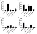
Serum hormone levels of testosterone (A), estradiol (B), LH (C), and FSH (D) of control, PCOS, and TMP-treated PCOS (150, 300, and 600 mg/kg BW) groups. Data are presented as mean ± SEM. *p < 0.05, compared to the control group, #p < 0.05, compared to the PCOS group.
3.3. Effect of TMP seed extract on organ weights and gross morphology in PCOS rats
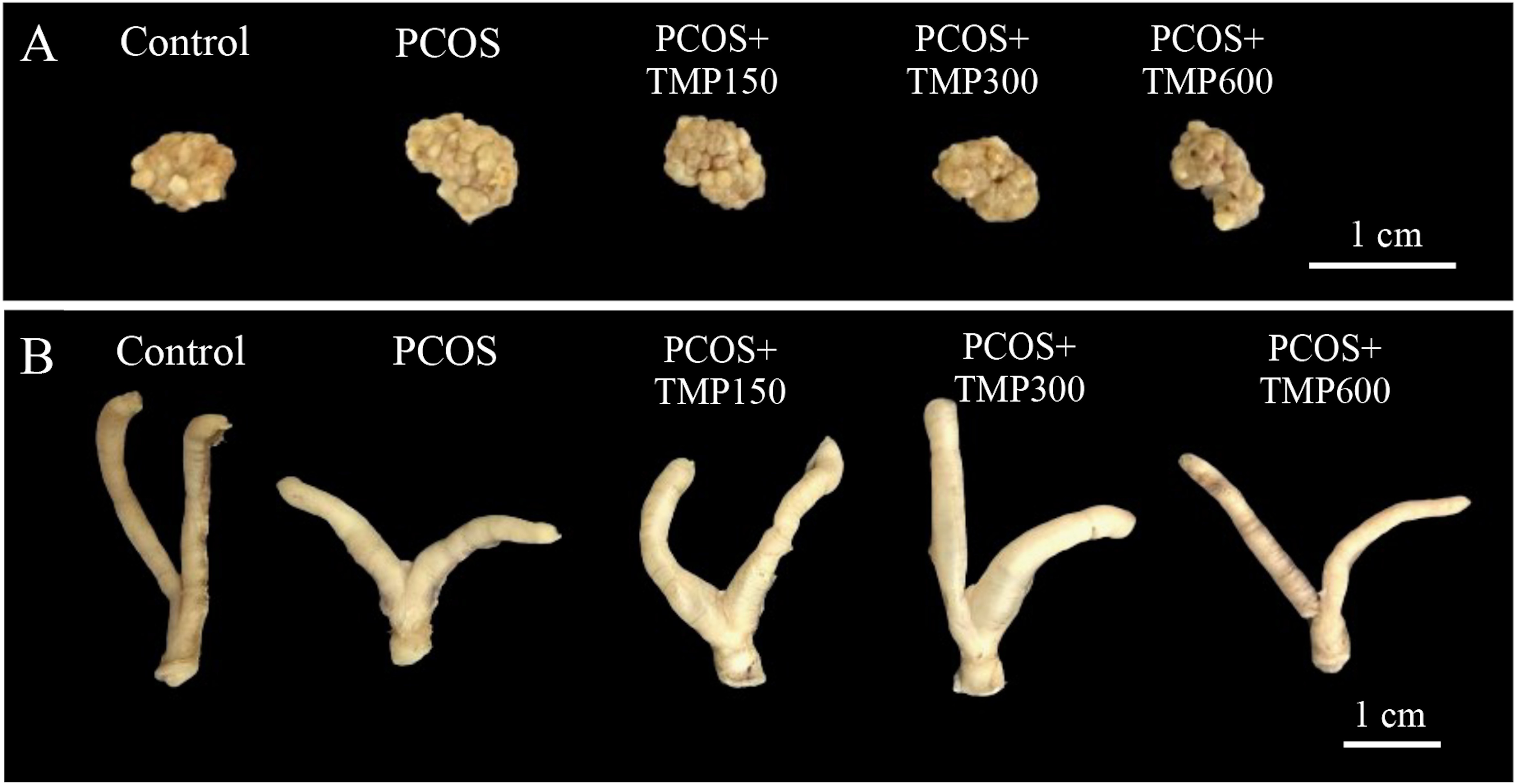
The results showed that PCOS rats exhibited a significant increase in body weight compared with control (*p < 0.05). A treatment with TMP seed extract at 300 mg/kg BW significantly decreased body weight compared to the PCOS group (#p < 0.05). Although PCOS+TMP150 and TMP600 groups showed no statistical difference, those body weights tended to be decreased as compared with the PCOS group (Table 1). The ovarian absolute and relative weight was significantly higher in the PCOS group compared to controls (*p < 0.05). However, TMP treatment did not improve ovarian weight parameters as compared to PCOS group. In contrast, those weights of uterus were improved in TMP300 and TMP600 treated groups (*p < 0.05) as shown in the Table 1, which was related to their morphology and size (Figure 2).
Comparisons of body, ovarian, and uterine weights among control, PCOS, and TMP-treated PCOS groups (150, 300, and 600 mg/kg BW, respectively).
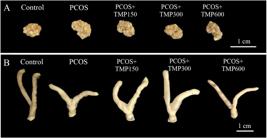
Representative photographs showing the morphology and size of ovary (A) and uterus (B) observed in control, PCOS, and TMP-treated PCOS groups (150, 300, and 600 mg/kg BW).
3.4. TMP seed extract improved the polycystic ovary
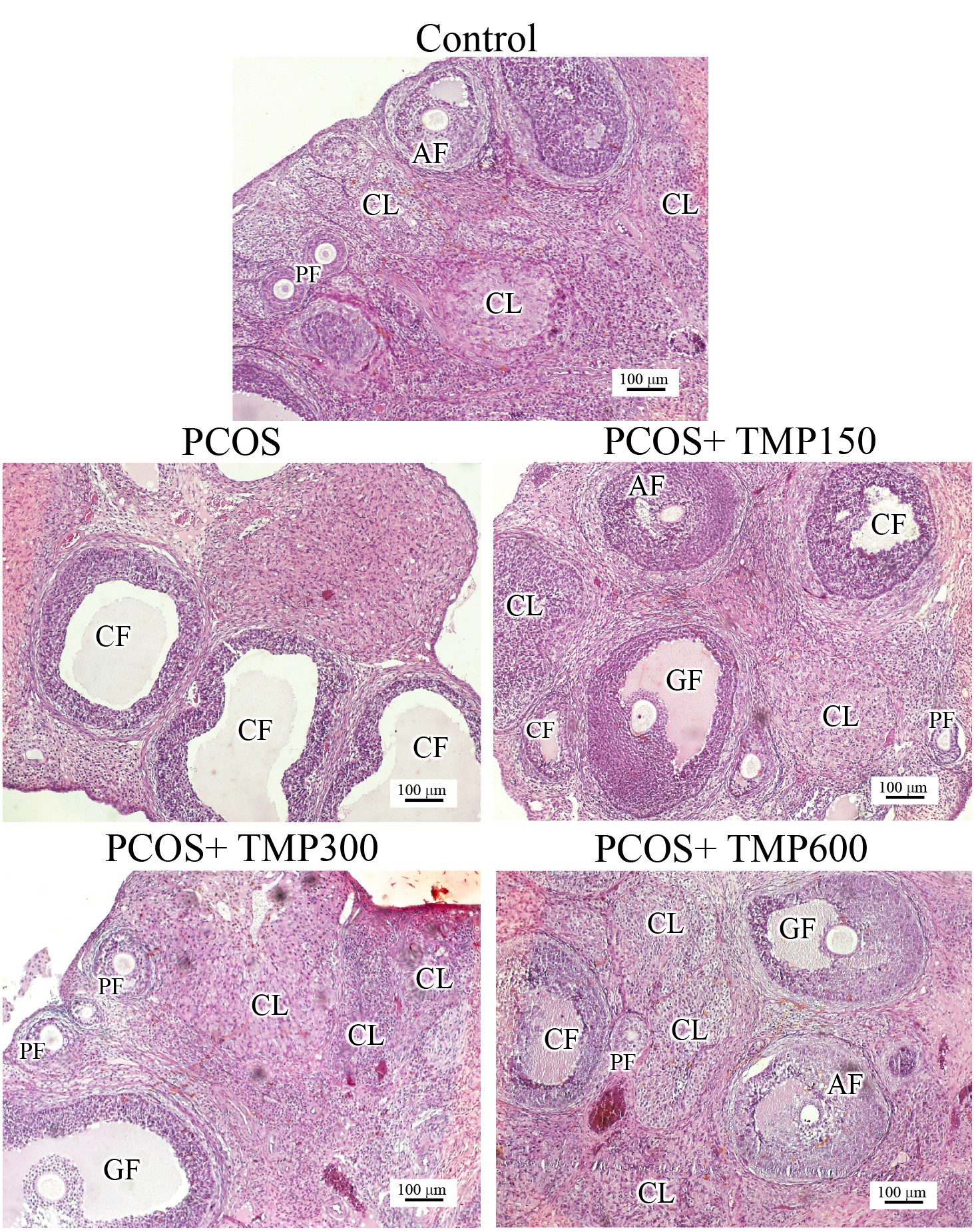
It was observed that the ovaries of PCOS rats exhibited numerous large cystic follicles with no mature or Graafian follicle as compared to control (Figure 3). Among the TMP-treated groups, the PCOS+TMP300 group showed histological recovery of active ovary, characterized by preantral and antral follicles and multiple corpus lutea. In addition, the PCOS+TMP150 and PCOS+TMP600 also showed improvement of the ovary compared to that of the untreated PCOS rats as demonstrated in the Figure 3.
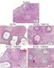
Representative photomicrographs showing the histology of the ovary in control, PCOS, and TMP-treated PCOS groups (150, 300, and 600 mg/kg BW). PF; preantral follicle, AN; antral follicle, CL; corpus luteum, CF; cystic follicle, GF; Graafian (mature) follicle.
3.5. TMP seed extract altered the expression of protein markers in ovarian PCOS rats

The expressions of AR, ERα, and TNF-α in the PCOS ovary were markedly increased compared to the control (Figure 4A), which were corroborated with their relative intensities (Figures 4B–D). It was found that no alteration in those protein expressions was observed in the PCOS+TMP150 group. In contrast, the PCOS+TMP300 group showed a significant decrease of AR, ERα, and TNF-α expressions compared to those of the PCOS group (Figures 4B–D). Interestingly, the expression of AR and ERα was also significantly decreased in the PCOS+TMP600 group.
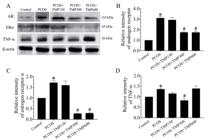
Western blot analysis of AR, ERα, and TNF-α expressions in ovary lysate (A) and the relative intensities (B–D) compared among control, PCOS, and TMP-treated PCOS groups (150, 300, and 600 mg/kg BW). β-actin was used as an internal control. Data are presented as mean ± SEM. *p< 0.05, compared to the control group; #p< 0.05, compared to the PCOS group.
3.6. Effects of TMP seed extract on fertility outcomes in PCOS rats

Figure 5 showed the fertility outcome and number of implantation sites compared among the control, PCOS, and TMP-treated groups. No implantation sites were observed in PCOS uterine horns (Figure 5A). It was found that TMP seed extract could promote embryo implantation, similar to control animals (Figure 5A). The figure 5B showed the restoring fertility index in all TMP-treated groups as compared to PCOS rats (0%). The fertility index was 80%, 75%, and 40% in the PCOS rats treated with TMP150, 300, and 600 mg/kg BW, respectively (Figure 5B). Indeed, the number of implantation sites per a pregnant female of TMP-treated groups remained comparable to the control group but was not observed in PCOS animals (Figure 5C).
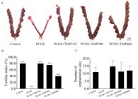
Representative photographs of rat uteri showing implantation sites on gestational day 13 in control, PCOS, and TMP-treated PCOS (150, 300, and 600 mg/kgBW) groups (A). Percent of the fertility index (B) and number of implantation sites per a pregnant female (C).
4. Discussion
The present study aimed to elucidate the therapeutic potential of Thai Mucuna pruriens (TMP) seed extract in a letrozole-induced polycystic ovary syndrome (PCOS) rat model, focusing on its endocrine, histological, molecular, and fertility outcomes. Letrozole-induced PCOS is characterized by persistent hyperandrogenism, anovulation, and altered gonadotropin secretion, simulating the core characteristics of the human syndrome (Balen et al., 2016; Rosenfield and Ehrmann, 2016; Dey et al., 2023). Similar to previous studies, untreated PCOS rats in this study also developed estrous acyclicity, elevated testosterone and luteinizing hormone (LH), and suppressed estradiol and follicle-stimulating hormone (FSH), alongside increased body and ovarian weights, as well as classic polycystic ovarian histopathology (Kafali et al., 2004; Patil and Kulkarni, 2025). TMP seed administration, especially at the 300 mg/kg dose, produced a meaningful reversal of these PCOS phenotypes. Restoration of estrous cyclicity, normalization of hormonal profiles, and histological ovarian recovery all points to TMP’s capacity to reactivate folliculogenesis and steroidogenesis. The marked increase in estradiol and FSH, concomitant with reduced LH, suggests a balanced hypothalamic-pituitary-ovarian axis and is highlighted by the presence of multiple corpora lutea, indicative of ovulation (Walters et al., 2015). These findings were consistent with previous reports of medicinal plants such as Curcuma longa and Terminalia chebula, which have demonstrated endocrine and histological normalization in PCOS models (Akter et al., 2023; Kalimuthu et al., 2025), but the recent results provided novel evidence specifically for TMP in this issue. At the molecular level, TMP extract at 300 mg/kg significantly reduced ovarian androgen receptor (AR), estrogen receptor alpha (ERα), and tumor necrosis factor-alpha (TNF-α) expressions. The normalization of AR is especially pertinent given the established link between androgen excess, AR overexpression, and follicular arrest in PCOS (Bunsueb et al., 2020; Rosenfield and Ehrmann, 2016; Wang et al., 2015). Similarly, ERα normalization supports improved estrogen signaling, crucial for folliculogenesis, as disruption in ERα/ERβ equilibrium underlies impaired follicle development (Xu et al., 2021; Kamal et al., 2022). Importantly, the decrease in TNF-α reflects an amelioration of intraovarian inflammation, which may restore granulosa cell aromatase activity and estrogen biosynthesis (Rezvanfar et al., 2015). Chronic low-grade inflammation is a key pathological feature of PCOS, and elevated TNF-α has been shown to impair folliculogenesis by promoting granulosa cell apoptosis and disrupting follicle maturation, as well as to inhibit steroidogenesis through suppression of aromatase activity, leading to reduced estradiol synthesis and persistent hyperandrogenism (Duleba and Dokras, 2012; Rezvanfar et al., 2015). In the present study, the reduction of ovarian TNF-α expression in the TMP300 group was accompanied by improved hormonal balance, ovarian morphology, and fertility indices, supporting the role of inflammation modulation in TMP-mediated reproductive recovery. The differential efficacy between TMP300 and TMP600 indicates a non-linear dose-dependent response. Plant-derived phytochemicals are known to exhibit biphasic or hormetic effects, in which moderate doses exert optimal anti-inflammatory and endocrine-regulatory actions, whereas higher doses may induce counter-regulatory responses. In the present study, TMP300 achieved more complete normalization of ovarian TNF-α expression, hormonal profiles, and fertility outcomes than TMP600, suggesting that optimal reproductive recovery depends on balanced endocrine–inflammatory regulation rather than increasing dose alone (Xi et al., 2022). This might be supported by the incomplete endocrine and molecular normalization, persistent TNF-α elevation, and comparatively lower fertility index in the high-dose group. It was noted that the increased uterine mass in TMP600-treated rats, although partially restored toward control levels, was in line with the dissociated ovarian and uterine responses reported under conditions of persistent inflammation (Kaneko et al., 2004). The restoration of fertility, documented by recovered implantation rates and fertility indices in TMP150 and especially TMP300 groups, underscores the clinical relevance of TMP for fertility management in PCOS. Persistent subfertility in the TMP600 cohort further highlights that effective PCOS therapy requires not only ovulation induction but also coordinated hormonal and inflammatory modulation (Lalitkumar et al., 2005). Taken together, these findings indicated that TMP exerts integrated endocrine, anti-inflammatory, and ovarian receptor-modulating effects, with the 300 mg/kg dose achieving superior normalization across all parameters. This expands on two previous studies reported in other Mucuna species (Ojo et al., 2014; Sogbesan et al., 2022), by providing the first comprehensive demonstration of TMP seeds in a well-established PCOS model. In addition, changes in uterine weight may also provide insight into the endocrine responsiveness of reproductive tissues. Letrozole-induced PCOS markedly reduced uterine weight, consistent with impaired estrogenic stimulation, as uterine weight is widely recognized as an in vivo index of estrogenic activity in rodent models (Odum et al., 1997). TMP administration increased uterine weight, suggesting restoration of estrogen responsiveness. Notably, uterine weight in the TMP300 group exceeded control values, which may indicate enhanced uterine responsiveness to estrogenic stimulation rather than simple normalization, whereas TMP600 restored uterine weight toward control levels. Therefore, although uterine weight gain coincided with improved reproductive outcomes, this response should be interpreted with caution in the absence of uterine histological assessment. Limitations of the present study include the lack of mechanistic exploration beyond the endpoints tested and not assessing long-term reproductive or metabolic sequelae. Future investigation needs to clarify the active phytochemical constituents, dose-response relationships, and potential metabolic benefits of TMP.
5. Conclusion
The present study provided robust experimental evidence that TMP seed extract ameliorated reproductive, endocrine, and inflammatory disturbances characteristic of PCOS rats. The 300 mg/kg dose was most effective, restoring estrous cyclicity, correcting hormonal imbalances, reducing androgen and inflammatory signaling, and fully recovering ovarian function and fertility capacity. TMP (300 mg/kg) is a promising phytotherapeutic for further investigation as an alternative in PCOS management.
Acknowledgements
Sudtida Bunsueb was supported by Invitation Research (Grant Number IN65247), Faculty of Medicine, Khon Kaen University, Thailand.
Data Availability Statement
All data supporting the findings of this study are available in the Zenodo repository and can be accessed via DOI: https://doi.org/10.5281/zenodo.17698668
References
-
AKTER, T., ZAHAN, M.S., NAWAL, N., RAHMAN, M.H., TANJUM, T.N., ARAFAT, K.I., MONI, A., ISLAM, M.N. and UDDIN, M.J., 2023. Potentials of curcumin against polycystic ovary syndrome: pharmacological insights and therapeutic promises. Heliyon, vol. 9, no. 6, pp. e16957. https://doi.org/10.1016/j.heliyon.2023.e16957 PMid:37346347.
» https://doi.org/10.1016/j.heliyon.2023.e16957 -
AWOUNFACK, C.F., MVONDO, M.A., ZINGUE, S., ATEBA, S.B., DJIOGUE, S., MEGNEKOU, R., TANTOH NDINTEH, D. and NJAMEN, D., 2018. Myrianthus arboreus P. Beauv (Cecropiaceae) extracts accelerates sexual maturation and increases fertility index and gestational rate in female Wistar rats. Medicines, vol. 5, no. 3, pp. 73. https://doi.org/10.3390/medicines5030073 PMid:29986496.
» https://doi.org/10.3390/medicines5030073 -
BAEK, H.S., PARK, M.K., KIM, H.M., IM, J.M., SEO, H.S., PARK, H.J. and NAH, S.S., 2022. Reproductive and developmental toxicity screening test of new TiO2 GST in Sprague-Dawley rats. Environmental Analysis, Health and Toxicology, vol. 37, no. 3, pp. e2022018. https://doi.org/10.5620/eaht.2022018 PMid:36262062.
» https://doi.org/10.5620/eaht.2022018 -
BALEN, A.H., MORLEY, L.C., MISSO, M., FRANKS, S., LEGRO, R.S., WIJEYARATNE, C.N., STENER-VICTORIN, E., FAUSER, B.C., NORMAN, R.J. and TEEDE, H., 2016. The management of anovulatory infertility in women with polycystic ovary syndrome: an analysis of the evidence to support the development of global WHO guidance. Human Reproduction Update, vol. 22, no. 6, pp. 687-708. https://doi.org/10.1093/humupd/dmw025 PMid:27511809.
» https://doi.org/10.1093/humupd/dmw025 -
BUNSUEB, S., ARUN, S., CHAIYAMOON, A., WU, A.T. and IAMSAARD, S., 2020. Overexpression of tyrosine phosphorylated proteins in reproductive tissues of polycystic ovary syndrome rats induced by letrozole. Asian Pacific Journal of Reproduction, vol. 9, no. 6, pp. 275-282. https://doi.org/10.4103/2305-0500.298775
» https://doi.org/10.4103/2305-0500.298775 -
BUNSUEB, S. and IAMSAARD, S., 2025. Supplementary – Representative Estrous Cycle Patterns. Zenodo. https://doi.org/10.5281/zenodo.17698668
» https://doi.org/10.5281/zenodo.17698668 -
CHOOWONG-IN, P., SATTAYASAI, J., BOONCHOONG, P., POODENDAEN, C., WU, A.T., TANGSRISAKDA, N., SAWATPANICH, T., ARUN, S., UABUNDIT, N. and IAMSAARD, S., 2022. Protective effects of Thai Mucuna pruriens (L.) DC. var. pruriens seeds on sexual behaviors and essential reproductive markers in chronic unpredictable mild stress mice. Journal of Traditional and Complementary Medicine, vol. 12, no. 4, pp. 402-413. https://doi.org/10.1016/j.jtcme.2021.12.001 PMid:35747354.
» https://doi.org/10.1016/j.jtcme.2021.12.001 -
DEY, R., BHATTACHARYA, K., BASAK, A.K., PAUL, N., BANDYOPADHYAY, R., CHAUDHURI, G.R., PURKAIT, M.P., BHATTACHARJEE, A., BOSE, C., SHUKLA, N., BHADURI, R., ROY, S.S. and SYAMAL, A.K., 2023. Inflammatory perspectives of polycystic ovary syndrome: role of specific mediators and markers. Middle East Fertility Society Journal, vol. 28, no. 1, pp. 33. https://doi.org/10.1186/s43043-023-00158-2
» https://doi.org/10.1186/s43043-023-00158-2 -
DULEBA, A.J. and DOKRAS, A., 2012. Is PCOS an inflammatory process? Fertility and Sterility, vol. 97, no. 1, pp. 7-12. https://doi.org/10.1016/j.fertnstert.2011.11.023 PMid:22192135.
» https://doi.org/10.1016/j.fertnstert.2011.11.023 -
IAMSAARD, S., ARUN, S., BURAWAT, J., YANNASITHINON, S., TONGPAN, S., BUNSUEB, S., LAPYUNEYONG, N., CHOOWONG-IN, P., TANGSRISAKDA, N., CHAIMONTRI, C. and SUKHORUM, W., 2020. Evaluation of antioxidant capacity and reproductive toxicity of aqueous extract of Thai Mucuna pruriens seeds. Journal of Integrative Medicine, vol. 18, no. 3, pp. 265-273. https://doi.org/10.1016/j.joim.2020.03.003 PMid:32249078.
» https://doi.org/10.1016/j.joim.2020.03.003 -
IMAGEJ, 2025 [viewed 25 November 2025]. ImageJ: Image processing and analysis in Java [online]. Available from: https://imagej.net/ij/
» https://imagej.net/ij/ -
KAEWNIN, J., VALLIBHAKARA, O., ARJ-ONG VALLIBHAKARA, S., WATTANAKRAI, P., BUTSRIPOOM, B., SOMSOOK, E. and SOPHONSRITSUK, A., 2017. Prevalence of polycystic ovary syndrome in Thai University adolescents. Gynecological Endocrinology : The Official Journal of the International Society of Gynecological Endocrinology, vol. 34, no. 6, pp. 476-480. https://doi.org/10.1080/09513590.2017.1409716 PMid:29202617.
» https://doi.org/10.1080/09513590.2017.1409716 -
KAFALI, H., IRIADAM, M., OZARDALI, I. and DEMIR, N., 2004. Letrozole-induced polycystic ovaries in the rat: a new model for cystic ovarian disease. Archives of Medical Research, vol. 35, no. 2, pp. 103-108. https://doi.org/10.1016/j.arcmed.2003.10.005 PMid:15010188.
» https://doi.org/10.1016/j.arcmed.2003.10.005 -
KALIMUTHU, V., CHANDRAN MANIMEGALAI, S., VENKATESAN, R., KRISHNAMOORTHY, S.P., DEY, N., RAMESH, T. and BALAMUTHU, K., 2025. Exploring the therapeutic potential of Terminalia chebula Retz. in alleviating the complications of letrozole-induced PCOS in rat model. Reproductive Sciences, vol. 32, no. 3, pp. 836-853. https://doi.org/10.1007/s43032-025-01813-x PMid:39939489.
» https://doi.org/10.1007/s43032-025-01813-x -
KAMAL, D.A.M., IBRAHIM, S.F., UGUSMAN, A. and MOKHTAR, M.H., 2022. Kelulut honey regulates sex steroid receptors in a polycystic ovary syndrome rat model. International Journal of Molecular Sciences, vol. 23, no. 23, pp. 14757. https://doi.org/10.3390/ijms232314757 PMid:36499085.
» https://doi.org/10.3390/ijms232314757 -
KANEKO, K., AOKI, H., FURUICHI, T., HATORI, S., TANIMOTO, H. and KAWAKAMI, S., 2004. Influence of uterine inflammation on the estrous cycle in rats. The Journal of Reproduction and Development, vol. 50, no. 3, pp. 361-367. https://doi.org/10.1262/jrd.50.361 PMid:15226601.
» https://doi.org/10.1262/jrd.50.361 -
LALITKUMAR, P.G., SENGUPTA, J. and GHOSH, D., 2005. Endometrial tumor necrosis factor alpha (TNFα) is a likely mediator of early luteal phase mifepristone-mediated negative effector action on the preimplantation embryo. Reproduction : The Official Journal of the Society for the Study of Fertility, vol. 129, no. 3, pp. 323-335. https://doi.org/10.1530/rep.1.00433 PMid:15749959.
» https://doi.org/10.1530/rep.1.00433 -
LAPYUNEYONG, N., TANGSRISAKDA, N., CHOOWONG-IN, P., CHAISIWAMONGKOL, K., UABUNDIT, N., SAWATPANICH, T., ARUN, S., WU, A.T. and IAMSAARD, S., 2022. Seed extract of Thai Mucuna pruriens reduced male reproductive damage in rats induced by chronic stress. Pharmaceutical Biology, vol. 60, no. 1, pp. 374-383. https://doi.org/10.1080/13880209.2022.2034896 PMid:35180038.
» https://doi.org/10.1080/13880209.2022.2034896 -
LEGRO, R.S., BRZYSKI, R.G., DIAMOND, M.P., COUTIFARIS, C., SCHLAFF, W.D., CASSON, P., CHRISTMAN, G.M., HUANG, H., YAN, Q., ALVERO, R., HAISENLEDER, D.J., BARNHART, K.T., BATES, G.W., USADI, R., LUCIDI, S., BAKER, V., TRUSSELL, J.C., KRAWETZ, S.A., SNYDER, P., OHL, D., SANTORO, N., EISENBERG, E. and ZHANG, H., 2014. Letrozole versus clomiphene for infertility in the polycystic ovary syndrome. The New England Journal of Medicine, vol. 371, no. 2, pp. 119-129. https://doi.org/10.1056/NEJMoa1313517 PMid:25006718.
» https://doi.org/10.1056/NEJMoa1313517 -
LORD, J.M., FLIGHT, I.H. and NORMAN, R.J., 2003. Metformin in polycystic ovary syndrome: systematic review and meta-analysis. BMJ (Clinical Research Ed.), vol. 327, no. 7421, pp. 951-953. https://doi.org/10.1136/bmj.327.7421.951 PMid:14576245.
» https://doi.org/10.1136/bmj.327.7421.951 -
MCLEAN, A.C., VALENZUELA, N., FAI, S. and BENNETT, S.A.L., 2012. Performing vaginal lavage, crystal violet staining, and vaginal cytological evaluation for mouse estrous cycle staging identification. Journal of Visualized Experiments, vol. 67, no. 67, pp. e4389. https://doi.org/10.3791/4389 PMid:23007862.
» https://doi.org/10.3791/4389 -
MURRI, M., LUQUE-RAMÍREZ, M., INSENSER, M., OJEDA-OJEDA, M. and ESCOBAR-MORREALE, H.F., 2013. Circulating markers of oxidative stress and polycystic ovary syndrome (PCOS): a systematic review and meta-analysis. Human Reproduction Update, vol. 19, no. 3, pp. 268-288. https://doi.org/10.1093/humupd/dms059 PMid:23303572.
» https://doi.org/10.1093/humupd/dms059 -
MVONDO, M.A., MZEMDEM TSOPLFACK, F.I., AWOUNFACK, C.F. and NJAMEN, D., 2020. The leaf aqueous extract of Myrianthus arboreus P. Beauv. (Cecropiaceae) improved letrozole-induced polycystic ovarian syndrome associated conditions and infertility in female Wistar rats. BMC Complementary Medicine and Therapies, vol. 20, no. 1, pp. 275. https://doi.org/10.1186/s12906-020-03070-8 PMid:32917200.
» https://doi.org/10.1186/s12906-020-03070-8 -
ODUM, J., LEFEVRE, P.A., TITTENSOR, S., PATON, D., ROUTLEDGE, E.J., BERESFORD, N.A., SUMPTER, J.P. and ASHBY, J., 1997. The rodent uterotrophic assay: critical protocol features, studies with nonyl phenols, and comparison with a yeast estrogenicity assay. Regulatory Toxicology and Pharmacology : RTP, vol. 25, no. 2, pp. 176-188. https://doi.org/10.1006/rtph.1997.1100 PMid:9185893.
» https://doi.org/10.1006/rtph.1997.1100 -
OJO, T.N., GBOTOLORUN, S.C. and OREMOSU, A.A., 2014. Fertility enhancing potential of Mucuna pruriens seeds in female Sprague-Dawley rats. British Journal of Medicine and Medical Research, vol. 4, no. 16, pp. 3148-3157. https://doi.org/10.9734/BJMMR/2014/8070
» https://doi.org/10.9734/BJMMR/2014/8070 -
PALOMBA, S., ORIO, F. and ZULLO, F., 2006. Ovulation induction in women with polycystic ovary syndrome. Fertility and Sterility, vol. 86, suppl. 1, pp. S26-S27. https://doi.org/10.1016/j.fertnstert.2006.03.018 PMid:16798282.
» https://doi.org/10.1016/j.fertnstert.2006.03.018 -
PATIL, S.B. and KULKARNI, Y.A., 2025. An insight into experimental animal models for polycystic ovarian syndrome and associated disorders. Reproductive Sciences, vol. 32, no. 12, pp. 3861-3876. https://doi.org/10.1007/s43032-025-02004-4 PMid:41258987.
» https://doi.org/10.1007/s43032-025-02004-4 -
REZVANFAR, M.A., SAEEDI, S., MANSOORI, P., SAADAT, S., GOOSHEH, M., SHOJAEI SAADI, H.A. and ABDOLLAHI, M., 2015. Dual targeting of TNF-α and free radical toxic stress as a promising strategy to manage experimental polycystic ovary. Pharmaceutical Biology, vol. 54, no. 1, pp. 80-90. https://doi.org/10.3109/13880209.2015.1014922 PMid:25955958.
» https://doi.org/10.3109/13880209.2015.1014922 -
ROSENFIELD, R.L. and EHRMANN, D.A., 2016. The pathogenesis of polycystic ovary syndrome (PCOS): the hypothesis of PCOS as functional ovarian hyperandrogenism revisited. Endocrine Reviews, vol. 37, no. 5, pp. 467-520. https://doi.org/10.1210/er.2015-1104 PMid:27459230.
» https://doi.org/10.1210/er.2015-1104 -
RUDNICKA, E., SUCHTA, K., GRYMOWICZ, M., CALIK-KSEPKA, A., SMOLARCZYK, K., DUSZEWSKA, A.M., SMOLARCZYK, R. and MECZEKALSKI, B., 2021. Chronic low grade inflammation in pathogenesis of PCOS. International Journal of Molecular Sciences, vol. 22, no. 7, pp. 3789. https://doi.org/10.3390/ijms22073789 PMid:33917519.
» https://doi.org/10.3390/ijms22073789 - SOGBESAN, Z.A., GBOTOLORUN, S.C. and OSINUBI, A.A.A., 2022. Effect of aqueous and ethanolic extract of Mucuna pruriens on ovarian histology in letrozole-induced polycystic ovarian syndrome in Sprague-Dawley rats: a preliminary study. Journal of Experimental and Clinical Anatomy, vol. 19, no. 1, pp. 23-31.
-
SU, P., CHEN, C. and SUN, Y., 2025. Physiopathology of polycystic ovary syndrome in endocrinology, metabolism and inflammation. Journal of Ovarian Research, vol. 18, no. 1, pp. 34. https://doi.org/10.1186/s13048-025-01621-6 PMid:39980043.
» https://doi.org/10.1186/s13048-025-01621-6 -
TANGSRISAKDA, N., KAMOLLERD, T., TAOTO, C., BUNSUEB, S., CHAIMONTRI, C., CHOOWONG-IN, P., LAPYUNEYONG, N., WU, A.T., THUKHAMMEE, W., WATTANATHORN, J., ARUN, S., SAWATPANICH, T. and IAMSAARD, S., 2022. Seed extract of Thai Mucuna pruriens (L.) DC. var. pruriens enhances sexual performance and improves male reproductive damages in ethanol-induced rats. Journal of Ethnopharmacology, vol. 292, pp. 115219. https://doi.org/10.1016/j.jep.2022.115219 PMid:35339625.
» https://doi.org/10.1016/j.jep.2022.115219 -
WANG, F., PAN, J., LIU, Y., MENG, Q., LV, P., QU, F., DING, G., KLAUSEN, C., LEUNG, P.C.K., CHAN, H.C., YAO, W., ZHOU, C., SHI, B., ZHANG, J., SHENG, J. and HUANG, H., 2015. Alternative splicing of the androgen receptor in polycystic ovary syndrome. Proceedings of the National Academy of Sciences of the United States of America, vol. 112, no. 15, pp. 4743-4748. https://doi.org/10.1073/pnas.1418216112 PMid:25825716.
» https://doi.org/10.1073/pnas.1418216112 -
WALTERS, K.A., 2015. Role of androgens in normal and pathological ovarian function. Reproduction : The Official Journal of the Society for the Study of Fertility, vol. 149, no. 4, pp. R193-R218. https://doi.org/10.1530/REP-14-0517 PMid:25516989.
» https://doi.org/10.1530/REP-14-0517 -
WORLD HEALTH ORGANIZATION – WHO, 2025 [viewed 25 November 2025]. Polycystic ovary syndrome. World Health Organization, Fact sheet, 7 February 2025 [online]. Available from: https://www.who.int/news-room/fact-sheets/detail/polycystic-ovary-syndrome
» https://www.who.int/news-room/fact-sheets/detail/polycystic-ovary-syndrome -
XI, X., WANG, J., QIN, Y., YOU, Y., HUANG, W. and ZHAN, J., 2022. The biphasic effect of flavonoids on oxidative stress and cell proliferation in breast cancer cells. Antioxidants, vol. 11, no. 4, pp. 622. https://doi.org/10.3390/antiox11040622 PMid:35453307.
» https://doi.org/10.3390/antiox11040622 -
XU, X.-L., DENG, S.-L., LIAN, Z.-X. and YU, K., 2021. Estrogen receptors in polycystic ovary syndrome. Cells, vol. 10, no. 2, pp. 459. https://doi.org/10.3390/cells10020459 PMid:33669960.
» https://doi.org/10.3390/cells10020459
Edited by
-
Editor:
Takako Matsumura Tundisi