Trabecular patterns and pixel intensity in ameloblastomas and odontogenic keratocysts: a case series using panoramic radiography
Abstract
Background Ameloblastomas (AM) and Odontogenic Keratocysts (OKC) are benign odontogenic lesions with potential for growth and local expansion and are considered aggressive in many cases. Despite being histologically distinct, they exhibit similar imaging characteristics, complicating radiographic diagnosis.
Aim This is an exploratory and descriptive case series study which aims to reevaluate and detail six cases diagnosed as AM and OKC, focusing on the trabecular characterization of the lesions through fractal dimension (FD) and pixel intensity (PI) analyses on panoramic radiographs (PR).
Results Radiographic features were reassessed, and diagnoses were confirmed for each case. Additionally, two analyses were conducted to characterize the bone pattern of the lesions. FD and PI were calculated using Image J® software. All cases were located in the mandible. The mean age for the AM patients (n=3) was 35.33 years, with a sex distribution of 2 females and 1 male. In one case, the lesion appeared as a radiolucent-unilocular image. The mean FD and PI values were 1.24 and 74.67, respectively. The mean age for OKC patients (n=3) was 32.33 years, with a predominance of males (2:1). The mean FD and PI values were 1.33 and 98.75, respectively.
Conclusion The results indicate that the FD values in this sample were most similar between the two pathologies, preventing differentiation based on FD alone. However, the PI values suggested that the AM lesions were less dense than the OKC lesions.
Keywords
Odontogenic cysts; Ameloblastoma; Radiography, panoramic; Fractals
Introduction
Ameloblastoma (AM) and odontogenic keratocysts (OKC) are relatively common benign odontogenic lesions that can demonstrate aggressive biological behavior, characterized by high recurrence rates1-5. These lesions are diagnosed based on well-defined microscopic criteria2. Consequently, their radiographic descriptions must be as detailed and accurate as possible, as this information is crucial for diagnosis, surgical planning, and treatment. Often, odontogenic lesions, including AM and OKC, exhibit similar imaging characteristics, leading to their consideration together when formulating diagnostic hypotheses during radiographic evaluation6-8.
Advanced imaging techniques, such as quantifying bone microarchitecture and pixel intensity (PI) analysis, have become essential in improving the accuracy of imaging interpretations6-10. Fractal analysis (FA), a mathematical method applied in various disciplines, has shown promise in diagnosing and managing conditions ranging from osteoporosis to lung cancer9,10. In dentistry, FA is primarily utilized to quantify trabecular bone architecture for diagnostic and surgical purposes, with results expressed as fractal dimension (FD) values11-13. Furthermore, PI analysis, which assesses image density using a grayscale, is valuable for estimating bone mass and identifying lytic lesions7. However, to date, there are no published case reports that utilize these imaging techniques to aid or guide the radiographic evaluation of odontogenic lesions, including AM and OKC14.
Recognizing the importance of detailed imaging evaluations as a vital tool to support the microscopic diagnosis of odontogenic lesions, we present a series of nine cases microscopically diagnosed as AM and OKC. This study employs FD and PI analyses to characterize the trabecular patterns of these lesions in panoramic radiographs.
Case Reports
Clinical-Radiographic Characteristics
Panoramic radiographs (PR) and histopathological reports from the Department of Oral Diagnosis and Pathology at FO-UFRJ were accessed for the evaluation of nine cases microscopically diagnosed as OKC (5 patients) and AM (4 patients), correlating with imaging exams included in this descriptive case series. Among the patients diagnosed with OKC — 4 were male and 1 female — the average age was 40 years, while the patients with AM (2 were male and 2 female) had an average age of 37 years (Table 1).
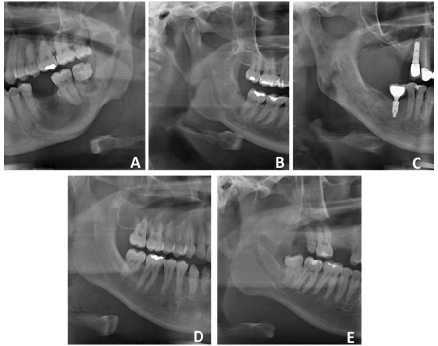
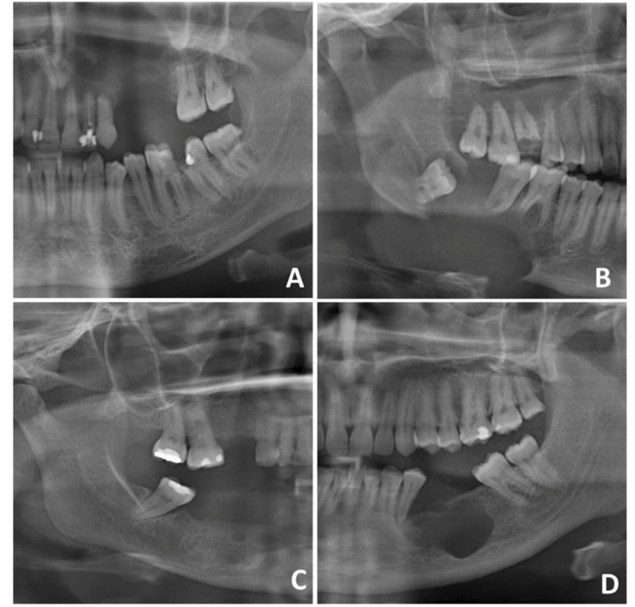
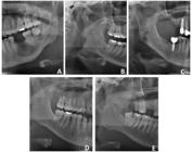
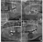
The radiographic characteristics of the lesions were analyzed, focusing on location, margins, contours, and relationship with adjacent structures. Since the analyses were performed on panoramic radiographs, the largest diameter of the lesion was not measured due to the technical limitations of the examination. Any linear measurement on panoramic radiographs cannot be considered reliable15. Three cases of OKC affected the mandible and 2 cases affected the maxilla (Figure 1); three cases of AM occurred in the mandible and only 1 in the maxilla (Figure 2). Of the 9 cases presented, 8 were multilocular and only 1 was unilocular. In 4 cases, root resorption was observed in adjacent teeth, with 2 cases of OKC and 2 cases of AM. The mandibular canal showed cortical rupture in 1 case of AM and deviation in its path in 3 cases, including 2 cases of OKC and 1 case of AM.
Fractal Analysis Characteristics and Pixel Intensity
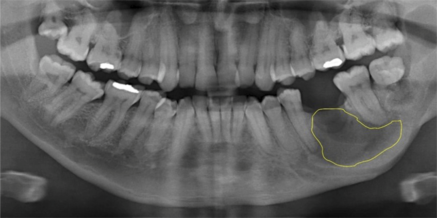
Additionally, trabecular characterization of the lesions was performed using PR. The free Image JTM software was used to calculate FD and PI values for each lesion. The first step in image analysis involved the selection of regions of interest (ROIs) for evaluation. In this study, ROIs were manually outlined by an experienced oral radiologist to cover the largest possible area of the lesion, excluding structures that could interfere with the analysis, such as dental roots, mandibular canals, and cortical bone16 (Figure 3). As eligibility criteria, images that showed better definitions were included, and those that presented overlapping anatomical structures were excluded. Thus, FD and PI analyses were performed only on lesions in the mandible, since lesions in the maxilla did not allow manual delimitation of the ROI.
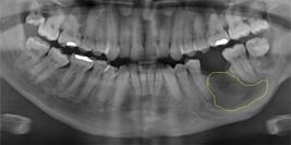
Panoramic radiograph showing the ROI manually delimited using Image J® software, in a lesion diagnosed as Odontogenic Keratocyst.
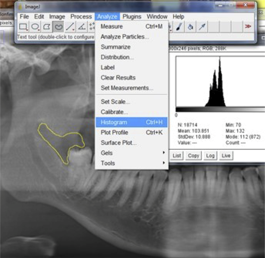
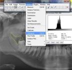
Each ROI was smoothed during image processing using a Gaussian filter (sigma = 35 pixels). This helps correct large density variations and eliminate discrepancies between similarly intense pixels, resulting in a highly blurred image. The blurred image was then subtracted from the original, and gray values were added to each pixel to prevent certain structures from standing out disproportionately. The image was then binarized with a brightness threshold of 128 and segmented into components that visually resemble trabeculae. Noise reduction followed, and the image was transformed into a one-pixel-wide line (skeletonization), enhancing texture and patterns for FD analysis (Figure 4). The FD value was calculated using the box-counting method, while the PI was measured using the Histogram tool (Figure 5)17. Both processes were performed with Image J® software.

Digital analysis of trabecular bone morphology. (A) Region of interest in trabecular bone covering the largest area of the lesion. (B) Result after blurring this region. (C) Result after subtracting (B) from (A) and adding 128. (D) Binary versions of image C. (E) Skeletonized trabecular pattern.
Discussion
OKC and AM are odontogenic lesions that can have similar clinical and imaging characteristics, but they exhibit important differences regarding their biological behaviors and treatment options. Therefore, additional information from imaging exams that enables a better radiographic interpretation of these lesions is essential for contributing to a more accurate and rapid diagnosis, which is established through microscopic evaluation and correlation with clinical-radiographic characteristics.
In this study, all patients underwent digital panoramic radiographs (PR). Although it is a two-dimensional (2D) exam, PR provides a comprehensive view of the gnathic bones and is commonly used as a diagnostic tool. A previous study assessed the impact of differences between PR and cone-beam computed tomography (CBCT) on radiographic characteristics and differential diagnosis, concluding that while CBCT offered advantages over PR, it did not significantly improve diagnostic accuracy8. Another study compared PR with CBCT for the diagnosis of AM, OKC, and dentigerous cysts (DC), finding no statistically significant difference in overall diagnostic accuracy18.
The findings of this study, consistent with several others, indicate that the posterior region of the mandible is the most common location for both AM and OKC4,5,19. Regarding patient demographics, a slight male predilection was observed, with a peak incidence in the second and third decades of life, aligning with the data from the present study. Studies have suggested that the growth of these lesions can lead to root resorption in adjacent teeth, with a higher incidence of resorption associated with AM compared to OKC, likely due to the greater aggressiveness of AM18,20. In this case series report, root resorption was observed in a ratio of 2:5 for OKC lesions and 3:4 for AM lesions. Notably, these lesions were extensive (approximately 40 mm in diameter) and were in contact with dental roots. Additionally, lesion growth resulted in disruption of the cortical of the mandibular canal (case 7-AM), the floor of the maxillary sinus (case 3-AM), and deviation of the mandibular canal path in three cases: two of OKC (cases 1 and 2) and one of AM (case 4).
Despite their distinct biological behaviors, OKC and AM can appear identical radiographically, making imaging exams crucial for the analysis of bone structures in dental clinical practice. Therefore, additional resources that enhance the objectivity and accuracy of these exams are highly desirable. Fractal dimension (FD) has been widely used to assess patterns of bone complexity in various conditions. In 2006, Tosoni et al.14 used pixel intensity (PI) and FD values to compare mandibular regions of normal, osteopenic, and osteoporotic women, detecting osteoporotic changes in trabecular bone in PR. More recently, studies have evaluated bone healing in periapical regions of teeth with apical periodontitis using FD before and after endodontic treatment. These studies found significant differences in FD values, suggesting that fractal analysis (FA) could be a valuable tool for monitoring such lesions21-23.
In the present study, the average FD values were slightly lower for AM lesions compared to OKC lesions. However, the values were quite similar in an individual analysis, making it difficult to differentiate between the two types of lesions in this sample. In contrast, the average PI values showed a more pronounced difference, indicating that AM lesions were less dense than OKC lesions. A case report published in 2022 highlighted an AM that mimicked apical periodontitis, emphasizing the need for a thorough investigation of incipient periapical lesions19. In line with this, case 6 in the present study was radiographically identified as a unilocular radiolucency with approximately 8.5 mm in diameter in an edentulous region. The patient showed no clinical symptoms and was referred based on a radiographic finding that could have been misdiagnosed as a residual cyst. Similarly, case 2 was a radiographic finding in the area corresponding to tooth 32, initially suspected to be a site of post-surgical bone repair. These examples illustrate the critical role of imaging exams in diagnosis and emphasize that small lesions or those resembling post-surgical healing or other benign conditions should not be overlooked in routine clinical evaluations by dental professionals.
In this study, the analyses were performed with ROIs manually delimited in the largest area of the lesions, excluding structures that could affect the analyses, such as dental roots, mandibular canals, and cortical bone16. In PR, the maxillary region presents many overlapping structures, making it a limitation to perform a manual ROI for FD and PI analyses on lesions in this location. Furthermore, this series of cases underscores the potential of using fractal dimension (FD) and pixel intensity (PI) analyses in panoramic radiographs as complementary tools in the assessment of gnathic bone lesions, particularly for distinguishing between ameloblastomas (AM) and odontogenic keratocysts (OKC). The findings align with existing literature, confirming that these lesions tend to have similar fractal patterns, while differences in pixel intensity may provide clues for differentiation. However, further studies with larger cohorts are essential to validate these findings and to establish robust imaging criteria for the early and accurate diagnosis of gnathic bone lesions. Expanding research in this area could significantly improve diagnostic precision, ultimately benefiting patient outcomes through earlier intervention and targeted treatment strategies.
The findings of this case series are consistent with the existing literature, particularly concerning the typical locations of AM and OKC lesions. In this sample, the average FD values did not differ between AM and OKC cases, while the average PI values suggested that AM lesions displayed less dense imaging characteristics compared to OKC. Further research with larger sample sizes is recommended to more accurately evaluate the diagnostic utility of these imaging parameters.
Acknowledgments
Financial support: Carlos Chagas Filho Foundation for Research Support of the State of Rio de Janeiro (FAPERJ). Project number: E-26/200.299/2023
References
-
1 Monteiro L, Santiago C, Amaral BD, Al-Mossallami A, Albuquerque R, Lopes C. An observational retrospective study of odontogenic cyst´s and tumours over an 18-year period in a Portuguese population according to the new WHO Head and Neck Tumour classification. Med Oral Patol Oral Cir Bucal. 2021 Jul;26(4):e482-e493. doi: 10.4317/medoral.24337.
» https://doi.org/10.4317/medoral.24337 -
2 Soluk-Tekkesin M, Wright JM. The World Health Organization Classification of Odontogenic Lesions: a summary of the changes of the 2022 (5th) edition. Turk Patoloji Derg. 2022;38(2):168-84. doi: 10.5146/tjpath.2022.01573.
» https://doi.org/10.5146/tjpath.2022.01573 -
3 Ahire MS, Tupkari JV, Chettiankandy TJ, Thakur A, Agrawal RR. Odontogenic tumors: a 35-year retrospective study of 250 cases in an Indian (Maharashtra) teaching institute. Indian J Cancer. 2018 Jul-Sep;55(3):265-72. doi: 10.4103/ijc.IJC_145_18.
» https://doi.org/10.4103/ijc.IJC_145_18 -
4 Sharma I, Venkatesh D, Bawa G, Vaseemuddin S, Joseph A, Sangtani JK. Epidemiological and clinicopathological analysis of 92 odontogenic tumors: a 5-year retrospective study. J Contemp Dent Pract. 2017 Nov;18(11):1056-60. doi: 10.5005/jp-journals-10024-2175.
» https://doi.org/10.5005/jp-journals-10024-2175 -
5 Nalabolu GRK, Mohiddin A, Hiremath SKS, Manyam R, Bharath TS, Raju PR. Epidemiological study of odontogenic tumours: an institutional experience. J Infect Public Health. 2017 May-Jun;10(3):324-30. doi: 10.1016/j.jiph.2016.05.014. Epub 2016 Jul 15.
» https://doi.org/10.1016/j.jiph.2016.05.014 -
6 Gomes JPP, Ogawa CM, Silveira RV, Castellano G, De Rosa CS, Yasuda CL, et al. Magnetic resonance imaging texture analysis to differentiate ameloblastoma from odontogenic keratocyst. Sci Rep. 2022 Nov;12(1):20047. doi: 10.1038/s41598-022-20802-7.
» https://doi.org/10.1038/s41598-022-20802-7 -
7 Robinson RA. Diagnosing the most common odontogenic cystic and osseous lesions of the jaws for the practicing pathologist. Mod Pathol. 2017 Jan;30(s1):S96-S103. doi: 10.1038/modpathol.2016.191.
» https://doi.org/10.1038/modpathol.2016.191 -
8 Lim LZ, Padilla RJ, Reside GJ, Tyndall DA. Comparing panoramic radiographs and cone beam computed tomography: Impact on radiographic features and differential diagnoses. Oral Surg Oral Med Oral Pathol Oral Radiol. 2018 Apr 17:S2212-4403(18)30888-5. doi: 10.1016/j.oooo.2018.03.019.
» https://doi.org/10.1016/j.oooo.2018.03.019 -
9 Mys K, Stockmans F, Vereecke E, van Lenthe GH. Quantification of bone microstructure in the wrist using cone-beam computed tomography. Bone. 2018 Sep;114:206-14. doi: 10.1016/j.bone.2018.06.006.
» https://doi.org/10.1016/j.bone.2018.06.006 -
10 Lennon FE, Cianci GC, Cipriani NA, Hensing TA, Zhang HJ, Chen CT, et al. Lung cancer-a fractal viewpoint. Nat Rev Clin Oncol. 2015 Nov;12(11):664-75. doi: 10.1038/nrclinonc.2015.108.
» https://doi.org/10.1038/nrclinonc.2015.108 -
11 Kato CN, Barra SG, Tavares NP, Amaral TM, Brasileiro CB, Mesquita RA, et al. Use of fractal analysis in dental images: a systematic review. Dentomaxillofac Radiol. 2020 Feb;49(2):20180457. doi: 10.1259/dmfr.20180457. Epub 2019 Aug 20.
» https://doi.org/10.1259/dmfr.20180457 -
12 Jung JH, Huh KH, Yong TH, Kang JH, Kim JE, Yi WJ, et al. Differentiation of osteosarcoma from osteomyelitis using microarchitectural analysis on panoramic radiographs. Sci Rep. 2022 Jul;12(1):12339. doi: 10.1038/s41598-022-16504-9.
» https://doi.org/10.1038/s41598-022-16504-9 -
13 Basavarajappa S, Konddajji Ramachandra V, Kumar S. Fractal dimension and lacunarity analysis of mandibular bone on digital panoramic radiographs of tobacco users. J Dent Res Dent Clin Dent Prospects. 2021 Spring;15(2):140-6. doi: 10.34172/joddd.2021.024.
» https://doi.org/10.34172/joddd.2021.024 -
14 Tosoni GM, Lurie AG, Cowan AE, Burleson JA. Pixel intensity and fractal analyses: detecting osteoporosis in perimenopausal and postmenopausal women by using digital panoramic images. Oral Surg Oral Med Oral Pathol Oral Radiol Endod. 2006 Aug;102(2):235-41. doi: 10.1016/j.tripleo.2005.08.020.
» https://doi.org/10.1016/j.tripleo.2005.08.020 -
15 Tassoker M, Akin D, Aydin Kabakci AD, Sener S. Comparison of cone-beam computed tomography and panoramic radiography for mandibular morphometry. Folia Morphol (Warsz). 2019;78(4):862-70. doi: 10.5603/FM.a2019.0031.
» https://doi.org/10.5603/FM.a2019.0031 -
16 Muraoka H, Kaneda T, Kondo T, Sawada E, Tokunaga S. Diagnostic efficacy of apparent diffusion coefficient, texture features, and their combination for differential diagnosis of odontogenic cysts and tumors. Oral Surg Oral Med Oral Pathol Oral Radiol. 2023 Jun;135(6):928-33. doi: 10.1016/j.oooo.2023.01.008.
» https://doi.org/10.1016/j.oooo.2023.01.008 -
17 Arrepia BF, Rocha TG, Medeiros AS, Ferreira MD, Fonseca-Gonçalves A, Visconti MA. The mandibular bone structure in children by fractal dimension and its correlation with pixel intensity values: a pilot study. Oral Radiol. 2023 Oct;39(4):771-8. doi: 10.1007/s11282-023-00693-8.
» https://doi.org/10.1007/s11282-023-00693-8 -
18 Cardoso LB, Lopes IA, Ikuta CRS, Capelozza ALA. Study between panoramic radiography and cone beam-computed tomography in the diagnosis of ameloblastoma, odontogenic keratocyst, and dentigerous cyst. J Craniofac Surg. 2020 Sep;31(6):1747-52. doi: 10.1097/SCS.0000000000006538.
» https://doi.org/10.1097/SCS.0000000000006538 -
19 Soimu G, Netto JD, Miranda ÁM, Perez DE, Armada L, Pires FR. Ameloblastomas mimicking apical periodontitis: a case series. Med Oral Patol Oral Cir Bucal. 2022 Jul;27(4):e397-e402. doi: 10.4317/medoral.25338.
» https://doi.org/10.4317/medoral.25338 -
20 Kitisubkanchana J, Reduwan NH, Poomsawat S, Pornprasertsuk-Damrongsri S, Wongchuensoontorn C. Odontogenic keratocyst and ameloblastoma: radiographic evaluation. Oral Radiol. 2021 Jan;37(1):55-65. doi: 10.1007/s11282-020-00425-2. Epub 2020 Feb 6. Erratum in: Oral Radiol. 2021 Oct;37(4):715-17. doi: 10.1007/s11282-021-00551-5.
» https://doi.org/10.1007/s11282-020-00425-2 -
21 Ugur Aydin Z, Toptas O, Göller Bulut D, Akay N, Kara T, Akbulut N. Effects of root-end filling on the fractal dimension of the periapical bone after periapical surgery: retrospective study. Clin Oral Investig. 2019 Sep;23(9):3645-51. doi: 10.1007/s00784-019-02967-0.
» https://doi.org/10.1007/s00784-019-02967-0 -
22 Ince Yusufoglu S, Ugur Aydin Z, Tulumbaci F, Bayrak S. Evaluation of different Apexification treatments of teeth with immature apices and apical periodontitis on the fractal dimensions of trabecular bone. Aust Endod J. 2021 Aug;47(2):163-9. doi: 10.1111/aej.12441. Epub 2020 Sep 9.
» https://doi.org/10.1111/aej.12441 -
23 Tosun S, Karataslioglu E, Tulgar MM, Derindag G. Fractal analysis and periapical index evaluation of multivisit nonsurgical endodontic retreatment: a retrospective study. Oral Surg Oral Med Oral Pathol Oral Radiol. 2022 Feb;133(2):245-51. doi: 10.1016/j.oooo.2021.08.016. Epub 2021 Aug 30.
» https://doi.org/10.1016/j.oooo.2021.08.016
-
Ethics Statement:
This study was approved by the Research Ethics Committee of the Clementino Fraga Filho University Hospital, under aproval number 5.597.208.
-
Data availability:
Datasets related to this article will be available to the corresponding author upon request.
Edited by
-
Editor:
Dr. Altair A. Del Bel Cury
Datasets related to this article will be available to the corresponding author upon request.