Physical and color stability of PMMA provisional crowns made by conventional vs digital methods: in situ study
Abstract
Aim This cross sectional in situ study aimed to evaluate the physical properties and color stability of polymethylmethacrylate (PMMA)-based resins used in provisional crowns.
Methods Twelve cubic specimens with 5mm edges of conventional self-, heat-polymerized acrylic resins, 3D-printed, and CAD-CAM milled PMMA resin were fabricated. Surface roughness, Knoop microhardness, wettability, and color stability were evaluated. Utilizing a split-mouth design, 8 cubes were randomly placed in a palatal intraoral appliance worn by 12 volunteers. Specimens were exposed to erosion extra-orally with 15 mL of cola-based soft drink (Coca-Cola®) three times daily for 5 minutes over 7 days. Statistical analysis was performed using one-way repeated measures ANOVA and Bonferroni multiple comparison analysis (α = 0.05).
Results PMMA resins exhibit comparable surface roughness and wettability under non-erosive and erosive conditions (p>0.05). However, self-polymerized and 3D-printed acrylic resins exhibited significant differences in microhardness after exposure to the erosive environment (p<0.05). CAD-CAM milled acrylic resins showed superior color stability compared to the other resins (p <0.05). The simulated erosive environment significantly alters the physical properties and color of provisional crown resins.
Conclusions These findings demonstrate that heat-polymerized and CAD-CAM acrylic resins exhibit superior behavior under non-erosive and erosive conditions, which may indicate better clinical performance, especially when indicated for longer-term use.
Keywords
Dental prosthesis; Computer-aided design; Tooth erosion; Surface properties
Introduction
Temporary fixed prostheses are essential in dentistry, used for dental rehabilitation during the curative and transitional phases until the final prosthesis is made1. Polymethylmethacrylate (PMMA) resin is one of the most used materials due to its versatility and accessibility. However, it has clinical limitations such as higher roughness compared to other materials, like indirect composite resin2. Fabrication of temporary fixed prostheses in PMMA can be conducted using direct or indirect methods, each with its own characteristics and challenges, particularly when exposed to the oral environment3.
With advancements in Digital Dentistry, new workflows have been developed for PMMA-based resins used in temporary crown fabrication4. In the additive manufacturing digital workflow, the prosthetic component is designed using CAD software and typically employs Digital Light Processing (DLP) technology to 3D-print, polymerizing liquid photosensitive resin using ultraviolet (UV) light5. Another approach is subtractive CAD-CAM milling, which involves creating a digital model for the CNC milling machine to mill pre-polymerized PMMA blocks6. Studies indicate that resins from the additive digital workflow exhibit lower flexural strength and hardness compared to those used in conventional and subtractive processes4. Moreover, printed resins show higher wear and microhardness, yet 3D-printed crowns demonstrate superior mechanical properties compared to CAD-CAM milled PMMA resin, albeit with inferior physical properties7,8.
Dental erosion, characterized by acidic dissolution of dental tissue due to intrinsic (i.e., gastroesophageal reflux) and extrinsic factors (drug use, consumption of acidic beverages), results in irreversible loss of dental mineralization and can negatively impact restorative materials, including PMMA prostheses9. This erosive environment has significant implications for the durability and functionality of restorations, leading to changes in the mechanical and aesthetic properties of provisional crowns9,10.
The intrinsic factors related to materials, such as chemical variations in resin substrates and differences in manufacturing techniques (including polymerization and post-curing), influence the physical properties and mechanical behavior of provisional crowns, affecting their clinical performance11,12. Specifically, clinical parameters of PMMA resins, such as roughness and color stability, are contentious and affected by oral environment conditions8,10. Therefore, this study aimed to evaluate the physical properties of provisional crown resins made using digital (additive and subtractive) and conventional (direct and indirect) techniques under erosive conditions.
Material and Methods
Experimental design
This double-blinded in situ study employed a split-mouth design with the following study factors: (1) the type of substrate used for provisional crowns, comprising four levels (conventional self- and heat-polymerized polymethyl methacrylate (PMMA), pre-polymerized PMMA blocks (CAD-CAM milled), and 3D-printed resin; and (2) the erosive challenge, through exposure to the beverage Coca-Cola® at two levels: no exposure (T0) and exposure (T1). The response variables evaluated included surface roughness (Ra, μm), Knoop microhardness (KHN, kgf/mm2), wettability, and color stability.
The study was approved by the Research Ethics Committee of the University Center of the Hermínio Ometto Foundation-FHO, registered under approval number 79382524.1.0000.5385. All participants provided written informed consent. A total of 12 individuals, all in good general and oral health, were selected. The inclusion criteria required participants to be between 20 and 30 years, presence of at least 30 natural teeth, absence of cavities or periodontal disease, and have a stimulated salivary flow rate within the normal range of 1.0 to 3.0 mL/min. The exclusion criteria included: history of gastroesophageal reflux, the use of medications that affect salivary flow, and the presence of a high-arched palate.
Preparation of experimental resin specimens
All specimens were fabricated in a cubic shape with 5 mm edges, following the manufacturers’ recommendations, regardless of the workflow, substrate, or experimental variable.
For the fabrication of the self-polymerized resin specimens, a prefabricated metal mold with cavities corresponding to the final specimen dimensions was used. The resin was proportioned and manipulated according to the manufacturer’s recommendations. For this purpose, a powder: liquid ratio of 3:1 was used, which was homogenized with a metal spatula in a glass mixing jar. The resin was inserted into the metal mold during its plastic phase and left for the polymerization time (10 min). After the material polymerized, the specimens were removed from the metal mold.
The heat-polymerized resin specimens were fabricated using the lost-wax technique. For this, wax cubes (Geo Classic Wax - opaque mint, Renfert, Germany) were created and cast in the same metal mold described previously. The wax cubes were invested in a flask using dental stone (GC Fujirock, Luven, Belgium). After the stone set, the wax was removed from the cavities using hot water to subsequently invest the PMMA resin. The resin was manipulated in its plastic phase according to the manufacturer’s recommendations and inserted into the previously isolated flask (Cel-Lac, SS White). The flask was then pressed (1000 kgf, for 2 hours) and subjected to the conventional polymerization cycle: the temperature was raised to 70°C and maintained for 30 minutes, then increased to 100°C for one and a half hours, after which the heat was turned off. The flask was left to cool in water (to 40°C) for approximately 20 minutes before devesting.
For the fabrication of the PMMA-based resin specimens by additive manufacturing (3D printing), a biocompatible resin in shade A2 (PriZma, Makertech Bio Prov, São Paulo, Brazil) (Table 1) and a 3D printer (Flashforge 3D Printer, Hunter model, China) were used. In this process, a metal mold was used to create a wax cube (Geo Classic Wax - opaque mint, Renfert, Germany) with 5 mm edges, which was coated with a developer spray (Spotcheck, SKD-S2) before being scanned by a desktop scanner (Up3d, UP300e model, 2020, China). The resulting image was converted into an STL file using the scanner’s software (Up 3D Dental Station). The final STL file was then sent to the 3D printer, which planned and executed the printing of the specimens.
After shaking the biocompatible liquid resin in shade A2, it was injected into the printer’s tank, which initiated the 3D printing process. The process included a curing time of 3 seconds, a base layer curing time of 15 seconds, with 85% light intensity and a layer height of 0.05 mm, totaling 112 printed layers to form the designed structure. Printing was performed using the stereolithography (DLP) method, with a wavelength of 500 nm and a power of 50 W (Anycubic Photon S, Shenzhen Anycubic Technology Co., Ltd., China). After printing, the specimens were carefully removed from the printer’s metal build platform, manually detached from the support structures, washed with isopropyl alcohol, and post-cured in a UV light chamber for 15 minutes (Anycubic Wash and Cure Machine, Shenzhen Anycubic Technology Co., Ltd., China) to complete the final curing step.
For the fabrication of the milled acrylic resin specimens, the same STL file used for the printed resin was employed, opting for the PMMA acrylic block for the CAD/CAM system (Primavita, São Paulo, Brazil) in shade A2 (Table 1). This block was milled by the Cerec Primemill milling machine (Dentsply Sirona, York, Pennsylvania, USA), using diamond burs Diamond 1.4 CS, 1.2 CS, and Bur 2.5 PMMA CS (Dentsply Sirona, Germany), at low speed and with constant irrigation, according to the planned dimensions for each analysis.
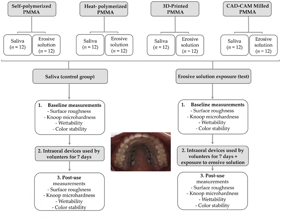
The composition and brand of all PMMA resins used in this study are presented in Table 1. After polymerization, the specimens were finished using a tungsten carbide bur and then sequentially smoothed with silicon carbide abrasive paper (#320, #400, #600, #800, and #1200). The specimens were then thoroughly rinsed with water. The dimensions of each specimen were measured with a digital micrometer (Model 293-421-20; minimum resolution: 0.001 mm; Mitutoyo, Kawasaki, Japan). After measurement, the specimens were ultrasonically cleaned in distilled water for 10 min They were then rinsed again, immersed in distilled water, and stored at 37°C for 48 h13. Subsequently, the specimens were transferred to a 100% humidity environment maintained at 7°C under refrigeration until further use14,15. To ensure consistency, all groups underwent the same finalization protocol, and all procedures were performed by a single researcher. Complete blinding of the study was not possible due to the distinct colors of the resins (66 and A2). However, we have ensured that the numerical coding of the samples was performed by a research assistant who was not involved in the study objectives or the evaluation of the results to minimize bias. The schematic diagram of the experimental design is detailed in Figure 1. Immediately prior (Baseline) and following the in situ phase (Pos-exposure to saliva/erosive), the specimens underwent a comprehensive evaluation of the following parameters:
Surface Roughness
Surface roughness was quantitatively assessed using a Surfcorder SE 1700 profilometer (Kosaka Laboratory Ltd., Tokyo, Japan). The instrument was calibrated to operate at a constant scanning speed of 0.5 mm/s, with a measurement length of 0.8 mm and a resolution of 0.0001 µm. The surface roughness values (Ra), expressed in micrometers (µm), were determined by calculating the arithmetic mean of three independent measurements taken at different locations on each specimen to ensure representative sampling16.
Knoop Microhardness
The evaluation of surface Knoop microhardness was conducted using a microdurometer (Shimadzu, model HMV-2000, Kyoto, Japan). A Knoop diamond indenter was used to create indentations on the specimen surface with a load of 25 grams for a duration of 5 s16. Five indentations were made on each specimen, spaced 500 µm apart to prevent interaction between the measurement sites. The Knoop microhardness (KHN) was calculated using the formula: , where (14228) is a constant, (c) is the applied load (25g), and (d) is the length (µm) of the longest diagonal of the indentation.
Wettability
The wettability was determined using the a Ramé-Hart 500 goniometer (Ramé-Hart Instrument Co., NJ, USA). Sessile drops of 15 µL of distilled water were dispensed onto the specimen surface using an automated dispensing system integrated with the goniometer (Ramé-Hart Automated Dispensing System; Ramé-Hart Instrument Co., NJ, USA). Images of the sessile drops were captured immediately after deposition, and the contact angles were automatically calculated using the Ramé-Hart DROPimage Standard software (Ramé-Hart Instrument Co., NJ, USA).
Color changes
Initial color measurements were carried out using a spectroradiometer (Easyshade Advance -VITA, Vita Zahnfabrik, Germany). Spectral reflectance data was collected for each specimen at wavelengths ranging from 380 to 780 nm at 5 nm intervals. Thereafter, these measurements were converted into CIELAB parameters. The device has previously demonstrated high validity and reproducibility in assessing the color of dental restoration materials17,18.
After collecting the baseline color, the specimens were placed in the intraoral devices used by volunteers, as described in detail in the next section. The color change (∆E) of each specimen was determined19 by comparing the measurements performed before and after exposure to saliva only or to saliva combined with an erosive solution (Coca-Cola). The L*, a* and b* values obtained were used to quantify the color changes, calculated using the following formula:
Where ,, and ; where i is referred to as the initial color measurement and f as the final color measurement.
In situ erosive challenge
For the in situ phase, a custom-made intraoral device similar to a modified Hawley retainer with eight cavities was fabricated to hold pre-labeled specimens20, mounted with sticky wax in a split-mouth design to reduce bias. Only the surface of the right hemiarch was exposed for the erosive challenge. The devices were disinfected with 4% chlorhexidine, stored in water, and adjusted for fit. Volunteers were instructed to clean the device three times a day, avoid eating or drinking while wearing it, and store it in a moist environment. The devices were worn continuously, except during meals and erosion procedures. For the erosive challenge, the intraoral device containing the specimens was removed from the mouth, and the cavities corresponding to the erosive group were immersed in 15 mL of Coca-Cola® for 5 minutes, three times daily for 7 days21. The other hemiarch of the device, with the specimens from the control group, was exposed exclusively to the volunteers’ natural saliva during use.
Statistical analysis
The data were assessed for normality using Shapiro–Wilk method. To compare the different restorative materials and to verify the influence of evaluation time (baseline, saliva or treatment) on the response variables (physical properties and color stability variables), one-way repeated measures ANOVA was performed. Multiple pairwise comparisons were examined with the Bonferroni post-hoc test. A mean difference significant at the 0.05 level was used for all tests. Statistical analyses were performed with statistical software (IBM SPSS 26).
Results
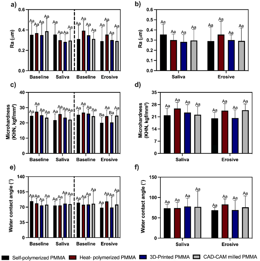
The results of the physical properties of the specimens, both prior to (baseline) and following the in situ phase for the specimens subjected and not subjected to erosive challenges, is presented in Figure 2. No statistically significant differences in surface roughness were observed based on the type of resin neither between the different erosive and non-erosive exposure solutions (p > 0.05). Regarding microhardness, no statistically significant differences were observed for resins exposed to non-erosive environment (saliva). Conversely, significant differences were observed for self-polymerized acrylic resin and 3D-printed acrylic resin (p < 0.05), which showed a reduction in microhardness after exposure to erosive environment compared to the heat-polymerized acrylic resin and CAD-CAM milled acrylic resin. In the presence or absence of an erosive challenge, wettability presented no significant differences between the different types of resin and at both saliva and erosive conditions exposure.
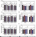
Physical properties by group (n = 12) and timepoints: (a) Specimens roughness (Ra) at baseline and after exposure to saliva and at baseline and after exposure to erosive environment; (b) Roughness (Ra) for specimens after exposure to saliva versus erosive environment; (c) Specimens microhardness at baseline and after exposure to saliva and at baseline and after exposure to erosive environment; (d) Microhardness for specimens after exposure to saliva versus erosive environment; (e) Specimens wettability at baseline and after exposure to saliva and at baseline and after exposure to erosive environment; (f) Wettability for specimens after exposure to saliva versus erosive environment. Capital letters compare the same surface at different moments. Lower case letters compare different surfaces within the same moment of evaluation (baseline, saliva, erosive environment).
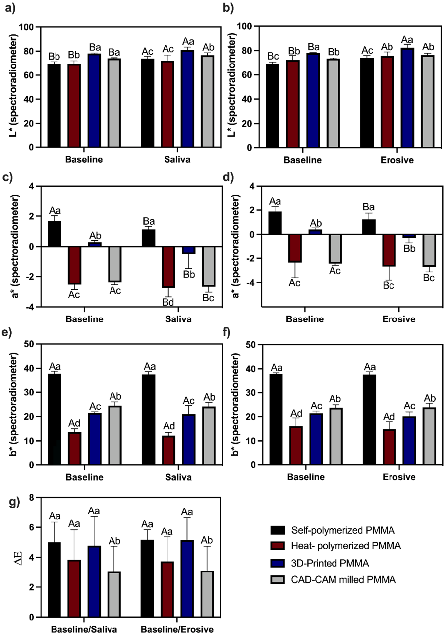
The statistical analysis of the luminosity values (L*) showed that the different materials have different degrees of luminosity (Figure 3a, 2b). Considering the color measurements for the baseline and after exposure to the oral environment with and without an erosive condition PMMA resins showed increased luminosity after saliva and erosive solution exposure compared with the baseline analysis. For a* values indicating red-green color components (Figure 3c, 2d), positive values characterizing the red component were higher for self-polymerized PMMA and 3D printed PMMA. After exposure to the oral environment with and without an erosive condition, the red-green color components showed significant differences compared with baseline analysis for all resin types (p < 0.05). Yellowish appearance represented by b* values was higher for self-polymerized PMMA, followed by CAD-CAM milled PMMA, 3D printed PMMA, and heat-polymerized PMMA surfaces (p < 0.05) (Figure 3e, 2f). Exposure to the oral environment with and without an erosive did not change the yellowish appearance of the evaluated surfaces. The results obtained for color change are observed in Figure 3g. As regards the ΔE values, CAD-CAM milled PMMA showed lower color changes compared to the other resin tested at both conditions erosive and non-erosive environment exposure.
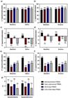
L*, a*, b*, and ΔE values by group (n = 12) and timepoints: (a–f) L*, a*, b* at baseline and after non-erosive/erosive exposure; (g) ΔE between baseline and post-exposure. Capital letters compare the same surface at different moments. Lower case letters compare different surfaces within the same moment of evaluation (baseline, saliva, erosive environment).
Discussion
The resistance of provisional crowns to erosion and chemical degradation is a critical determinant of their clinical performance, underscoring the importance of understanding resin behavior under erosive conditions to guide material selection. In this study, microhardness and color stability varied by resin type, and the simulated erosive environment affected both conventional and digital flow resins.
In this study, after the polishing protocol, surface roughness was consistently obtained across all experimental groups, ensuring consistent initial roughness values. Interestingly, it was observed that the exposure to a non-erosive oral environment resulted in no significant reduction in surface roughness across all resin types. The results for the erosive conditions can be explained by the exposure method used. The dropping of erosive solutions allowed only a short period of exposure, which was designed to simulate the brief, repetitive acidic challenges in the oral cavity. While this may not have been enough to change the roughness of the surfaces, it indicates a high initial resistance of the materials to such discrete erosive events.
Previous studies have established a strong correlation between surface roughness and other physical properties of PMMA acrylic resins, such as hardness22,23. CAD-CAM milled resins, known for their denser and more homogeneous structure, typically demonstrate superior resistance to chemical erosion and wear compared to other resins used in different fabrication workflows23. In this in situ study, self-polymerized PMMA and 3D-printed PMMA specimens exhibited a significant reduction in microhardness after exposure to erosive environment. The lower physical stability of these resins is likely attributable to their comparatively lower degree of conversion, resulting in a higher concentration of unreacted monomers and a less densely cross-linked matrix that is more susceptible to hydrolytic degradation in erosive environments. The stability of microhardness observed in all PMMA substrates following salivary exposure suggests a comparable resistance to chemical degradation within the oral environment. This integrity is attributed to the material’s intrinsic compositional stability. The polymer’s hydrolysis-resistant carbon-carbon structure, coupled with low water sorption and solubility, exhibits behaviors consistent between conventional and additive-manufactured PMMA, mitigating significant hydrolytic degradation. Furthermore, because these PMMA-based resins formulated for provisional restorations lack labile silicate fillers and susceptible coupling agents, primary chemical degradation pathways may be diminished, collectively preserving surface microhardness24.
Knoop microhardness is a key indicator of a material’s surface mechanical properties, reflecting its surface stability and resistance to near-surface plastic deformation. The stability of microhardness values in this study indicates that the tested PMMA substrates maintained their surface integrity against the erosive challenges applied25. This observation aligns with the hypothesis that the hydrolysis of ester radicals in PMMA monomers, which could potentially weaken the resin matrix, did not occur to a significant extent under the experimental conditions employed. This may be attributed to the inherent chemical stability of PMMA resins, which limits the extent of hydrolytic degradation even in acidic environments26. Furthermore, the stability of microhardness across resin types highlights the importance of considering specific experimental conditions, such as the duration and intensity of exposure to erosive agents. These factors play a critical role in determining the extent of PMMA substrate degradation and must be carefully considered to ensure clinically relevant conclusions.
The present findings indicated that wettability presented no significant differences between the resin types under either salivary or erosive conditions. This result aligns with the fundamental surface properties of methacrylate-based polymers. PMMA is characterized as a weakly polar substrate with low surface-free energy, which generally confers a baseline hydrophobicity and reduces its interaction with polar liquids26. The lack of significant variation observed across groups suggests that the different resin formulations, whether conventional or digitally manufactured, share this core physicochemical characteristic. Consequently, the similar wettability profiles indicate that all tested materials would be expected to exhibit comparable initial biological responses, such as salivary pellicle formation and biofilm adhesion, regardless of the chemical challenge. This surface property homogeneity is a favorable finding, as the inherent hydrophobicity of these PMMA resins may contribute to their clinical performance by minimizing fluid-mediated degradation.
Concerning color stability, CAD-CAM milled PMMA resins have shown lower color changes. Notably, 3D-printed and CAD-CAM milled PMMA resins have been established as a benchmark for superior surface topography27. Indeed, resins such as self-polymerized PMMA and 3D printed PMMA may show greater color changes due to the higher content of residual monomers28, as well as the presence of different types of pigment. These factors may be associated with both the lower color stability observed and the lower microhardness identified after exposure to the erosive environment. Finally, the results should be interpreted with caution, as they were obtained under controlled conditions with a specific erosive agent. Future in situ studies and clinical trials are needed to validate the findings and improve PMMA resin performance in more realistic oral environments.
Under the conditions of this study, heat-polymerized and CAD-CAM acrylic resins demonstrated superior color stability compared to self-polymerized and 3D-printed resins when exposed to erosive and salivary environments. As no significant differences in microhardness were found among the materials, their clinical performance for longer-term use may be primarily influenced by this enhanced resistance to discoloration.
Acknowledgments
None.
References
-
1 Burns DR, Beck DA, Nelson SK; Committee on Research in Fixed Prosthodontics of the Academy of Fixed Prosthodontics. A review of selected dental literature on contemporary provisional fixed prosthodontic treatment: report of the Committee on Research in Fixed Prosthodontics of the Academy of Fixed Prosthodontics. J Prosthet Dent. 2003 Nov;90(5):474-97. doi: 10.1016/s0022-3913(03)00259-2.
» https://doi.org/10.1016/s0022-3913(03)00259-2 -
2 Narde J, Ahmed N, Keskar V, Pandurangan KK. Evaluation of the colour stability and surface roughness of polymethylmethacrylate and indirect composites with and without ageing: an in-vitro study. Cureus. 2024 Aug;16(8):e68073. doi: 10.7759/cureus.68073.
» https://doi.org/10.7759/cureus.68073 -
3 Regish KM, Sharma D, Prithviraj DR. Techniques of fabrication of provisional restoration: an overview. Int J Dent. 2011;2011:134659. doi: 10.1155/2011/134659.
» https://doi.org/10.1155/2011/134659 -
4 Lourinho C, Salgado H, Correia A, Fonseca P. Mechanical properties of polymethyl methacrylate as denture base material: heat-polymerized vs. 3D-printed-systematic review and meta-analysis of in vitro studies. Biomedicines. 2022 Oct;10(10):2565. doi: 10.3390/biomedicines10102565.
» https://doi.org/10.3390/biomedicines10102565 -
5 Piedra-Cascón W, Sadeghpour M, Att W, Revilla-León M. A vat-polymerized 3-dimensionally printed dual-material occlusal device: A dental technique. J Prosthet Dent. 2021 Sep;126(3):271-5. doi: 10.1016/j.prosdent.2020.07.011. Epub 2020 Sep 18.
» https://doi.org/10.1016/j.prosdent.2020.07.011 -
6 Jalalian E, Younesi F, Golalipour S, Khorshidi S, Mahdavisaedabadi SH, Sayyari M. Assessment of marginal and internal adaptation in provisional crowns utilizing three distinct materials. J Contemp Dent Pract. 2023 Nov;24(11):853-8. doi: 10.5005/jp-journals-10024-3589.
» https://doi.org/10.5005/jp-journals-10024-3589 -
7 Jain S, Sayed ME, Shetty M, Alqahtani SM, Al Wadei MHD, Gupta SG, et al. Physical and mechanical properties of 3D-printed provisional crowns and fixed dental prosthesis resins compared to cad/cam milled and conventional provisional resins: a systematic review and meta-analysis. Polymers (Basel). 2022 Jun;14(13):2691. doi: 10.3390/polym14132691.
» https://doi.org/10.3390/polym14132691 -
8 Alzahrani SJ, Hajjaj MS, Azhari AA, Ahmed WM, Yeslam HE, Carvalho RM. Mechanical properties of three-dimensional printed provisional resin materials for crown and fixed dental prosthesis: a systematic review. Bioengineering (Basel). 2023 May;10(6):663. doi: 10.3390/bioengineering10060663.
» https://doi.org/10.3390/bioengineering10060663 -
9 Wayakanon P, Narakaew T, Wayakanon K. Effects of various beverages on characteristics of provisional restoration materials. Clin Exp Dent Res. 2024 Apr;10(2):e842. doi: 10.1002/cre2.842.
» https://doi.org/10.1002/cre2.842 -
10 Sayed ME, Jain S, Jokhadar HF, Alshahrani AA, AlResayes SS, Alqahtani SM, et al. Effect of smokeless tobacco on color stability and surface roughness of CAD/CAM milled, 3D printed, and conventional provisional crown and fixed dental prosthesis materials: an in vitro study. Technol Health Care. 2024;32(3):1697-711. doi: 10.3233/THC-230723. Retraction in: Technol Health Care. 2026 Jan;34(1):34-5. doi: 10.1177/09287329251390260.
» https://doi.org/10.3233/THC-230723 -
11 Bergamo ETP, Campos TMB, Piza MMT, Gutierrez E, Lopes ACO, Witek L, et al. Temporary materials used in prosthodontics: the effect of composition, fabrication mode, and aging on mechanical properties. J Mech Behav Biomed Mater. 2022 Sep;133:105333. doi: 10.1016/j.jmbbm.2022.105333.
» https://doi.org/10.1016/j.jmbbm.2022.105333 -
12 Ellakany P, Fouda SM, Mahrous AA, AlGhamdi MA, Aly NM. Influence of CAD/CAM milling and 3D-printing fabrication methods on the mechanical properties of 3-unit interim fixed dental prosthesis after thermo-mechanical aging process. Polymers (Basel). 2022 Sep;14(19):4103. doi: 10.3390/polym14194103.
» https://doi.org/10.3390/polym14194103 -
13 Iwaki M, Kanazawa M, Arakida T, Minakuchi S. Mechanical properties of a polymethyl methacrylate block for CAD/CAM dentures. J Oral Sci. 2020 Sep;62(4):420-2. doi: 10.2334/josnusd.19-0448.
» https://doi.org/10.2334/josnusd.19-0448 -
14 Unemori M, Matsuya Y, Matsuya S, Akashi A, Akamine A. Water absorption of poly(methyl methacrylate) containing 4-methacryloxyethyl trimellitic anhydride. Biomaterials. 2003 Apr;24(8):1381-7. doi: 10.1016/s0142-9612(02)00521-5.
» https://doi.org/10.1016/s0142-9612(02)00521-5 -
15 Henderson JY, Korioth TVP, Tantbirojn D, Versluis A. Failure load of milled, 3D-printed, and conventional chairside-dispensed interim 3-unit fixed dental prostheses. J Prosthet Dent. 2022 Feb;127(2):275.e1-275.e7. doi: 10.1016/j.prosdent.2021.11.005. Epub 2021 Dec 10.
» https://doi.org/10.1016/j.prosdent.2021.11.005 -
16 Morel LL, Almeida MVR, Santos KMD, Praseres MF, Gonçalves Girundi AL, Alexandrino LD, et al. Reinforcement of 3D-printed resins for denture base by adding aramid fibers: effect on mechanical, surface, and optical properties. J Prosthodont. 2024 Sep 24. doi: 10.1111/jopr.13957.
» https://doi.org/10.1111/jopr.13957 -
17 Schelkopf S, Dini C, Beline T, Wee AG, Barão VAR, Sukotjo C, et al. The effect of smoking and brushing on the color stability and stainability of different CAD/CAM restorative materials. Materials (Basel). 2022 Oct;15(19):6901. doi: 10.3390/ma15196901.
» https://doi.org/10.3390/ma15196901 -
18 Akl MA, Sim CPC, Nunn ME, Zeng LL, Hamza TA, Wee AG. Validation of two clinical color measuring instruments for use in dental research. J Dent. 2022 Oct;125:104223. doi: 10.1016/j.jdent.2022.104223.
» https://doi.org/10.1016/j.jdent.2022.104223 -
19 Paravina RD, Ghinea R, Herrera LJ, Bona AD, Igiel C, Linninger M, et al. Color difference thresholds in dentistry. J Esthet Restor Dent. 2015 Mar-Apr;27 Suppl 1:S1-9. doi: 10.1111/jerd.12149.
» https://doi.org/10.1111/jerd.12149 -
20 Lyros I, Tsolakis IA, Maroulakos MP, Fora E, Lykogeorgos T, Dalampira M, et al. Orthodontic retainers-a critical review. Children (Basel). 2023 Jan;10(2):230. doi: 10.3390/children10020230.
» https://doi.org/10.3390/children10020230 -
21 Rios D, Honório HM, Francisconi LF, Magalhães AC, Machado MAAM, Buzalaf MA. In situ effect of an erosive challenge on different restorative materials and on enamel adjacent to these materials. J Dent. 2008 Feb;36(2):152-7. doi: 10.1016/j.jdent.2007.11.013.
» https://doi.org/10.1016/j.jdent.2007.11.013 -
22 Wesemann C, Spies BC, Sterzenbach G, Beuer F, Kohal R, Wemken G, et al. Polymers for conventional, subtractive, and additive manufacturing of occlusal devices differ in hardness and flexural properties but not in wear resistance. Dent Mater. 2021 Mar;37(3):432-42. doi: 10.1016/j.dental.2020.11.020. Epub 2020 Dec 4
» https://doi.org/10.1016/j.dental.2020.11.020 -
23 Tinastepe N, Malkondu O, Kazazoglu E. Hardness and surface roughness of differently processed denture base acrylic resins after immersion in simulated gastric acid. J Prosthet Dent. 2023 Feb;129(2):364.e1-364.e9. doi: 10.1016/j.prosdent.2022.12.001.
» https://doi.org/10.1016/j.prosdent.2022.12.001 -
24 Altalla H, Alawawda O, Bayindir F. Three-dimensionally printed versus conventional heat-polymerized PMMA denture base properties: a systematic review and meta-analysis. J Prosthet Dent. 2025 Dec;134(6):2191.e1-2191.e12. doi: 10.1016/j.prosdent.2025.08.014.
» https://doi.org/10.1016/j.prosdent.2025.08.014 -
25 Yildirim M, Aykent F, Özdogan MS. Comparison of fracture strength, surface hardness, and color stain of conventionally fabricated, 3D printed, and CAD-CAM milled interim prosthodontic materials after thermocycling. J Adv Prosthodont. 2024 Apr;16(2):115-25. doi: 10.4047/jap.2024.16.2.115.
» https://doi.org/10.4047/jap.2024.16.2.115 -
26 Tseng CC, Lin PY, Kirankumar R, Chuang ZW, Wu IH, Hsieh S. Surface degradation effects of carbonated soft drink on a resin based dental compound. Heliyon. 2021 Mar;7(3):e06400. doi: 10.1016/j.heliyon.2021.e06400.
» https://doi.org/10.1016/j.heliyon.2021.e06400 -
27 Kumari MK, Mohapatra A, Rath S, Choudhury GK, Garhnayak M, Mohapatra A. A comparative analysis of bacterial adhesion on different long-term provisional fixed prostheses fabricated by CAD/CAM Milling, 3D printing, and heat cure technique: an in vivo study. J Pharm Bioallied Sci. 2024 Feb;16(Suppl 1):S918-S20. doi: 10.4103/jpbs.jpbs_1096_23.
» https://doi.org/10.4103/jpbs.jpbs_1096_23 -
28 Srinivasan M, Chien EC, Kalberer N, Alambiaga Caravaca AM, Castelleno AL, Kamnoedboon P, et al. Analysis of the residual monomer content in milled and 3D-printed removable CAD-CAM complete dentures: an in vitro study. J Dent. 2022 May;120:104094. doi: 10.1016/j.jdent.2022.104094.
» https://doi.org/10.1016/j.jdent.2022.104094
-
Data availability:
Datasets related to this article will be available upon request to the corresponding author.
Edited by
-
Editor:
Dr. Altair A. Del Bel Cury
Datasets related to this article will be available upon request to the corresponding author.