The Casado (2017) method for sex estimation of contemporary brazilian skulls: a validation study
Abstract
Casado (2017) described a low-cost and practical method for sex estimation of adult skulls using precision calipers to evaluate anatomical landmarks.
Aim This study aimed to validate this method in Brazilian skulls. Methods: Measurements of 202 skulls (118 male and 84 female) were collected, according to Casado (2017).
Results The results were very promising. Except for the external occipital protuberance, the anatomical landmarks were related to sex (p < 0.0001). The anatomical landmarks from both sexes were included, and 72.16% of the skulls were correctly classified. The individual percentage per sex was similar, with 71.40% accuracy in male skulls and 72.92% in female skulls.
Conclusion The method efficiently identified sex differences in anatomical landmarks of Brazilian skulls regardless of age and ancestry.
Keywords
Forensic sciences; Craniometry; Forensic anthropology; Skull; Sex differences
Introduction
The worldwide population increased from one to six billion over the past 200 years, especially in urban areas. The number of deaths caused by diseases, natural disasters, and violent crimes also increased, highlighting the importance of person identification using their bones1.
Currently Brazil has over 210 million inhabitants and high miscegenation, which provides heterogeneous characteristics in the population, hindering forensic anthropology2,3. Miscegenation provided heterogeneous characteristics in the Brazilian population, making forensic anthropology challenging in this population3. Moreover, studies indicate secular skull changes in other populations4-8 however, whether these changes affected the Brazilian population is unclear.
The efficacy of sex estimation using only anatomical landmarks is not a consensus9. However, the skull is considered one of the best tools for sex estimation in the lack of pelvic or postcranial elements. The skull is resistant to adverse conditions and can be easily submitted to quantitative evaluations3,10-12. In addition, the best way to achieve efficient sex estimation is by using many variables13-15.
The usual method to identify sex from adult skulls comprises a quantitative or visual analysis of skeletal traits using an ordinal scale; its accuracy relies on a trained evaluator. Imaging tests are also widely used to study sexual dimorphism with high accuracy16-18. However, the high-cost equipment hinders access in many forensic analysis institutes19,20.
Simple, low-cost, and accessible techniques (e.g., precision calipers) for sex estimation that requires little training have demonstrated high reliability21. Casado11 (2017) proposed a low-cost and easy-to-replicate method using precision calipers compared with computed tomography. In Brazil, low-cost methods for sex estimation are important due to the miscegenation and varied socioeconomic characteristics between regions. Therefore, this study aimed to test and validate the method proposed by Casado11 to identify sexual dimorphism in Brazilian skulls.
Materials and Methods
This cross-sectional study was conducted at the Skull Museum of the Federal University of São Paulo, Brazil. Ethical approval was obtained from the University’s Ethics and Research Committee (no.: 3.035.353/2018).
Sample
Dry adult skulls with complete biological profiles (sex, age, and ancestry) were included. Those with fragmentation or trauma that hindered or could alter the measurements were excluded. From the original sample (n = 403), only 202 skulls (118 male and 84 women) were selected for analysis (Table 1).
The skulls were from deaths documented between 1933 and 1970, representing changes in a modern population4-6,8,22.
A precision compass (Mitutoyo®, São Paulo, Brazil) was used to measure the linear distances between the anatomical landmarks to determine the external projection of each trace. A digital depth caliper (Digimess®, São Paulo, Brazil) was used to measure the depth of bony protuberances and capture the three-dimensional nature of an anatomical landmark11.
Procedures
Twelve measurements were performed on six anatomical landmarks: three sites of muscle attachment (external occipital protuberance, nuchal protuberance [right {R}, left {L}, and center], and mastoid process [R and L]) and three anatomical landmarks evaluated due to hormonal differences (glabella, supraorbital ridge [R and L], and frontal boss [R, L, and center])11. The skulls were fixed on specific support, and one evaluator performed all measurements. For each measurement, the evaluator moved the depth compass (i.e., the central arm) to find the most protruding point of each anatomical landmark to determine the external projection. The measurements were systematized and followed the order described in Table 2.
Statistical analysis
The Shapiro-Wilk test analyzed the data distribution. The Mann-Whitney verified the relationship between sex and skull anatomical landmarks measurements. The multivariate analysis using discriminant functions was applied to find which group the skulls belonged to (0 = male; 1 = female). The Chi-square test analyzed the association between the results and the expected distribution. The analyses were performed in GraphPad Prism®, and the significance was set at p < 0.05.
To set the cut-off point for sex estimation accuracy, we considered the mean, standard deviation, and value range of each sex. The cut-off point was used to estimate the sex for each measurement. Then, the estimative was divided by the correct number of skulls from each sex. The same calculus was made for the overall classification.
Results
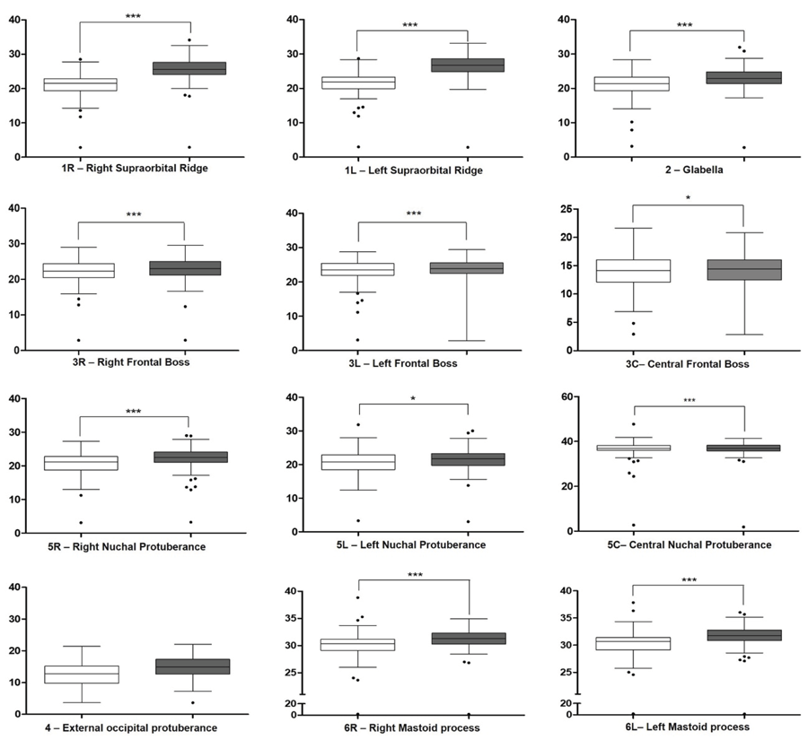
The sample was not normally distributed. Except for the external occipital protuberance, the anatomical landmarks were related to sex (Table 3, Figure 1).
Boxplot of Shapiro-Wilk distribution. White boxes represent male skulls and gray boxes represent female skulls. ***p < 0.001; *p < 0.05.
The measures differed between the sexes (p=0.0009), showing that the method effectively identified a sexual dimorphism in the skulls (Table 4).
In the general classification, we found an overall accuracy of 72.16%, including all anatomical landmarks. Moreover, the accuracy in each group was similar (71.40% accuracy in male and 72.92% in female skulls) (Table 5).
The accuracy values were separated by anatomical landmark and sex (Table 6); values varied between 64.30% and 82.20%. The anatomical landmarks that least identified the sexual dimorphism were the external occipital protuberance on male skulls (66.10%) and the left supraorbital ridge on female skulls (64.30%). Additionally, the ones that most identified it were the central nuchal protuberance (82.20%) on male skulls and the left frontal boss (82.10%) on female skulls.
Discussion
Although studies indicated secular changes in the skulls4-8, whether they affected the sexual dimorphism in Brazilian skulls is unclear. The evaluated adult skulls were similar to those analyzed by Casado11(2017). However, our sample included skulls from mixed-race people due to the miscegenation of the Brazilian population from immigration or migratory movements2,3. Thus, validating a method in Brazilian skulls must consider every ethnic group.
Although Brazilians have varied ethnic groups, their skulls exhibit similar sexual dimorphism. Studies stated that most differences exist within geographic populations, not between them23-25. However, Brazilian forensic anthropologists suggested that bones must be analyzed in each population to identify sexual differences, considering the notorious level of variation and miscegenation of contemporary human beings1,3,12,26.
The methods used to determine the external projection of anatomical landmarks can be subjective or objective. Subjective methods, such as visual inspection, relies on an experienced evaluator, taking time and being costly. Objective methods use measurements to quantify the anatomical landmarks and compare intra- and inter-populations. In the latter, the geometric analysis of photographs and imaging tests depend on specific software, computers, and specialized evaluators. Imaging tests are high-priced and far from reality in most forensic institutes.
In this study, the depth caliper measured and captured the three-dimensional nature of the anatomical landmarks of the skulls. Our results showed that using calipers is simple and effective and achieves values similar to traditional methods (visual inspection or imaging) in adult skulls, they are affordable, easy, and do not require specific training or long-time experience, making their use accessible to identify sexual dimorphism3,12,21,26-28. Although some studies separated the skulls into ethnic groups, this study considered all skulls as a single group.
In this study, the sex estimation accuracy was similar to the values found by Casado11(2017) (71.4% versus 69.6% for men; 72.9% versus 74.7% for women, respectively). The overall classification (72.20%) was also the same as presented by Casado11 (2017), suggesting high accuracy and reliability of the method. Walker25(2008) obtained similar results using visual inspection (78% accuracy for Native Americans and between 84% and 88% for Non-natives). In addition, Garvin et al.29 (2014) reported 82% accuracy for the overall sample and between 63% and 79% for each group, Krüger et al.30 (2015) found between 68% and 84% in the overall sample (without considering ancestry), and Godde et al.31 (2018) 80% accuracy in the overall sample.
Another method for sex estimation is the analysis of the geometry of anatomical landmarks in photographs. Nikita and Michopoulou13 (2018) reported accuracy of 85.10% and 94.60% in identifying male and female skulls, respectively. Also, Nikita32 (2019) reported 87% accuracy for the overall sample. Both studies used Greek skulls, a population with little miscegenation, possibly justifying the high accuracy. González-Colmenares et al.33 (2019) used the same technique in radiographs of Colombian skulls and found 88.60% accuracy. Other authors performed the same technique using computed tomography: Kimmerle et al.16 (2008) reported 77.80% accuracy; Bulut et al.34 (2016), 77.50%; and Čechová et al.35(2021), 72.80%. All values were similar to ours (72.20%), suggesting the reliability of our results and allowing method comparison.
Petaros et al.15 (2021), examined the volume of mastoid processes in Medieval Croatian skulls using visual inspection, precision calipers, and geometric analysis and obtained 67.1%, 69.4%, and 81.0% accuracy, respectively. This study was relevant to understand the contribution of each method. The more qualitative and quantitative information we can extract from the skulls, the greater the identification accuracy, greater population specificity susceptible to secular changes, and lesser challenge in the judicial sphere once judicial precedents require accurate and quantifiable methods to be presented in forensic testimony12. Therefore, we believe that the method proposed by Casado11(2017) has high admissibility in court.
In conclusion, although the method proposed by Casado was developed for American skulls, we found similar measure values in Brazilian skulls. Moreover, the method efficiently classified the sexual dimorphism of Brazilian skulls, regardless of age and ancestry.
References
-
1 Mello-Gentil T, Souza-Mello V. Contributions of anatomy to forensic sex estimation: focus on head and neck bones. Forensic Sci Res. 2021 Jul;7(1):11-23. doi: 10.1080/20961790.2021.1889136.
» https://doi.org/10.1080/20961790.2021.1889136 -
2 Cunha E, Lopez-Capp TT, Inojosa R, Marques SR, Moraes LOC, Liberti E, et al. The Brazilian identified human osteological collections. Forensic Sci Int. 2018 Aug;289:449.e1-449.e6. doi: 10.1016/j.forsciint.2018.05.040.
» https://doi.org/10.1016/j.forsciint.2018.05.040 - 3 Gomes SL, Cardozo AFC, Cavalcante DFB, Daruge Júnior E, Ulbricht V, Pereira Neto JS, et al. Sex determination by means of linear measurements and skull areas of brazilian adults. Rev Bras Odontol Leg 2020;7(3):87-96.
- 4 Jantz RL, Meadows Jantz L. Secular change in craniofacial morphology. Am J Hum Biol. 2000 May;12(3):327-38. doi: 10.1002/(SICI)1520-6300(200005/06)12:3<327::AID-AJHB3>3.0.CO;2-1.
-
5 Malina RM, Peña Reyes ME, Tan SK, Buschang PH, Little BB, Koziel S. Secular change in height, sitting height and leg length in rural Oaxaca, southern Mexico: 1968-2000. Ann Hum Biol. 2004 Nov-Dec;31(6):615-33. doi: 10.1080/03014460400018077.
» https://doi.org/10.1080/03014460400018077 - 6 Cridlin, S. An investigation of secular change in the craniofacial morphology of white and black males and females in the United States. In: American Journal of Physical Anthropology. New York: John Wiley & Sons; 2006. p.79.
-
7 Weisensee KE, Jantz RL. Secular changes in craniofacial morphology of the Portuguese using geometric morphometrics. Am J Phys Anthropol. 2011 Aug;145(4):548-59. doi: 10.1002/ajpa.21531.
» https://doi.org/10.1002/ajpa.21531 -
8 Spradley K, Stull KE, Hefner JT. Craniofacial secular change in recent mexican migrants. Hum Biol. 2016 Jan;88(1):15-29. doi: 10.13110/humanbiology.88.1.0015.
» https://doi.org/10.13110/humanbiology.88.1.0015 -
9 Spradley MK, Jantz RL. Sex estimation in forensic anthropology: skull versus postcranial elements. J Forensic Sci. 2011 Mar;56(2):289-96. doi: 10.1111/j.1556-4029.2010.01635.x.
» https://doi.org/10.1111/j.1556-4029.2010.01635.x - 10 Gupta AD, Banerjee A, Kumar A, Rao SR, Jose J. Discriminant function analysis of mastoid measurements in sex determination. J Life Sci 2012;4(1):1-5.
-
11 Casado AM. Quantifying sexual dimorphism in the human cranium: a preliminary analysis of a novel method. J Forensic Sci. 2017 Sep;62(5):1259-65. doi: 10.1111/1556-4029.13441.
» https://doi.org/10.1111/1556-4029.13441 - 12 Sassi C, Picapedra A, Álvarez-Vaz R, Martins Schmidt C, Ulbricht V, Daruge Júnior E, et al. Sex determination in a Brazilian sample from cranial morphometric parameters - a preliminary study. J Forensic Odontostomatol. 2020 May;38(1):8-17.
-
13 Nikita E, Michopoulou E. A quantitative approach for sex estimation based on cranial morphology. Am J Phys Anthropol. 2018 Mar;165(3):507-17. doi: 10.1002/ajpa.23376. Epub 2017 Dec 19.
» https://doi.org/10.1002/ajpa.23376 -
14 Sinhorini PA, Costa IAP, Lopez-Capp TT, Biazevic MGH, de Paiva LAS. Comparative analysis of four morphometric methods for sex estimation: A study conducted on human skulls. Leg Med (Tokyo). 2019 Jul;39:29-34. doi: 10.1016/j.legalmed.2019.06.001.
» https://doi.org/10.1016/j.legalmed.2019.06.001 -
15 Petaros A, Sholts SB, Cavka M, Šlaus M, Wärmländer SKTS. Sexual dimorphism in mastoid process volumes measured from 3D models of dry crania from mediaeval Croatia. Homo. 2021 Jun 25;72(2):113-27. doi: 10.1127/homo/2021/1243.
» https://doi.org/10.1127/homo/2021/1243 -
16 Kimmerle EH, Ross A, Slice D. Sexual dimorphism in America: geometric morphometric analysis of the craniofacial region. J Forensic Sci. 2008 Jan;53(1):54-7. doi: 10.1111/j.1556-4029.2007.00627.x.
» https://doi.org/10.1111/j.1556-4029.2007.00627.x -
17 Petaros A, Garvin HM, Sholts SB, Schlager S, Wärmländer SKTS. Sexual dimorphism and regional variation in human frontal bone inclination measured via digital 3D models. Leg Med (Tokyo). 2017 Nov;29:53-61. doi: 10.1016/j.legalmed.2017.10.001.
» https://doi.org/10.1016/j.legalmed.2017.10.001 -
18 Zhang Y, Schepartz LA. Three-dimensional geometric morphometric studies of modern human occipital variation. PLoS One. 2021 Jan;16(1):e0245445. doi: 10.1371/journal.pone.0245445.
» https://doi.org/10.1371/journal.pone.0245445 -
19 Garvin HM, Ruff CB. Sexual dimorphism in skeletal browridge and chin morphologies determined using a new quantitative method. Am J Phys Anthropol. 2012 Apr;147(4):661-70. doi: 10.1002/ajpa.22036.
» https://doi.org/10.1002/ajpa.22036 -
20 Lee M, Gerdau-Radonic K. Variation within physical and digital craniometrics. Forensic Sci Int. 2020 Jan;306:110092. doi: 10.1016/j.forsciint.2019.110092. Epub 2019 Nov 29.
» https://doi.org/10.1016/j.forsciint.2019.110092 -
21 Banerjee R. Commentary on: Casado AM. Quantifying sexual dimorphism in the human cranium: a preliminary analysis of a novel method. J Forensic Sci 2017;62(5):1259-65. J Forensic Sci. 2018 Nov;63(6):1932. doi: 10.1111/1556-4029.13910.
» https://doi.org/10.1111/1556-4029.13910 -
22 Jellinghaus K, Hoeland K, Hachmann C, Prescher A, Bohnert M, Jantz R. Cranial secular change from the nineteenth to the twentieth century in modern German individuals compared to modern Euro-American individuals. Int J Legal Med. 2018 Sep;132(5):1477-84. doi: 10.1007/s00414-018-1809-5.
» https://doi.org/10.1007/s00414-018-1809-5 - 23 Andreasen RO. A new perspective on the race debate. Br J Philos Sci 1998;49(2):199-225.
-
24 Roseman CC, Weaver TD. Multivariate apportionment of global human craniometric diversity. Am J Phys Anthropol. 2004 Nov;125(3):257-63. doi: 10.1002/ajpa.10424.
» https://doi.org/10.1002/ajpa.10424 - 25 Walker PL. Sexing skulls using discriminant function analysis of visually assessed traits. Am J Phys Anthropol. 2008 May;136(1):39-50. doi: 10.1002/ajpa.20776.
-
26 Leal MDOCD, Daruge Júnior E, Francesquini Júnior L, Costa ST, Delwing F, Jara Espejo MA, et al. Estimation of sex in Brazilian samples with cross-validation in populations of different regions. Forensic Sci Int: Rep. 2019 Nov;1:100030. doi: 10.1016/j.fsir.2019.100030.
» https://doi.org/10.1016/j.fsir.2019.100030 - 27 Fortes de Oliveira O, Lima Ribeiro Tinoco R, Daruge Júnior E, Silveira Dias Terada AS, Alves da Silva RH, Paranhos LR. Sexual dimorphism in Brazilian human skulls: discriminant function analysis. J Forensic Odontostomatol. 2012 Dec;30(2):26-33.
-
28 Ulbricht V, Schmidt CM, Groppo FC, Daruge Júnior E, Queluz DP, Francesquini Júnior L. Sex estimation in brazilian sample: qualitative or quantitative methodology? Braz J Oral Sci 2017;16:e17047. doi: 10.20396/bjos.v16i0.8650495.
» https://doi.org/10.20396/bjos.v16i0.8650495 -
29 Garvin HM, Sholts SB, Mosca LA. Sexual dimorphism in human cranial trait scores: effects of population, age, and body size. Am J Phys Anthropol. 2014 Jun;154(2):259-69. doi: 10.1002/ajpa.22502.
» https://doi.org/10.1002/ajpa.22502 -
30 Krüger GC, L'Abbé EN, Stull KE, Kenyhercz MW. Sexual dimorphism in cranial morphology among modern South Africans. Int J Legal Med. 2015 Jul;129(4):869-75. doi: 10.1007/s00414-014-1111-0. Epub 2014 Nov 14.
» https://doi.org/10.1007/s00414-014-1111-0 -
31 Godde K, Thompson MM, Hens SM. Sex estimation from cranial morphological traits: use of the methods across American Indians, modern North Americans, and ancient Egyptians. Homo. 2018 Sep;69(5):237-7. doi: 10.1016/j.jchb.2018.09.003.
» https://doi.org/10.1016/j.jchb.2018.09.003 -
32 Nikita E. Quantitative sex estimation based on cranial traits using r functions. J Forensic Sci. 2019 Jan;64(1):175-80. doi: 10.1111/1556-4029.13833. Epub 2018 Jun 1.
» https://doi.org/10.1111/1556-4029.13833 -
33 González-Colmenares G, Sanabria Medina C, Rojas-Sánchez MP, León K, Malpud A. Sex estimation from skull base radiographs in a contemporary Colombian population. J Forensic Leg Med. 2019 Feb;62:77-81. doi: 10.1016/j.jflm.2019.01.006.
» https://doi.org/10.1016/j.jflm.2019.01.006 -
34 Bulut O, Petaros A, Hizliol I, Wärmländer SK, Hekimoglu B. Sexual dimorphism in frontal bone roundness quantified by a novel 3D-based and landmark-free method. Forensic Sci Int. 2016 Apr;261:162.e1-5. doi: 10.1016/j.forsciint.2016.01.028.
» https://doi.org/10.1016/j.forsciint.2016.01.028 -
35 Cechová M, Dupej J, Bružek J, Bejdová Š, Velemínská J. A test of the Bulut et al (2016) landmark-free method of quantifying sex differences in frontal bone roundness in a contemporary Czech sample. J Forensic Sci. 2021 Mar;66(2):694-9. doi: 10.1111/1556-4029.14603. Epub 2020 Oct 26.
» https://doi.org/10.1111/1556-4029.14603
-
Ethical Approval:
The research protocol was submitted for evaluation and approved by the Ethical Committee of the Federal University of São Paulo São Paulo, Brazil (nº 3.035.353/2018). The stages of the study were carried out according to the approved guidelines.
-
Data availability:
Datasets related to this article will be available to the corresponding author upon request.
-
Funding:
This research did not receive any specific grant from funding agencies in the public, commercial, or not-for-profit sectors.
Edited by
-
Editor:
Dr. Altair A. Del Bel Cury
Datasets related to this article will be available to the corresponding author upon request.