
 Impact of the COVID-19 pandemic on oral lesions diagnosed through biopsy in pediatric patients
Abstract
Aim To compare the patterns of diagnosis for oral lesions and the number of biopsies performed in children aged 0 to 12 years before and during the COVID-19 pandemic in a Brazilian Oral Pathology Service.
Methods In this retrospective cross-sectional study, data were collected from patient records and biopsy forms, including demographic information, clinical diagnosis, lesion duration, localization, and symptomatology of oral lesions. Data were categorized into three periods: prepandemic, first pandemic, and second pandemic.
Results A total of 99 cases (43 boys and 56 girls) were included, with the highest mean age recorded during the first and second pandemic periods. Salivary gland pathology (n = 28, 28.1%), reactive lesions (n = 20, 20.2%), and developmental and inflammatory cysts (n = 18, 18.2%) were the most frequent diagnoses. Lesions presenting as bullae and vesicles were significantly associated with a duration of 1 to 6 months. A significant decrease in the number of oral biopsies performed was observed across the prepandemic (n = 46, 46.5%), first pandemic (n = 28, 28.3%), and second pandemic (n = 25, 25.3%) periods.
Conclusions Despite the disruption of healthcare services caused by the COVID-19 pandemic, there was no significant increase in the frequency of oral lesions in pediatric patients. However, the mean age of the patients increased during the pandemic periods, suggesting potential delays in diagnosis. Additionally, the total number of oral biopsies in children substantially decreased.
Keywords:
Biopsy; Mouth diseases; COVID-19; Child; Oral health
Introduction
In 2019, a novel respiratory syndromic disease was identified in China. As it spread globally, coronavirus disease 2019 (COVID-19) was declared a global emergency status pandemic in March 11, 2020. Over the next 39 months, the COVID-19 pandemic led to widespread mortality, as well as profound health, financial, and political consequences. The World Health Organization declared the end of the emergency status on May 5, 20231.
Mental and physical stressors have been shown to weaken immune system and contribute to depression, anxiety and behavioral changes, particularly in children and adolescents. During the COVID-19 pandemic, pediatric attendance at dental appointments decreased, mainly due to safety measures implemented to curb virus transmission2. Consequently, oral health in children received limited attention3. Although immediate and emergency dental consultations continued during the pandemic, typically addressing pain or trauma4, oral lesions in children are often painless, slow to develop5, and frequently overlooked. Biopsy of abnormal oral tissues require immediate attention6, but may have been neglected during the pandemic, highlighting potential gaps in the management and treatment of oral lesions diagnosed through biopsy during this period.
An oral biopsy procedure may be performed by a general dentist, an oral medicine specialist or a trained pediatric dentist5. Incisional biopsy is typically performed in suspected malignancy or extensive lesions, while excisional biopsy serves both diagnostic and therapeutic purposes for small benign lesions7. Histopathological examination, combined with clinical and imaging signs, remains a cornerstone for accurately diagnosing oral lesions5,7.
The prevalence of oral mucosal lesions in children is uncertain due to variability in research methodology, diagnostic criteria, and lesion description8. Common oral mucosal lesions diagnosed through biopsy in children are mucoceles, fibrous lesions and pyogenic granulomas. However, prevalence appears to vary based on the geographical location and study design5. Understanding the distribution, etiology, and epidemiology of oral cavity pathologies is crucial for promoting primary prevention, enabling early diagnosis, and ensuring appropriate treatment9.
Therefore, this retrospective cross-sectional study aimed to investigate the following hypotheses: “Has the frequency of oral lesions diagnosed by biopsy in children aged 0 to 12 years increased during the COVID-19 pandemic? Has the number of oral biopsies in children decreased during the pandemic?”.
Material and Methods
This study adhered to the principles outlined in the Declaration of Helsinki and received approval from the Research Ethics Committee of Universidade Federal de Alfenas (#4.832.585).
The research was conducted at an Oral Pathology service located in the southeastern region of Brazil, spanning the period before and during the COVID-19 pandemic. Data were obtained from patient records and biopsy forms. The study included children aged 0 to 12 years who underwent incisional or excisional oral cavity biopsies and were diagnosed at the Oral Pathology service between July 2018 and May 2023.
Inclusion criteria encompassed cases of oral cavity lesions affecting children aged 0 to 12 years, divided into the pre-pandemic period (July 2018-March 2020), and the pandemic period, which was further subdivided into the first pandemic period (March 2020-October 2021), and the second pandemic period (November 2021-May 2023). Epidemiological data, including gender, age, and skin color; alongside clinical data such as primary lesion type, lesion color, location, symptomatology, and evolution, were recorded using a standardized data sheet.
Histopathological diagnoses were categorized into six groups: (1) developmental and inflammatory cysts, (2) dental follicles, (3) epithelial HPV-related lesions, (4) reactive lesions, (5) salivary gland pathologies, (6) indeterminate and (7) miscellaneous cases. The first group included dentigerous cysts, epidermoid cysts, odontogenic keratocysts, periapical cysts, and nonspecific inflammatory odontogenic cysts. The second group comprised exclusively dental follicle diagnoses. The third group included squamous papilloma and verruca vulgaris. Reactive lesions encompassed pyogenic granulomas and fibrous hyperplasia. The fifth group was restricted to mucus extravasation phenomena (mucocele or ranula). Cases requiring clinical and imaging correlation to finalize a diagnosis were categorized as indeterminate. The miscellaneous group included diagnoses such as periapical granuloma, odontoma, and granular cell tumors that could not be classified elsewhere.
Descriptive statistical analysis of demographic data was performed across the prepandemic, first pandemic, and second pandemic periods. Quantitative data were analyzed using the Shapiro-Wilk test and Levene’s test. Subsequently, a one-way ANOVA, complemented by a bootstrapped post hoc Tukey test (1,000 resamples; 95% CI BCa) was employed to identify differences in mean age across periods10. Qualitative data (e.g., demographic characteristics, time period, diagnosis, lesion attributes, evolution, site, and symptomatology) were analyzed using the Chi-Square test. A binomial test was conducted to compare the proportion of biopsies performed during each pandemic period, assuming an expected ratio of 0.5 for the total of biopsies. All statistical analyses were performed at a 5% significance level using JASP software version 0.18.3, and graphical plots were generated using GraphPad software version 9.4.
Results
A total of 99 patients (43 boys and 56 girls), aged 0 to 12 years, diagnosed between July 2018 and May 2023 were included in this study. Table 1 presents the age distribution across the evaluated periods. ANOVA revealed statistically significant differences between groups (F = 6.62; p = 0.02). Significant differences in mean age were observed between the prepandemic period and the first pandemic period [(∆M = -1.6; 95% CI Bca (-2.78, -0.25); p = 0.01)] and the second pandemic period [(∆M = -1.75; 95% CI Bca (-2.64, -0.73); p = 0.01)]. No significant difference in age was found between the first and second pandemic periods [(∆M = -0.12; 95% CI Bca (-1.42, - 0.95); p = 0.97)] (Figure 1).
Regarding demographic characteristics, 51.5% of the patients were Caucasian (n = 51), 27.3% were Brown (n = 27), 8.1% were Black (n = 8), and skin color was not reported in 13.1% of cases (n = 13). Most lesions were located in soft tissues (68.7%, n = 68), followed by intraosseous lesions (30.3%, n = 30), and one case (1.0%) involved both soft tissue and intraosseous components. Symptomatology was reported in 4.0% of cases (n = 4), denied in 76.8% (n = 76), and not reported in 19.2% (n = 19) cases. Variables such as demographic data (gender and skin color), diagnosis, clinical evolution, lesion location, and symptomatology were not statistically significantly associated with the variable period (p > 0.05).
Out of 99 biopsies, 46 (46.5%) were performed during the prepandemic period (p = 0.547). However, there was a significant reduction in biopsies during the first pandemic period, with 28 cases (28.3%; p < 0.0001), followed by a further decrease during the second pandemic period, with 25 cases (25.3%; p < 0.0001) (Figure 2).
Biopsies performed during the prepandemic period and the first and second pandemic periods (n = 99).
Conversely, clinical evolution and primary clinical lesion showed a statistically significant association (Chi-square test: X2 = 42.68; df = 12; p < 0.0001) (Table 2). Bubbles and vesicles were significantly associated with evolution times of 1 and 6 months (standardized residual = 2.66), while the absence of a clinical lesion was predominantly diagnosed during radiographic or tomographic examinations (standardized residual = 5.41). The frequency of histopathological diagnoses is detailed in Table 3, with no significant differences observed across the evaluated periods.
Discussion
This study aimed to compare the diagnoses of oral lesions in children aged 0 to 12 years before and during the COVID-19 pandemic. The main findings include: (1) no increase in the frequency of lesions across the analyzed periods, (2) a lower mean age of patients in the prepandemic period compared to the first and the second pandemic periods, (3) no significant differences in oral histopathological diagnoses among the periods, and (4) a reduction in the number of oral biopsies performed during the pandemic periods.
The restriction of elective and preventive dental appointments during the early pandemic was anticipated to exacerbate oral health problems in children over time11,12, particularly by the second pandemic period. In this study, bubbles and vesicles were observed more frequently with durations between one and six months, suggesting that some lesions emerged during the first pandemic period and progressed over months before being biopsied.
A recent scoping review of 34 studies analyzing 40,522 biopsy reports identified mucoceles, fibrous lesions, dental follicles, and HPV-related lesions as the most frequent diagnoses. On the American continent, mucoceles ranked first, fibrous lesions fourth, pyogenic granuloma seventh, and HPV-related lesions ninth5. Similarly, this study, conducted in a single service in Southeast Brazil, identified salivary gland pathologies, reactive lesions (e.g. fibrous lesions and pyogenic granulomas) and developmental and inflammatory cysts as the most frequent, followed by epithelial HPV-related lesions. These findings align with the global patterns reported by Hong et al.5.
In Brazil, a study examining the pandemic’s impact on oral soft tissue biopsies within the National Health System revealed a 75.6% decrease in the Southeast region in 2020. Among children and adolescents, biopsy rates dropped by over 75%13. Similarly, the current study found a significant reduction in biopsies among children aged 0 to 12 years during the pandemic, as illustrated in Figure 2. This decline underscores the disrupted pediatric oral healthcare system and its impact on diagnosing oral lesions through biopsy during the pandemic.
Diagnosing and managing oral lesions in children often falls outside the routine scope of pediatric dentistry. Conversely, specialists in oral medicine and pathology may lack experience treating children5. Effective communication and collaboration between pediatric dentists and these specialists are crucial to preventing delays in diagnosis and management. Biopsy, a minor procedure typically performed under local anesthesia in dental settings7, allows for precise histopathological diagnosis, facilitating comprehensive care through a multidisciplinary approach to pediatric oral health.
A limitation of this retrospective study is the reliance on the accuracy of data recorded in medical files and biopsy forms. Incomplete patient information and insufficient clinical descriptions may have compromised the quality of the data presented.
In conclusion, The COVID-19 pandemic significantly impacted healthcare delivery, including pediatric oral healthcare. Contrary to expectations of an increase in oral health problems due to restricted access to dental appointments, this study found no significant rise in the frequency of oral lesions or change in histopathological diagnoses between the prepandemic and pandemic periods. However, a marked decrease in the number of oral biopsies among children aged 0 to 12 years was observed during the pandemic periods. Additionally, the mean age of children diagnosed with oral lesions was higher during the pandemic, indicating a potential delay in diagnosing oral conditions in younger children during this time.
These findings emphasize the critical role of effective communication and collaboration between pediatric dentists and oral medicine/oral pathology specialists. Interdisciplinary cooperation is essential for timely diagnosis and management of oral lesions, ensuring optimal oral health outcomes in pediatric populations. Future observational studies are necessary to provide additional data on oral lesions diagnosed by biopsy in children, contributing to a deeper understanding of the long-term effects of the pandemic on pediatric oral health.
Acknowledgments
The authors thank the National Council for Scientific and Technological Development (PIBIC/CNPq) – Brazil for funding this study (grant #119674/2021-1) and UNIFAL-MG (PRPPG Edital 07/2023).
References
-
1 World Health Organization. Copenhagen: Emergencies. Coronavirus disease (COVID-19) pandemic. [cited 2024 Mar 26]. Available from: https://www.who.int/europe/emergencies/situations/covid-19
» https://www.who.int/europe/emergencies/situations/covid-19 -
2 Oliveira JMD, Butini L, Pauletto P, Lehmkuhl KM, Stefani CM, Bolan M, et al. Mental health effects prevalence in children and adolescents during the COVID-19 pandemic: a systematic review. Worldviews Evid Based Nurs. 2022 Apr;19(2):130-7. doi: 10.1111/wvn.12566.
» https://doi.org/10.1111/wvn.12566 -
3 Okike I, Reid A, Woonsam K, Dickenson A. COVID-19 and the impact on child dental services in the UK. BMJ Paediatr Open. 2021 Jan;5(1):e000853. doi: 10.1136/bmjpo-2020-000853.
» https://doi.org/10.1136/bmjpo-2020-000853 -
4 Luo W, Lee GHM, Nalabothu P, Kumar H. Paediatric dental care during and post-COVID-19 era: changes and challenges ahead. Pediatr Dent J. 2021 Apr;31(1):33-42. doi: 10.1016/j.pdj.2021.01.003.
» https://doi.org/10.1016/j.pdj.2021.01.003 -
5 Hong CHL, Dean DR, Hull K, Hu SJ, Sim YF, Nadeau C, et al. World Workshop on Oral Medicine VII: relative frequency of oral mucosal lesions in children, a scoping review. Oral Dis. 2019 Jun;25 Suppl 1:193-203. doi: 10.1111/odi.13112.
» https://doi.org/10.1111/odi.13112 -
6 American Dental Association. Chicago: What Constitutes a Dental Emergency? Update 2021 Mar 30 [cited 2024 Mar 26]. Available from: https://www.ada.org/-/media/project/ada-organization/ada/ada-org/files/resources/coronavirus/covid-19-practice-resources/ada_covid19_dental_emergency_dds.pdf?rev=51b8c64b2b6e45f1a7edca6343be8985&hash=DB74D9EA759D90C491B3B0EE7D934A88
» https://www.ada.org/-/media/project/ada-organization/ada/ada-org/files/resources/coronavirus/covid-19-practice-resources/ada_covid19_dental_emergency_dds.pdf?rev=51b8c64b2b6e45f1a7edca6343be8985&hash=DB74D9EA759D90C491B3B0EE7D934A88 -
7 7. Shanti RM, Tanaka T, Stanton DC. Oral biopsy techniques. Dermatol Clin. 2020 Oct;38(4):421-7. doi: 10.1016/j.det.2020.05.003.
» https://doi.org/10.1016/j.det.2020.05.003 -
8 Majorana A, Bardellini E, Flocchini P, Amadori F, Conti G, Campus G. Oral mucosal lesions in children from 0 to 12 years old: ten years' experience. Oral Surg Oral Med Oral Pathol Oral Radiol Endod. 2010 Jul;110(1):e13-8. doi: 10.1016/j.tripleo.2010.02.025.
» https://doi.org/10.1016/j.tripleo.2010.02.025 -
9 Furlanetto DL, Crighton A, Topping GV. Differences in methodologies of measuring the prevalence of oral mucosal lesions in children and adolescents. Int J Paediatr Dent. 2006 Jan;16(1):31-9. doi: 10.1111/j.1365-263X.2006.00674.x.
» https://doi.org/10.1111/j.1365-263X.2006.00674.x -
10 Haukoos JS, Lewis RJ. Advanced statistics: bootstrapping confidence intervals for statistics with "difficult" distributions. Acad Emerg Med. 2005 Apr;12(4):360-5. doi: 10.1197/j.aem.2004.11.018.
» https://doi.org/10.1197/j.aem.2004.11.018 -
11 Üstün N, Akgöl BB, Bayram M. Influence of COVID-19 pandemic on paediatric dental attendance. Clin Oral Investig. 2021 Nov;25(11):6185-91. doi: 10.1007/s00784-021-03917-5.
» https://doi.org/10.1007/s00784-021-03917-5 -
12 Luzzi V, Ierardo G, Bossù M, Polimeni A. Paediatric oral health during and after the COVID-19 pandemic. Int J Paediatr Dent. 2021 Jan;31(1):20-6. doi: 10.1111/ipd.12737. Epub 2020 Oct 16.
» https://doi.org/10.1111/ipd.12737 -
13 Cunha AR, Antunes JLF, Martins MD, Petti S, Hugo FN. The impact of the COVID-19 pandemic on oral biopsies in the Brazilian National Health System. Oral Dis. 2022 Apr;28 Suppl 1(Suppl 1):925-8. doi: 10.1111/odi.13620. Epub 2020 Sep 22.
» https://doi.org/10.1111/odi.13620
-
Ethics approval statement:
The study protocol was approved by the Research Ethics Committee of Universidade Federal de Alfenas (#4.832.585).
-
Data availability:
The data that support the findings of this study are available from the corresponding author, Thaís Cristina Esteves-Pereira, upon reasonable request.
Edited by
-
Editor:
Dr. Altair A. Del Bel Cury
The data that support the findings of this study are available from the corresponding author, Thaís Cristina Esteves-Pereira, upon reasonable request.

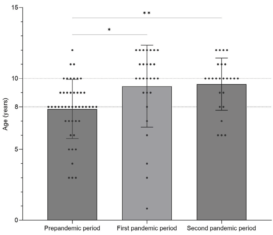 Legend: Scatter plot with a bar representing the ANOVA One-Way test complemented by Tukey test results for children’s age comparisons across periods. Asterisks represent statistically significant differences between groups (p < 0.05). Dots represent the samples.
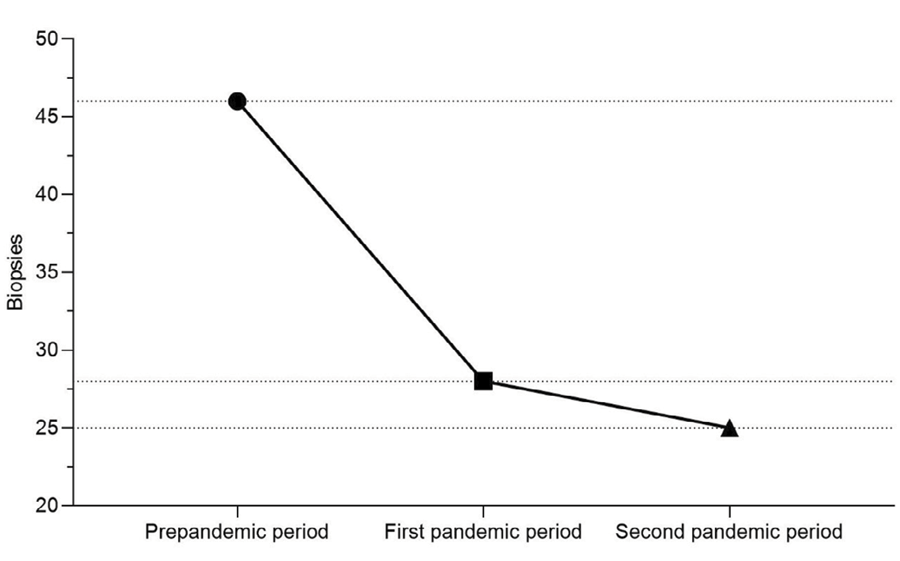 Legend: Scatter plot representing the number of biopsies performed in different periods.