Precision in mandibular implant overdentures: examining component height and implant number as biomechanical predictors
Abstract
Aim Selection of prosthetic attachment height and implant number can influence the supporting structures of implant mandibular overdentures (IMOs), including peri-implant tissue and prosthetic components. This study evaluated the biomechanical effects of single IMO (SIMO) or two IMO (TIMO) systems with different stud transmucosal heights (2, 3, or 4 mm) on cortical bone, mucosal tissue, implants, and prosthetic elements.
Methods Twelve three-dimensional finite element models were created using McNeel Rhinoceros 3D v7.0®, representing scenarios with 2 mm (SIMO-2mm, TIMO-2mm), 3 mm (SIMO-3mm, TIMO-3mm), and 4 mm (SIMO-4mm, TIMO-4mm) heights. Simulations included 100 N oblique loading (30º) on lower central incisors and 150 N axial loading on the first molars. Von Mises stress (implant, housing, stud), maximum and minimal principal stress (bone, tissue, prosthesis, nylon), and displacement were analyzed through numerical data and color-coded images.
Results Oblique loading resulted in higher stress, particularly in SIMO-4mm and TIMO-4mm. Nylon inserts in SIMO configurations experienced high compressive stress, especially in SIMO-4mm. TIMO configurations under oblique loading caused critical stress in peri-implant bone. Prosthetic tensile stress and displacement were greatest in SIMO-4mm under anterior loading. High transmucosal heights increased stress on housing, attachment, and prosthesis, particularly in systems with thin soft tissue.
Conclusion SIMO and TIMO rehabilitations under oblique loading showed high stress in ductile structures, with significant implications for SIMO-4mm due to stress concentration in nylon, prosthesis base, and displacement. Elevated transmucosal heights should be used cautiously in cases of limited soft tissue thickness.
Keywords
Dental implants; Finite element analysis; Mandible
Introduction
Rehabilitation of fully edentulous patients has always been a challenge in clinical practice1, especially using mandibular conventional complete dentures, where biomechanical (stability and retention)2,3 and bone reduction issues frequently arise4. These limitations posed can be effectively addressed through the advantages offered by dental implant-retained prosthodontics, such as implant mandibular overdentures (IMOs)3,5-7. Among these, expert consensus has identified the placement of 2 IMO (TIMO) as the treatment of choice for the edentulous mandibles, supported by multiple clinical studies demonstrating significant patient reported outcomes improvements over conventional dentures8. Although the TIMO has been considered the first treatment of choice for the edentulous mandible, the initial expenses and the available residual ridge make it unaffordable or even unfeasible for many elderly individuals9. Therefore, alternative treatment options with up to 10 years of follow-up have advocated for a single IMO (SIMO) as a viable rehabilitation plan10, combining high patient satisfaction with lower costs, reduced surgery time, and minimized comorbidities11,12. Among the alternatives (TIMO or SIMO), the primary benefit is the improvement of biomechanical drawbacks associated with conventional dentures. Although these benefits have been sparsely addressed in in silico studies13, evidence suggests that the stress patterns between ductile and non-ductile structures are similar for both SIMO and TIMO14-16.
The improvement of comfort and masticatory function have been reported by elderly patients as the main reasons for rehabilitation switch (TIMO or SIMO), when compared to lower conventional complete dentures2,3,9,10,17; thus, increasing the success rate of the treatment3,18. Mechanically, those benefits are mainly related to the retention component (i.e., attachment) which is mainly selected according to the clinical experience and the supracrestal tissue height11,19. Among attachment choices, the height may influence masticatory load dissipation and usability in long-term assessments6. A clinical study evaluating marginal bone loss around dental implants following TIMO installation highlighted the impact of attachment selection (specifically, stud height) on peri-implant bone health7. Notably, the data revealed that abutments with increased height (4 mm) posed a significant risk factor for marginal bone loss within a 12-month period. However, additional biomechanical data to substantiate and justify these findings remain limited. Similarly, while biomechanical information for TIMO is sparse, the SIMO rehabilitation design also lacks sufficient data, both clinically and in silico, regarding the effects of attachment height.
Altogether, the number of implants used for rehabilitation (SIMO or TIMO) and the attachment heights may provide a rationale for load dissipation along the cortical bone. Within this context, variations in attachment heights (1–5 mm) have been biomechanically assessed only for TIMO20,21. A previous in silico study informed that stress dissipation in TIMO treatment plan was enhanced by the stud compared to the ball system, when assessing different heights. In details, the ball system with 4 mm height and 2 mm mucosal tissue exercised more lateral forces on implant and authors suggest that attachment height should be carefully select21. However, data was solely provided for the ball system. Meanwhile, an in vitro study evaluated the dissipation of loads suffered by a TIMO with ball and locator retention system, and concluded that the inadequate choice of locator height can lead to the incidence of off-axis lateral loads21. However the biomechanical influence of inappropriate component choice for SIMO and TIMO rehabilitation components (cortical bone, mucosa, prosthesis, associated implant) remains insufficiently reported. Therefore, the objective of this study is to evaluate stress distribution using finite element analysis across the cortical bone, implant, housing, stud, nylon, mucosal tissue, and prosthesis. The analysis will consider different implant configurations (SIMO or TIMO) and attachment heights (2, 3, or 4 mm). The null hypotheses to be tested are that varying stud heights (2, 3, and 4 mm), implant number (SIMO and TIMO), and loading direction (axial and oblique) would result in biomechanical similarities between ductile and non-ductile structures.
Materials and Methods
This study was performed by using a generic 3-dimensional (3D) model of a human mandible obtained at the Center of Information Technology Renato Archer database. The digital computer-aided design (CAD) geometry of the mandible was identical for both rehabilitation structures (SIMO and TIMO), modeled with a Class III bone structure based on the Cawood & Howell classification4. This classification describes an alveolar ridge with a very rounded shape, adequate height, and sufficient thickness. The digital reconstruction was created using Rhinoceros 7.0 SR12 Software (McNell North America, Seattle, Washington, EUA). Pre-processing section was initiated by the mandibular structuration, specifically isolating a 2 mm thick cortical layer and extracting cancellous bone through Boolean difference operations16,22,23. The mucosal tissue, upper recovering the cortical bone was defined with 1,5mm thickness, aiming to reproduce a thicker mucosal tissue24. The implants (Titamax Ti/Ex; Neodent) placed (3.75 mm in diameter x 11 mm in length) described the first independent variable, either using single (SIMO) or two (TIMO) implants for the mandibular rehabilitation. Regarding the implant location for the TIMO groups, it was maintained 25 mm apart, representing the distance between the canine tooth15. Meanwhile, it was positioned in the midline region for the SIMO setting14. The attached component to the implant had a stud design (Equator; Titamax Ti/Ex; Neodent) with specific 3 transmucosal heights, defining the other independent variables: 2 mm (SIMO- or TIMO-2mm), 3 mm (SIMO- or TIMO-3mm), and 4 mm (SIMO- or TIMO-4mm). The geometry applied for the individual implants and the associated stud attachments were modeled following the engineering comercial drawings. Moreover, the implants were rigidly attached along the bone interface and bonded to simulate 100% osseointegration14,15. Regarding the prosthesis reconstruction it was designed a length of teeth until the second molar and the base thickness was reduced according the attachment height increased.
After defining the geometries and assembling the solids of the 6 models (SIMO-2mm, IMO-3mm, IMO-4mm, TIMO-2mm, TIMO-3mm, and TIMO-4mm), they were exported to HyperWorks 19.0 Software (HyperWorks 19.0, Altair, Troy, Michigan, EUA) in step format. All 3D meshes were built using at least 3 elements in each dimension in order to obtain accurate results with a higher degree of convergence14. Sequentially, the material properties, fixation, and boundary conditions were established to perform the linear elastic analysis. Specifically, 2 contact area were defined as sliding, first between prosthesis and mucosal tissue, while the second between attachment and nylon, aiming to reproduce a clinical situation23,25. The other contact areas were defined by freeze and the posterior region of the mandible was kept fixed to simulate anatomical condition for the mandible16.
The structures of interest in this study was cortical bone (Young Modulus = 13.700 MPa and Possion´s ratio = 0.35), trabecular bone (Young Modulus = 1.370 MPa and Possion´s ratio = 0.3), implants/ housing/ attachment (Young Modulus = 103.400 MPa and Possion´s ratio = 0.35), IMO (Young Modulus = 8.300 MPa and Possion´s ratio = 0.28), nylon (Young Modulus = 2.400 Mpa and Possion´s ratio = 0.39), and mucosal tissue (Young Modulus = 340 MPa and Possion´s ratio = 0.45)26,27. The cortical and cancellous bone were modeled as isotropic, linearly elastic, and homogeneous, simulating a clinical scenario with total osseointegration16. For boundary conditions, rigid fixation was applied to the posterior regions of the mucosa, cortical, and cancellous bone to prevent displacement in the x, y, and z Cartesian planes. The hemi-mandible models were mirrored to assume symmetry between the two sides14.
To reproduce a clinical scenario, a third independent variable was added, an oblique load of 100-N (50-N in each incisor) distributed in 73 nodes was applied in the mesial of the central incisor, with an angulation of 3014. According to the actual state-of-art, occlusal loads in edentulous patients vary between 100-N to 200-N and the anterior load applied will represent a critical scenario of unfavorable biomechanical distribution14,27. To enhance data collection, an axial load of 150 N (75 N on each first molar) was applied to the central sulcus of the first molar, distributed across 125 nodes. The application of this load was designed to reproduce a clinical situation in patients rehabilitated with a bilateral balanced occlusion, simulating the loads suffered in the masticatory process15.
Mathematical equations (processing analysis) were performed using Optistruct solver in all models in order to understand the stress distribution mechanism. The command analysis was set, according to the material. First, the models were assessed based on the prosthesis displacement in milimiters15. The second analysis (von Mises equivalent stress) was conducted for ductile materials (implants, stud attachment, and housing)14,15. Nevertheless, as the cortical bone is a brittle material (nonductile structure), the maximum principal stresses were obtained to better understand the influence of different prostheses designs on the peri-implant bone15. This equation was also applied for the prosthesis. The minimum principal stresses were recorded for the nylon and mucosa to investigate its compression15. Thereafter, stresses were plotted numerically, color-coded by stress maps, and compared among all the 12 groups (factor 1: implant number; factor 2: attachment height; factor 3: loading location).
Results
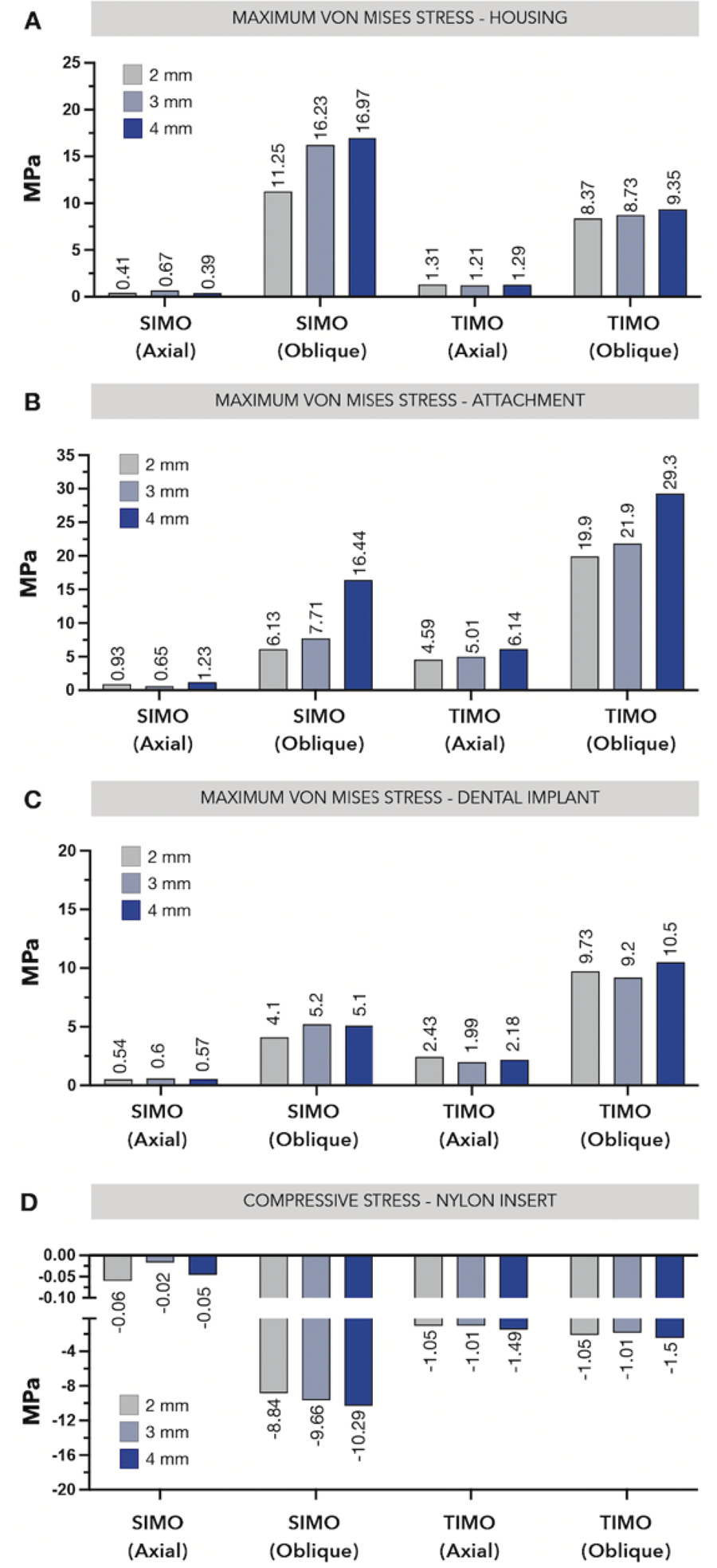
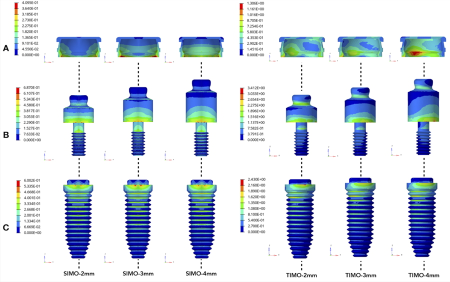
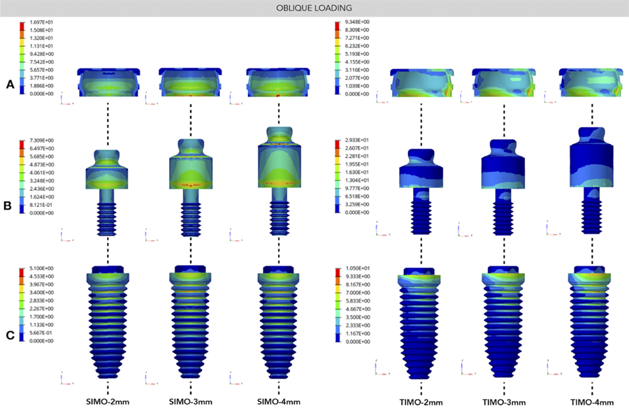
Ductile materials (housing, attachment, and implant) behaved differently depending on the load type, number of implants, and attachment height. The housing (Figure 1a), which is the component attached to the prosthesis base, exhibited higher von Mises equivalent stress under oblique loading conditions (8.37–16.97 MPa) compared to axial loading (0.39–1.31 MPa), regardless of the number of implants (TIMO or SIMO) (Figure 1a). When considering variations in attachment height, the highest stress values were observed in SIMO-4mm (16.97 MPa) and TIMO-4mm (9.35 MPa) (Figure 1a). In contrast, the groups with SIMOs generally exhibited stress values below 1 MPa under axial loading (Figure 1A). Stress maps for axial loading in the housing (Figure 2a) displayed a well-rounded distribution, whereas oblique loading (Figure 3a) revealed a vestibular stress profile directed toward the center of the component, aligned with the loading direction.
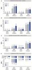
Stress distribution in ductile materials and in the nylon insert: equivalent von Mises stress in the (A) Housing, (B) Attachment, and (C) Implant. Minimum Principal Stress in the (D) nylon insert.
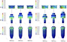
Color-Coded stress maps showing the von Mises stress distribution, when applying the axial loading, in the (A) Housing, (B) Attachment, and (C) Implant.
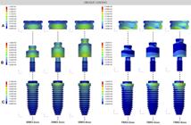
Color-Coded stress maps showing the von Mises stress distribution, when applying the oblique loading, in the (A) Housing, (B) Attachment, and (C) Implant.
When assessing the attachment (Figure 1b), it is evident that the loading direction produced a stress profile similar to that observed in the housing. Oblique loading (6.13–29.3 MPa) resulted in higher stress values than axial loading (0.65–6.14 MPa) (Figure 1b). The highest stress levels were associated with components with greater transmucosal height, particularly SIMO-4mm (16.4 MPa) and TIMO-4mm (29.3 MPa) (Figure 1b). Notably, all groups, except SIMO under oblique loading, showed higher stress values in the attachment (0.65–29.3 MPa) compared to the housing (0.39–9.35 MPa) (Figure 1b). Stress maps under axial loading were minimal (Figure 2b) compared to the under oblique loading (Figure 3b). The last (Figure 3b) revealed that stress was concentrated primarily between the attachment and the implant platform for both TIMO and SIMO groups. However, in the SIMO groups (Figure 3b), the neck of the component was also significantly stressed, particularly in SIMO-4mm.
The implant was the most distal ductile structure from the point of loading application (Figure 1c). Stress values, measured in MPa, were found to be relatively consistent across different transmucosal heights within the groups (Figure 1c). For the SIMO axial load, stress ranged from 0.54 to 0.6 MPa, and for the SIMO oblique load, from 4.1 to 5.2 MPa (Figure 1c). Similarly, TIMO axial loading resulted in stress values between 1.99 and 2.43 MPa, while TIMO oblique loading ranged from 9.2 to 10.5 MPa (Figure 1c). As observed in the previously mentioned structures (housing and attachment), oblique loading consistently produced the highest stress values, regardless of the number of implants. However, among all the ductile structures analyzed, the dental implant exhibited the lowest stress values (Figure 1c). In the TIMO groups, whether subjected to posterior (Figure 2c) and anterior (Figure 3c) loading, stress was primarily concentrated on the implant platform and the first 3 threads. For the SIMO groups, the stress extended further downward, nearly reaching the bottom of the implant, regardless of the loading direction.
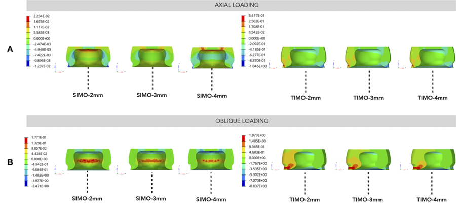
The compressive stress in the nylon insert showed comparable values (-1.01 to -1.5 MPa) across the TIMO groups under both axial and oblique loading conditions (Figure 1d). The highest stress values (-8.84 to -10.29 MPa) were recorded in the SIMO oblique groups, with the SIMO-4mm group exhibiting the peak minimum principal stress of -10.29 Mpa (Figure 1d). These findings for the SIMO axial groups align closely with the stress values observed in the housing (Figure 1d). Intrusion of the prosthesis was possible to be identified in the stress-map for the axial loading for SIMO and TIMO (Figure 4a), that displayed a stress concentration in the upper side of the nylon, irrespective the attachment height. When it was considered the oblique loading (Figure 4b) for SIMO, the stress switched for the center of the component, suggesting a lateral movement.
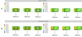
Color-Coded stress maps showing the Minimal Principal stress distribution in the nylon insert, when applying: (A) the axial and (B) oblique loading.
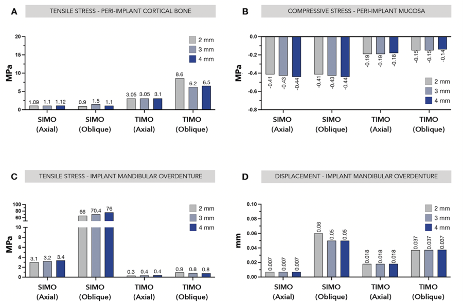
Peri-implant cortical bone was critically compromised for the anterior oblique loading (Figure 5a) in the TIMO groups (6.2-8.6 MPa), irrespective of the transmucosal height. The SIMO groups had almost equivalent values (0.9-1.5 MPa) (Figure 5a). The mucosal bearing area under compression had higher values for the SIMO configuration (Figure 5b), irrespective the loading direction, and the transmucosal height had minimal interference with values ranging from -0.41 to -0.44 MPa. Regarding the tensile stress in the prosthesis (Figure 5c), it was jeopardized by the anterior oblique loading, especially in the SIMO groups with higher values as the transmucosal height increased (SIMO-2mm: 66 MPa SIMO-3mm:70.4 MPa SIMO-4mm: 76 MPa). The prosthesis movement was assessed by its displacement (Figure 5d), this data was only affected by the direction of loading, being higher in the anterior load specially for SIMO (0.05-0.06 mm) when compared with TIMO (0.37 mm).
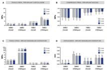
Biomechanical Behavior: (A) Maximum principal stress in peri-implant cortical bone, (B) maximum principal stress in the implant mandibular overdenture, (C) minimum principal stress in the mucosa, and (D) displacement of the prosthesis.
Discussion
The present investigation explored the mechanical behavior of various mandibular rehabilitation settings, emphasizing the influence of load type, number of implants, and attachment height on stress distribution and structural response. The findings highlighted distinct patterns in stress magnitude and distribution across both ductile structures (housing, attachment, and implant) and non-ductile structures (nylon inserts, prosthesis, and peri-implant tissues such as cortical bone and mucosa). Oblique loading consistently generated higher stress levels compared to axial loading across all structures, with notable variations depending on implant number (TIMO vs. SIMO) and the transmucosal profile of the attachments. These findings led to the rejection of all three null hypotheses regarding implant number, transmucosal height, and loading direction. The results for the housing revealed SIMO with higher transmucosal profile (SIMO-3mm and SIMO-4mm) had the higher stress concentration between 16.23 and 16.97 Mpa, respectively. Possible justifications might be related to the proximity to the loading application14,16, the reduction of the prosthesis base as the attachement height increased12, and freedom for lateral movements11.
The applicatin of 100-N distributed (30º off axis) in 73 nodes in the central incisor for SIMO might be a critical scenario related to the anterior food bitting14. Since, the oblique load represents the inclined biting force, based on the law of solid mechanics. This implies that when a force is applied to an inclined plane, it is divided into two componentes, being one parallel and one perpendicular to the inclined plane. Such a scenario might be particularly critical in SIMO rehabilitations, especially in cases with a high transmucosal profile (SIMO-3 and SIMO-4) and soft tissue thickness below 2 mm. In line with this assumption, placing a higher component may compromise the resin layer, leading to poor bonding at the housing interface. Consequently, the likelihood of SIMO fractures increases due to high occlusal loads, as well as the initiation and propagation of cracks in areas of the prosthesis with localized stress12. This finding aligns with the observed high tensile stress in the SIMO resin, particularly under oblique loading.
Moreover, the limitation of movement associated with the stud component may function as a stress absorber, redistributing stress between the housing and the nylon insert or vice versa11. The last assumption may also explain the tenfold increase in stress observed in the nylon insert for the SIMO group under oblique loading. The excessive stress observed in scenarios with oblique loading, a high transmucosal profile, and SIMO components may suggest the need for short-term maintenance appointments or the incorporation of reinforcement frameworks in the prosthetic base9,12.
The stud attachment was the ductile structure with the upmost values of von Mises stress. This output goes along with previous studies14,15. This can be attributed to the location of the load on the fulcrum formed by the two components, as well as the lever arm created in conjunction with the implant. For all models examined, an increase in attachment height resulted in higher stress values across all areas. In the case of a removable prosthesis, the vertical prosthetic space, considering both the implant and peri-implant tissue, can be mechanically divided into two lever arms: the distance from the implant base to the attachment and the distance from the attachment to the occlusal plane. Increasing the first lever arm or attachment height generates greater off-axial bending forces, leading to a simultaneous rise in stress values. Additionally, the concurrent reduction in prosthesis space and base thickness increases the flexibility of the overdenture, causing larger deflections and movements, reduced tissue contact, and a subsequent increase in the observed stresses13. Therefore, an optimal treatment plan is crucial to minimize the risk of functional complications. This includes evaluating the available prosthetic space to select the appropriate prosthetic rehabilitation and choosing the correct components.
The groups (SIMO and TIMO) evaluated under axial loading (150 N applied in the first molars distributed across 125 nodes) displayed lower values of loading for the housing, attachment implant, and nylon. Those data might be related to the distance between the loading and the components under evaluation. This assumption is particularly consistent for the SIMO group. When evaluating prosthesis displacement, it was observed that the movement of the rehabilitation is more closely related to the number of implants than to the transmucosal height. Because of the vertical and horizontal displacement pattern observed in the studied rehabilitation, the number of nylon can reduce the movement, especially in the TIMO groups15. The instability observed in the SIMO groups may require additional attention, including closer clinical monitoring of the patient and more frequent maintenance appointments as previously noted17. Therefore, the mucosal support in SIMO is more evidente due to the freedom in rotation and less components to turn the system more rigid12,18.
A previous clinical study evaluating TIMO with standard and mini implants found that marginal bone loss around implants was influenced by the height of the stud abutments. Specifically, abutments with greater height (4 mm) were identified as a risk factor for marginal bone loss within a 12-month period7. However, in our study, transmucosal height had minimal impact, except in the case of TIMO-2mm under oblique loading. These findings should be interpreted with caution, as bone loss is a multifactorial outcome affected by factors such as proper prosthesis adaptation, consistent daily overdenture insertion practices, and parallel implant positioning3,7,10,14,15. Moreover, previous biomechanical studies using similar methodologies did not consider cortical bone as an independent variable, which limits direct comparisons20,21. To enhance understanding, it is suggested that higher soft tissue height be considered to aid clinicians in selecting appropriate abutments. Studies have shown that implants with prosthetic abutments <2 mm are associated with greater marginal bone loss compared to those with abutments ≥2 mm19. Therefore, further research is needed to evaluate the impact of varying soft tissue heights and their influence on clinical outcomes.
Previous clinical study assessing TIMO with standard and mini implants observed that marginal bone loss around the implants had influence by the stud height7. Specifically, it was noticed that abutments with greater height (4 mm) presented as a risk factor for marginal bone loss in a period of 12 months. However, regarding our data the transmucosal height had minimal influence, only for the TIMO-2mm under oblique loading. The data should be evaluated with caution since the bone loss is a complex outcome, and may be compromised by the the proper adaptation of the prosthesis itself, daily proper practice of the overdenture insertion, and parallel implant position. Regarding previous data, the biomechanical studies with the same methodology had not assessed the cortical bone as an independent variable.
Limitations of the present study design included geometric variations among commercially available components and implants. Although the anatomic features of the mandible were standardized, small variations in bone regulation to place the implants may have influenced the results, specifically the cortical bone. Moreover, a single implant connection type was used to fabricate the SIMO and TIMO models. Another limitation was that lack of dynamic loading used which may not accurately reflect physiological forces or excentric movements in bilateral balanced occlusion14. Using a prospective clinical design to address biological differences could represent the long-term performance under fatigue cyclic loading3,12,17,18.
Based on the findings of this finite element study, the following conclusions were drawn:
-
Rehabilitations with SIMO and TIMO behaved biomechanically different, according to the loading direction. The oblique loading exhibited a high maximum von Mises stress, especially for a high transmucosal profile (SIMO-4mm and TIMO-4mm).
-
Models with SIMO displayed a high compressive stress in the mucosa, mainly because the lever arm is increased compared with TIMO. Also, SIMO under oblique loading may require attention when transposing the scenario clinically, since presented high stress in the nylon, prosthesis base, and displacement.
-
Selecting a high transmucosal component might be critical when having low soft tissue thickness (1.5 mm). The concern may be related to the high stress observed in the housing, attachment, and prosthesis.
References
-
1 Alvarez-Arenal A, Gonzalez-Gonzalez I, deLlanos-Lanchares H, Brizuela-Velasco A, Martin-Fernandez E, Ellacuria-Echebarria J. Influence of implant positions and occlusal forces on peri-implant bone stress in mandibular two-implant overdentures: a 3-Dimensional finite element analysis. J Oral Implantol. 2017 Dec;43(6):419-28. doi: 10.1563/aaid-joi-D-17-00170.
» https://doi.org/10.1563/aaid-joi-D-17-00170 -
2 Marcello-Machado RM, Faot F, Schuster AJ, Bielemann AM, Nascimento GG, Del Bel Cury AA. How fast can treatment with overdentures improve the masticatory function and OHRQoL of atrophic edentulous patients? A 1-year longitudinal clinical study. Clin Oral Implants Res. 2018 Feb;29(2):215-26. doi: 10.1111/clr.13101. Epub 2017 Dec 7.
» https://doi.org/10.1111/clr.13101 -
3 Schuster AJ, Marcello-Machado RM, Bielemann AM, Nascimento GG, Pinto Lde R, Del Bel Cury AA, et al. Short-term quality of life change perceived by patients after transition to mandibular overdentures. Braz Oral Res. 2017 Mar;31:e5. doi: 10.1590/1807-3107bor-2017.vol31.0005.
» https://doi.org/10.1590/1807-3107bor-2017.vol31.0005 -
4 Cawood JI, Howell RA. A classification of the edentulous jaws. Int J Oral Maxillofac Surg. 1988 Aug;17(4):232-6. doi: 10.1016/s0901-5027(88)80047-x.
» https://doi.org/10.1016/s0901-5027(88)80047-x -
5 Cardoso RG, Melo LA, Barbosa GA, Calderon PD, Germano AR, Mestriner Junior W, et al. Impact of mandibular conventional denture and overdenture on quality of life and masticatory efficiency. Braz Oral Res. 2016 Oct;30(1):e102. doi: 10.1590/1807-3107BOR-2016.vol30.0102.
» https://doi.org/10.1590/1807-3107BOR-2016.vol30.0102 -
6 Mifsud DP, Cortes ARG, Zarb MJ, Attard NJ. Maintenance and risk factors for fractures of overdentures using immediately loaded conventional diameter or mini implants with Locator abutments: A cohort study. Clin Implant Dent Relat Res. 2020 Dec;22(6):706-12. doi: 10.1111/cid.12952.
» https://doi.org/10.1111/cid.12952 -
7 Mifsud DP, Sammut EJ, Degiorgio J, Borg P, Cortes ARG, Attard NJ. Immediately loaded mini-implants supporting mandibular overdentures: A one-year comparative prospective cohort study. Clin Implant Dent Relat Res. 2020 Aug;22(4):507-13. doi: 10.1111/cid.12930.
» https://doi.org/10.1111/cid.12930 -
8 Thomason JM, Feine J, Exley C, Moynihan P, Müller F, Naert I, et al. Mandibular two implant-supported overdentures as the first choice standard of care for edentulous patients--the York Consensus Statement. Br Dent J. 2009 Aug;207(4):185-6. doi: 10.1038/sj.bdj.2009.728.
» https://doi.org/10.1038/sj.bdj.2009.728 -
9 Waltenberger L, Reissmann DR, Blender S, Fritzer E, Heydecke G, Kappel S, et al. Impact of loading protocol of a mandibular single implant-supported complete denture on oral health-related quality of life over a period of 5 years: a randomized controlled trial. J Dent. 2024 Mar;142:104769. doi: 10.1016/j.jdent.2023.104769. Epub 2023 Nov 4.
» https://doi.org/10.1016/j.jdent.2023.104769 -
10 Coutinho PC, Nogueira TE, Leles CR. Single-implant mandibular overdentures: clinical, radiographic, and patient-reported outcomes after a 5-year follow-up. J Prosthet Dent. 2022 Nov;128(5):949-55. doi: 10.1016/j.prosdent.2021.01.007. Epub 2021 Feb 25.
» https://doi.org/10.1016/j.prosdent.2021.01.007 -
11 Trang BNH, Kanazawa M, Murakami N, Wakabayashi N, Hada T, Sahaprom N, et al. Stress distribution of one-piece and two-piece mini-Implant overdentures with various attachment systems and diameters: A finite element analysis. J Prosthodont Res. 2023 Jul;67(3):430-6. doi: 10.2186/jpr.JPR_D_22_00108. Epub 2022 Dec 12.
» https://doi.org/10.2186/jpr.JPR_D_22_00108 -
12 de Paula MS, Cardoso JB, de Menezes EEG, Nogueira TE, McKenna G, Leles CR. A prospective cohort on the incidence of fractures in single-implant mandibular overdentures. J Dent. 2020 Dec;103:103521. doi: 10.1016/j.jdent.2020.103521.
» https://doi.org/10.1016/j.jdent.2020.103521 - 13 Ebadian B, Farzin M, Talebi S, Khodaeian N. Evaluation of stress distribution of implant-retained mandibular overdenture with different vertical restorative spaces: a finite element analysis. Dent Res J (Isfahan). 2012 Nov;9(6):741-7.
-
14 Borges GA, Presotto AGC, Caldas RA, Pisani MX, Mesquita MF. Is one dental mini-implant biomechanically appropriate for the retention of a mandibular overdenture? A comparison with Morse taper and external hexagon platforms. J Prosthet Dent. 2021 Mar;125(3):491-9. doi: 10.1016/j.prosdent.2020.01.038. Epub 2020 Apr 8.
» https://doi.org/10.1016/j.prosdent.2020.01.038 -
15 Pisani MX, Presotto AGC, Mesquita MF, Barão VAR, Kemmoku DT, Del Bel Cury AA. Biomechanical behavior of 2-implant- and single-implant-retained mandibular overdentures with conventional or mini implants. J Prosthet Dent. 2018 Sep;120(3):421-30. doi: 10.1016/j.prosdent.2017.12.012.
» https://doi.org/10.1016/j.prosdent.2017.12.012 -
16 Liu J, Pan S, Dong J, Mo Z, Fan Y, Feng H. Influence of implant number on the biomechanical behaviour of mandibular implant-retained/supported overdentures: a three-dimensional finite element analysis. J Dent. 2013 Mar;41(3):241-9. doi: 10.1016/j.jdent.2012.11.008. Epub 2012 Nov 14.
» https://doi.org/10.1016/j.jdent.2012.11.008 -
17 Hauck KE, Trentin MS, Skiba THI, Shibli JA, De Carli JP. Clinical and satisfaction outcomes of using one or two dental implants for mandibular overdentures: preliminary short-term follow-up of a randomized clinical trial. Int J Implant Dent. 2021 Feb;7(1):10. doi: 10.1186/s40729-020-00286-8.
» https://doi.org/10.1186/s40729-020-00286-8 -
18 Bryant SR, Walton JN, MacEntee MI. A 5-year randomized trial to compare 1 or 2 implants for implant overdentures. J Dent Res. 2015 Jan;94(1):36-43. doi: 10.1177/0022034514554224. Epub 2014 Oct 27.
» https://doi.org/10.1177/0022034514554224 -
19 Quispe-López N, Guadilla Y, Gómez-Polo C, López-Valverde N, Flores-Fraile J, Montero J. The influence of implant depth, abutment height and mucosal phenotype on peri-implant bone levels: a 2-year clinical trial. J Dent. 2024 Sep;148:105264. doi: 10.1016/j.jdent.2024.105264.
» https://doi.org/10.1016/j.jdent.2024.105264 -
20 Khurana N, Rodrigues S, Shenoy S, Saldanha S, Pai U, Shetty T, et al. A comparative evaluation of stress distribution with two attachment systems of varying heights in a mandibular implant-supported overdenture: a three-dimensional finite element analysis. J Prosthodont. 2019 Feb;28(2):e795-e805. doi: 10.1111/jopr.12966. Epub 2018 Sep 6.
» https://doi.org/10.1111/jopr.12966 -
21 Ying Z, Gonda T, Maeda Y. Influence of attachment height and shape on lateral force transmission in implant overdenture treatment. Int J Prosthodont. 2017 November/December;30(6):586-91. doi: 10.11607/ijp.5400.
» https://doi.org/10.11607/ijp.5400 -
22 El-Anwar MI, El-Taftazany EA, Hamed HA, ElHay MAA. Influence of number of implants and attachment type on stress distribution in mandibular implant-retained overdentures: finite element analysis. Open Access Maced J Med Sci. 2017 Mar;5(2):244-9. doi: 10.3889/oamjms.2017.047.
» https://doi.org/10.3889/oamjms.2017.047 -
23 Ozan O, Ramoglu S. Effect of implant height differences on different attachment types and peri-implant bone in mandibular two-implant overdentures: 3D finite element study. J Oral Implantol. 2015 Jun;41(3):e50-9. doi: 10.1563/AAID-JOI-D-13-00239. Epub 2014 Jan 28.
» https://doi.org/10.1563/AAID-JOI-D-13-00239 -
24 Unsal GS, Erbasar GNH, Aykent F, Ozyilmaz OY, Ozdogan MS. Evaluation of stress distribution on mandibular implant-supported overdentures with different bone heights and attachment types: a 3D finite element analysis. J Oral Implantol. 2019 Oct;45(5):363-70. doi: 10.1563/aaid-joi-D-19-00076.
» https://doi.org/10.1563/aaid-joi-D-19-00076 -
25 Daas M, Dubois G, Bonnet AS, Lipinski P, Rignon-Bret C. A complete finite element model of a mandibular implant-retained overdenture with two implants: comparison between rigid and resilient attachment configurations. Med Eng Phys. 2008 Mar;30(2):218-25. doi: 10.1016/j.medengphy.2007.02.005. Epub 2007 Mar 26.
» https://doi.org/10.1016/j.medengphy.2007.02.005 -
26 Barão VA, Assunção WG, Tabata LF, de Sousa EA, Rocha EP. Effect of different mucosa thickness and resiliency on stress distribution of implant-retained overdentures-2D FEA. Comput Methods Programs Biomed. 2008 Nov;92(2):213-23. doi: 10.1016/j.cmpb.2008.07.009.
» https://doi.org/10.1016/j.cmpb.2008.07.009 -
27 Liu X, Pang F, Li Y, Jia H, Cui X, Yue Y, et al. Effects of different positions and angles of implants in maxillary edentulous jaw on surrounding bone stress under dynamic loading: a three-dimensional finite element analysis. Comput Math Methods Med. 2019 Dec;2019:8074096. doi: 10.1155/2019/8074096.
» https://doi.org/10.1155/2019/8074096
-
Data availability:
Datasets related to this article will be available to the corresponding author upon request.
-
Funding sources:
This study was funded by Universidade Estadual de Campinas (UNICAMP) (postdoctoral schoolarship to G.A.B.).
Edited by
-
Editor:
Dr. Altair A. Del Bel Cury
Datasets related to this article will be available to the corresponding author upon request.