 Gamma-terpinene counteracts in vivo dinitrochlorobenzene-induced atopic dermatitis in mice
Abstract
Atopic dermatitis is a chronic skin condition characterized by impaired barrier function and underlying inflammation. Monoterpenes, a diverse class of secondary metabolites commonly found in essential oils, have been extensively documented for their potent anti-inflammatory properties. Therefore, this study evaluated the effects of gamma-terpinene (GT) on dinitrochlorobenzene (DNCB)-induced atopic dermatitis in a murine model. Macroscopic and histopathological analyses were conducted to assess skin lesions and ear edema, along with the assessment of serum immunoglobulin E (IgE), lactate dehydrogenase (LDH), and inflammatory cytokines (IL-4, IL-6, IL-1β, and TNF-α) in skin tissues. Histopathological analysis revealed a reduction in skin thickness, spongiosis, inflammatory cell infiltration, along with the presence of blisters and vesicles in both the epidermis and dermis. Additionally, cellular disarrangement and dermal fibrosis were observed. GT significantly decreased ear edema, serum IgE, and LDH levels, along with the expression of tissue IL-4, IL-6, IL-1β, and TNF-α. Therefore, the potential anti-inflammatory response induced by GT in atopic dermatitis suggests its potential for the development of GT-based therapies for chronic inflammatory skin disorders.
Keywords:
Cytokines; Dermatological; Dinitrochlorobenzene; Monoterpenes; Skin pharmacology
INTRODUCTION
Atopic dermatitis (AD) is a chronic and relapsing inflammatory skin disorder characterized by impaired skin barrier function. It is one of the most prevalent allergic diseases, affecting approximately 10–30% of the global population, with a higher incidence in children (Ku et al., 2017). Clinical symptoms include impaired skin barrier, erythema, edema, dryness, urlers and itching, all of which significantly affect the patient's quality of life and interfere with daily activities such as work, study, and sleep (Kang et al., 2021). Additionally, AD is characterized by immune dysregulation, evidenced by elevated serum immunoglobulin E (IgE) levels and infiltration of immune cells, predominantly T helper (Th) cells, leading to increased IgE production. This is attributed to an imbalance between Th1 and Th2 cell populations, with a predominance of cytokines such as IL-4 and IL-13 (Weidinger et al., 2018). Patients with AD experience a significant decline in quality of life, with effects on both physical and psychosocial well-being. These may include depression, anxiety, sleep disturbances, reduced productivity at work, and impairment in daily activities (Gochnauer et al., 2017). AD pathogenesis remains poorly understood. However, AD is associated with a compromised skin barrier, resulting in increased transepidermal water loss and subsequent cutaneous dehydration. This process triggers an inflammatory response characterized by the production of various cytokines, including interleukins, interferon-γ, tumor necrosis factor-α, and thymic stromal lymphopoietin (Damiani et al., 2019). When exposed to external stimuli, such as allergens and chemicals, the skin responds by activating specific receptors on sensory fibers, including protease-activated receptors (PAR) and MAS-related G protein–coupled receptors (Mrgpr). This activation increases calcium influx and triggers the release of neuropeptides from the skin, which have pro-inflammatory effects, such as vasodilation, plasma leakage, and leukocyte infiltration (Legat, 2021). Skin neuropeptides, such as substance P (SP), play a role in the pathogenesis of various skin diseases, including AD. Substance P (SP) can be released by nerve fibers, mast cells, monocytes, keratinocytes, and eosinophils, where it activates the neurokinin-1 receptor (NK1-R) on mast cells, triggering degranulation and the release of inflammatory mediators, including TNF-α, leukotriene B4 (LTB4), IL-1α, IL-1β, nerve growth factor (NGF), IL-6, prostaglandins, and histamine. These mediators contribute to vasodilation, plasma extravasation, and increased skin inflammation (Hosokawa, Takeuchi, Furue, 2009). The first line of treatment targets the skin and includes routine care, such as the use of emollients and topical therapies. Topical corticosteroids are commonly used to reduce inflammation; however, they are associated with local side effects, including skin atrophy, hypopigmentation, hypertrichosis, telangiectasia, rosacea, perioral dermatitis, purpura, impaired wound healing, and exacerbation of skin infections, acne, and stretch marks (Coondoo et al., 2014).
Systemic treatments for atopic dermatitis (AD) include immunosuppressants and oral corticosteroids. The most effective immunosuppressants for AD management are cyclosporine, azathioprine, mycophenolate mofetil, and methotrexate. However, their use should generally be restricted to a duration of approximately six months due to potential adverse effects. Additionally, renal and hepatic function should be carefully monitored to mitigate the risk of toxicity (Silverberg, 2017). Over the past decade, significant advances have been made in elucidating the molecular mechanisms underlying atopic dermatitis (AD), facilitating the development of novel targeted therapeutic strategies for disease management. The Food and Drug Administration recently approved Janus kinase inhibitors (JAKis) for adults with moderate to severe and refractory AD who do not respond adequately to other systemic treatments. These drugs represent a novel therapeutic approach and may provide an alternative treatment option for patients dependent on conventional immunosuppressants. However, the efficacy of these emerging pharmacological strategies must be carefully evaluated in light of the potential adverse effects observed in clinical trials (Rick et al., 2023). Consequently, there is a pressing need to explore therapeutic options for atopic dermatitis that offer both safety and efficacy while minimizing adverse effects.
Monoterpenes constitute about 90% of chemical composition of essential oils. These compounds exhibit a diversity of chemical structure and pharmacological activities, such as: antioxidant, anti-inflammatory, analgesic, antimicrobial, and antifungal (Dehsheikh et al., 2020). Gamma-terpinene (GT) (1-isopropyl-4-methyl-1,4-cyclohexadiene), with molecular formula C10H16, is a major monoterpene found in the essential oils of several plant species. GT exhibits a range of biological activities, including anti-inflammatory and antinociceptive properties, as observed in Bunium persicum (Boiss) (Hajhashemi, Sajjadi, & Zomorodkia, 2011). Similarly, the essential oil of Chamaecyparis obtusa (Siebold & Zucc.) was reported to inhibit the development of DNCB-induced AD-like lesions in BALB/c mice by suppressing the overproduction of serum IgE and Th1/Th2 cytokines (Joo et al., 2010). Interestingly, Ramalho et al. (2015) demonstrated that GT reduces inflammatory parameters, including the production of pro-inflammatory cytokines (TNF-α and IL-1β), as well as neutrophil migration in a carrageenan-induced peritonitis model. Subsequently, the same group reported that GT inhibited the production of IL-1β and IL-6 in LPS-stimulated macrophages, further supporting its anti-inflammatory properties (Ramalho et al., 2016). Regarding the toxicity of GT, a previous study demonstrated that rats orally administered GT at doses up to 2 g/kg exhibited no apparent signs of toxicity after 14 days of observation (Passos et al., 2015), indicating that GT is safe and non-toxic at this dose. Recently, Souza et al. (2024) demonstrated that GT exhibits low toxicity on endothelial (L-929 (CC50 = 333.3 µM)) and fibroblast SVEC 4-10 (CC50 = 366.7 µM) cells. Consistent with existing literature, it is suggested that GT is safe and may contribute positively to the development of new drugs due to its favorable safety profile. Moreover, these findings indicate that GT exhibits potent anti-inflammatory properties, making it a promising natural therapeutic option for AD.
Given the lack of studies on GT in the context of atopic dermatitis (AD) and the need for more effective, economically viable treatments with fewer adverse effects, this study aimed to evaluate the effects of GT in a DNCB-induced AD model in mice and explore its underlying mechanisms. Additionally, the effects of GT during both the acute and chronic phases of AD were compared to those of dexamethasone, a commonly used steroidal anti-inflammatory drug (Lugović-Mihić et al., 2023)
MATERIAL AND METHODS
Obtaining the GT
GT was purchased from Sigma-Aldrich (Merck KGaA, Darmstadt, Germany). The doses of 100 and 200 mg/kg of the GT were used based on a study by Ramalho et al. (2015). GT was mixed with in 2% Tween 80 in 0.9% NaCl (10 mL/kg) and the treatment was performed orally (v.o.).
Animals
Male Swiss mice (Mus musculus), weighing 25-30 g, were divided into 5 groups of 8 animals (n=40) and obtained from the Experimental Animal Facility of the Medicinal Plants Research Centre (NPPM) at the Federal University of Piauí (UFPI). The mice were kept at 24 ± 1°C with a 12-hour light/dark cycle and access to water and food was provided ad libitum. Euthanasia procedures were performed using an overdose of anaesthetic (thiopental sodium, 150 mg/kg and lidocaine 10 mg/kg, intraperitoneally). All experimental protocols were submitted to and approved by the Ethics Committee on Animal Experimentation (CEUA) of the Federal University of Piauí (Teresina, Brazil) on November 9, 2018, approval number 517/2018. This study adheres to the ARRIVE Guidelines for reporting animal research.
Induction of atopic dermatitis by DNCB
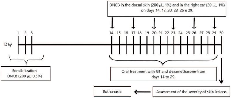
AD was induced by dinitrochlorobenzene (DNCB) as previously described (Park et al., 2014). Initially, the dorsal skin of animals was shaved under anesthesia (i.p.) with ketamine (100 mg/kg) and xylazine (10 mg/kg), 200 μL of 0.5% DNCB (acetone/oil (Gallo® olive oil) in a 3:1 ratio) was applied during the first 3 experimental days for sensitization. On days 14, 17, 20, 23, 26, and 29, AD was induced by applying 1% DNCB (acetone/oil in a 3:1 ratio) on the dorsal skin (200 μL) and right ear (20 μL) of the animals.
The animals were distributed into different treatment groups; Group I (SHAM group) was treated with the vehicle (acetone/oil in a 3:1) and saline; group II (negative control) was sensitized with dinitrochlorobenzene (DNCB) and treated with saline; animals of groups III (GT group 100 mg/kg + DNCB) and IV (GT group 200 mg/kg + DNCB) were sensitized with DNCB and treated with GT at doses of 100 and 200 mg/kg, respectively; and group V (dexamethasone (used as a positive control) + DNCB group) was sensitized with DNCB and treated with dexamethasone (3mg/kg).
Treatments were administered daily via oral gavage from days 14 to 29. On 30 th day of the experimental protocol several analyses were performed, including, macroscopic analysis of skin lesions, histological analysis of inflammatory cell infiltration, evaluation of DNCB-induced ear edema, measurement of serum levels of immunoglobulin E (IgE) and lactate dehydrogenase (LDH), and determination of tissue cytokine levels (TNF-α, IL-1β, IL-4, and IL-6).
The detailed scheme of the AD induction protocol is illustrated in Figure 1.
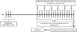
The time regimen for the induction of atopic dermatitis (AD) in mice using 2,4-dinitrochlorobenzene (DNCB) was as follows: DNCB sensitization or treatment with vehicle (acetone/oil (Gallo® olive oil) in a 3:1 ratio) was applied topically to the dorsal skin and behind the right ear. Animals taken out of the experiment on day 13. GT or dexamethasone were administered orally every day starting from day 14 through day 29. Mices were euthanized on the 30th day of experimental protocol.
Assessment of the severity of skin lesions
On day 30, the severity of dermatitis on the dorsal skin was evaluated based on four indicators of skin lesions, which were described as follows: (1) erythema/hemorrhage, (2) edema, (3) excoria-tion/erosion, and (4) desquamation/resection. The above symptoms were rated as follows: 0 (no symptoms), 1 (mild), 2 (moderate) and 3 (severe) and the sum of the scores used to analyze the severity of the lesions (Park et al., 2014).
Histological analysis
Histopathological analysis was performed for the sham, saline, GT 100 mg/kg, GT 200 mg/kg and dexamethasone 3 mg/kg groups. Briefl y, skin samples were collected, processed, and stained with hematoxylin and eosin for evaluation. Histological parameters used to evaluate the samples included shin thickness and characteristic of cells (keratinocytes), presence of edema and infl ammatory cells in the epidermis, while in the dermis, fi brosis, sebaceous glands, and hair follicles were observed, along with aggregates of infl ammatory cells within the dermal papillae. These parameters were evaluated and classifi ed as absent, mild (small aggregates in a small proportion of less than 30% of the microscopic fi elds using a magnifi cation of 400×), moderate (aggregates observed in 30-60% of the microscopic fi elds observed at 400× magnifi cation) or intense (aggregates seen in more than 60% of the microscopic fi elds observed at ×400 magnifi cation).
DNCB-induced ear edema
DNCB-induced ear edema is an experimental model used to study the pathogenesis of contact dermatitis. In this model, the DNCB sensitization of animals' ears produces ear edema accompanied by significant infiltration of inflammatory cells in the dermis, including mast cells and eosinophils, thereby characterizing the histopathological features of atopic dermatitis (Fujii et al., 2009). Furthermore, the presence of pro-inflammatory cytokines and IgE production contributes to increased skin thickness and exacerbates the deterioration of skin tissue observed in atopic dermatitis.
On the 30th day, after euthanasia, the ears were excised according to the method outlined by Park et al. (2014). Edema was assessed by measuring the weight difference between the ears and expressed in milligrams (mg).
Determination of serum Immunoglobulin E (IgE) levels
Blood samples were collected from the orbital plexus of the animals and centrifuged at 3,500 rpm for 10 minutes. Serum total IgE levels were determined using the IgE Turbiquest® system, which operates on the principle of immunoturbidimetry, in an automatic analyzer (VITROS 4600). The measurement was expressed in U/L and performed according to the manufacturer's instructions.
Measurement of serum lactate dehydrogenase (LDH) levels
For LDH measurement, blood samples were collected from the retro-orbital plexus of the animals and centrifuged at 3,500 rpm for 10 minutes. Analysis was performed using a chemical method with Vitros Chemistry Products LDH Slides in an automatic analyzer (VITROS 4600). Serum LDH levels were measured in U/L
Determination of tissue levels of TNF-α, IL-1β, IL-4 and IL-6 by ELISA
The enzyme immunoassay was performed using 100 μL of each reaction component at room temperature, 25±5°C. The standard kit (R&D Systems, Inc.) was used to evaluate IL-4, IL-6, IL-1β, and TNF-α levels according to the manufacturer's instructions. The capture antibodies were diluted in PBS and the other antibodies, substrates and samples were diluted in phosphate buffer supplemented with BSA 1% (PBS pH 7.4 + Bovine Serum Albumin. For a concentration of 10 mM, the following were added: 9.5 g of BSA, 8.38 g of sodium chloride, 0.21 g of potassium phosphate (monobasic), 1.20 g of sodium phosphate (dibasic), 0.21 g of potassium chloride, and 1 L of distilled water. The constituents were added then to distilled water and stirred until a homogeneous solution was formed. The pH of the mixture was verified using a pH meter and adjusted to 7.4. Tissue from the skin lesions was homogenized at 10% (i.e., 1 mL of buffer per 100 mg of tissue), and the homogenate was prepared in ice-cold buffer mentioned above.
The plates were washed three times with washing solution (PBS pH 7.4 + 0.05% Tween 20) at each step. For the ELISA analysis, each well of the plate was coated with the optimal concentration of the capture antibody by overnight incubation (room temperature 25±5 ºC). After blocking with BSA for 1 h, followed by washing, samples and standards (in triplicate) were added to the wells and incubated for 2 hours. After washing, the biotinylated detection antibody was added and incubated for 2 h. After incubation and washing, streptavidin was added and incubated for 20 minutes (followed by washing). Chromogenic substrate TMB + H2O2 (tetramethylbenzidine + hydrogen peroxide) was added and incubated for 20 minutes at room temperature, protected from light. Then, 100 μL of the stop solution (1M H2SO4) was added to each well and the absorbance was measured at 450 nm using microplate spectrophotometer and the data were calculated from the curve of the standards in serial concentration and results were expressed in pg/mL.
Statistical analysis
All date were expressed as mean ± standard deviation (SD) and were submitted to one-way analysis of variance, and post hoc Tukey's multiple comparison tests. Macroscopic analysis (severity of skin lesions induced by DNCB) was performed using the Kruskal-Wallis’ test. Differences were considered statistically signifi cant for p<0.05. Statistical analyses were performed using the GraphPad Prism® 6.0 software (La Jolla, CA, USA).
RESULTS
Effect of GT on the severity of DNCB-induced skin lesions
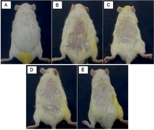
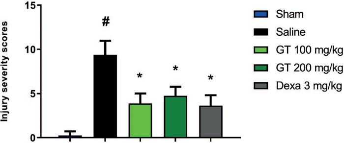
All mice sensitized with DNCB developed AD, with lesion formation on the dorsal region where the inducting agent was administered (Figure 2). The vehicle group (9.4±0.71) (Figure 2B) showed higher lesion expression because it did not receive treatment after AD induction and demonstrated signifi cantly higher skin lesion severity when compared with the Sham group (Figure 2A) (0.2±0.25). Oral treatment with GT at doses of 100 mg/kg (3.4±0.62) and 200 mg/kg (4.1±0.48) (Figures 2C, 2D, respectively) reduced the severity of DNCB-induced skin lesions. Animals treated with dexamethasone (3 mg/kg) (postive control) showed signifi cant decrease in the severity of lesions compared to vehicle group (3.6±0.68). Conversely, no notable difference was observed in the GT groups and dexamethasone group (positive control) (Figure 2E, Figure 3).
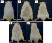
Effect of GT on the clinical characteristics of DNCB-induced skin lesions. Effect of GT (100 mg/kg and 200 mg/kg, po) and dexamethasone (3 mg/kg, po) on the clinical characteristics of skin lesions induced by DNCB in mice (n =8), 30th day of experimental protocol. Section A - Sham group: Received only the vehicle on the dorsal skin (acetone/olive oil, 3:1) and oral treatment with saline solution; Section B - Vehicle group: Received DNCB on the dorsal skin and oral treatment with saline solution; Section C – GT treated group (100 mg/ kg, v.o.); (D) GT treated group (200 mg/kg, v.o.); (E) Dexamethasone treated group (3 mg/kg, v.o.).
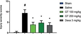
Effect of GT on the severity of skin lesions in DNCB-induced AD in mice. Effect of GT on the severity of skin lesions induced by DNCB in mice (n=8), 30th day of experimental protocol. Animals were treated (v.o.) with vehicle (saline), GT (100 and 200 mg/kg), or dexamethasone (dexa 3 mg/kg). Values are expressed as mean ± SD of injury severity scores. *p<0.05 vs Saline (DNCB), #p<0.05 vs Sham (ANOVA, Kruskal-Wallis).
Effect of GT on histopathological parameters of DNCB-induced skin lesions
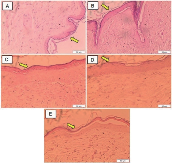
To evaluate the effect of GT treatment on AD, histological analysis using hematoxylin and eosin (HE) staining was performed using skin tissue to identify differences in physiological structures. A thin skin was observed, with two layers of keratinocytes, dermis with fibroblasts without inflammatory infiltrate, presence of sebaceous glands and hair in the naïve animals (Sham) (Figure 4A). The animals in the vehicle group (Figure 4B) exhibited thickened skin with spongiosis (intercellular edema) and increased keratinization, along with cellular disarrangement in the epidermis. Vesicle and blister formation were observed in both the epidermis and dermis (dermal papillae), accompanied by degenerating keratinocytes and infiltration of lymphocytes and neutrophils, eosinophils coupled with dermal fibrosis demonstrating the development of an inflammatory process after sensitization with DNCB.
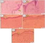
Histological analysis of DNCB-induced skin lesions. Effect of GT (100mg/kg and 200mg/kg, po) and dexamethasone (3mg/kg, po) on skin lesions induced by DNCB (acetone/olive oil 3:1) in mice (n=8), 30th day of experimental protocol. Sham group: Received only the vehicle on the dorsal skin and oral treatment with saline solution (A); Vehicle group: Received DNCB on the dorsal skin and oral treatment with saline solution (B); Received DNCB on the dorsal skin and oral treatment with GT (100 mg/kg) (C); Received DNCB on the dorsal skin and oral treatment with GT (200 mg/kg) (D); Received DNCB on the dorsal skin and oral treatment with dexamethasone (3mg/kg) (E). Tissues were stained with hematoxylin and eosin and images were captured using a 40X objective lens. Yellow arrows indicate the epidermal layer.
Animals treated with 100 mg/kg of GT (Figure 4C) showed thinner skin with thickening of the keratinized layer, the presence of intracellular blisters in isolated areas of the epidermis and dermis, and minimal inflammatory cell infiltration. Animals treated with 200 mg/kg of GT (Figure 4D) displayed the thinnest skin, with thickening confined to the keratinized layer, the presence of fibers in the dermal layer, and isolated inflammatory cells. Animals treated with dexamethasone (3 mg/kg) (Figure 4E) showed thin skin, an epidermis with rare blisters in some cells, and the presence of neutrophils and lymphocytes in scattered areas of the basal layer, with a preserved dermis.
Ear edema
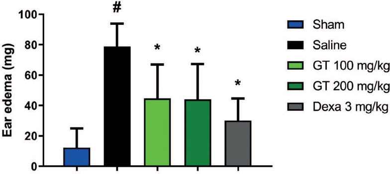
Topical application of DNCB on the ear of animals in vehicle group (78.8±5.39) promoted a marked increase of edema when compared with animals from Sham group (12.2±4.50). Edema formation significantly decreased with oral treatment of GT at 100 mg/kg (44.6±7.89), and 200 mg/kg (44.0±8.22) when compared with vehicle group. Treatment with dexamethasone (3 mg/kg) also significantly reduced edema formation (30.1±5.12) and no differences were observed compared with the animals treated with GT (100 or 200 mg/kg) (Figure 5).
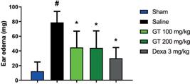
Effect of GT on DNCB-induced ear edema. Effect of GT on DNCB-induced ear edema in mice (n=8), 30th day of the experimental protocol. Animals were treated (v.o.) with vehicle (saline), GT (100 and 200 mg/kg), or dexamethasone (dexa 3 mg/kg). Results are expressed as mean ± SD of ear mass (mg) on the 30th day of the experimental protocol. *p<0.05 vs Saline (DNCB), #p<0.05 vs. Sham (one-way ANOVA and Tukey’s post-test).
Total serial Immunoglobulin E (IgE)
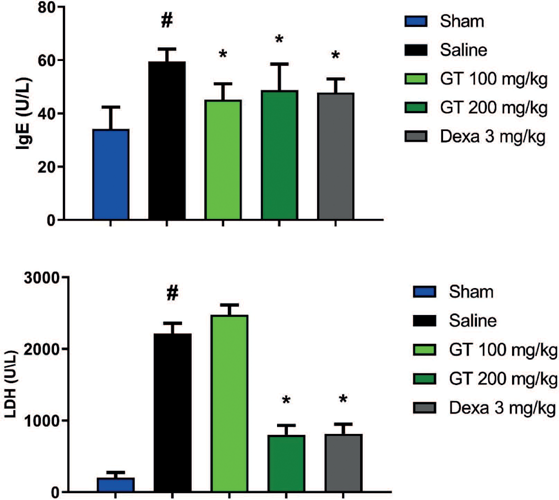
The animals in the vehicle group (59.6±1.62) exhibited high levels of IgE when compared to the animals in the Sham group (34.2±3.07). Mice treated with GT (100 and 200 mg/kg) showed significantly reduced serum IgE levels (45.2±2.10 and 48.8 ±3.46, respectively) compared with the vehicle group. Dexamethasone (3 mg/kg) significantly decreased the serum IgE level (47.9±1.93). Moreover, no statistically significant differences were observed between the GT (100 or 200 mg/kg) and dexamethasone groups (Figure 6A).
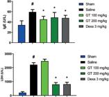
Effect of GT on serum total IgE levels (A) and LDH levels (B) in DNCB-induced AD in mice. Effect of GT on serum total IgE levels in mice (n=7-8) with AD induced by DNCB, 30th day of the experimental protocol. Animals were treated (v.o.) with vehicle (saline), GT (GT 100 mg/kg and GT 200 mg/kg) and dexamethasone (dexa 3 mg/kg). Values are expressed as mean ± SD of serum total IgE levels. *p<0.05 vs Saline (DNCB), #p<0.05 vs Sham (one way ANOVA and Tukey post-test).
Serum Lactate Dehydrogenase (LDH)
Animals of vehicle group displayed high LDH serum levels (2214.0±147.56) when compared to animals of Sham group (398.2±18.60). GT at 200 mg/kg markedly decreased the serum LDH levels (750.8±26.57) when compared with the vehicle group. The same was observed with the dexamethasone (3 mg/kg) group (762.0±41.50), which showed significant reduction in serum LDH. The 100 mg/kg dose (2393.0±65.96) did not alter serum LDH levels compared to the vehicle group. No significant difference in LDH levels was observed between the GT (200 mg/kg) and dexamethasone (3 mg/kg) groups. (Figure 6B).
Levels of IL-1β, IL-4, IL-6 and TNF-α in homogenate cutaneous lesions
DNCB treatment induced increase tissue levels of TNF-α in animals of vehicle group (763.9±83.77) when compared to Sham group (290.8±14.43). Mice treated with GT at doses of 100 or 200 mg/kg (208.4±37.35 and 287.4±59.08, respectively) exhibited a significant reduction (p<0.05) in TNF-α levels compared to the vehicle group. The dexamethasone (3 mg/kg) group (290.4±31.69) also showed a significant reduction in TNF-α levels (p<0.05) when compared to the vehicle group (Figure 7A).
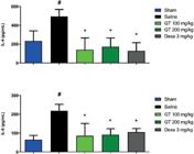
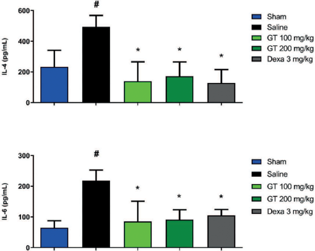
Effect of GT on the concentrations of TNF-α (A), IL-1β (B), IL-4 (C) and IL-6 (D) in the skin tissue homogenate from DNCB-induced AD cutaneous lesions in mice, 30th day of experimental protocol. Mice were treated (v.o.) with vehicle (saline), GT (100 and 200 mg/kg), or dexamethasone (dexa 3 mg/kg). Results are expressed as mean ± SD *p<0.05 vs Saline (DNCB), #p<0.05 vs Sham (one-way ANOVA and Tukey’s post-test).
DNCB treatment increased tissue levels of IL-1β (505.9±21.91) when compared to the Sham group (208.9±29.49). GT at 100 mg/kg (236.1±55.88) also reduced tissue levels of IL-1β. On the other hand, treatment with GT at 200 mg/kg (300.5±36.99) reduced IL-1β levels, although this reduction was not statistically significant when compared to the vehicle group. The result also shows that dexamethasone (3 mg/kg) (288.0±61.98) significantly reduced the expression of IL-1β (Figure 7B).
Animals treated with DNCB (494.4±36.98) exhibited elevated tissue levels of IL-4 compared to the Sham group (233.1±48.20). In mice treated with GT at 100 or 200 mg/kg (139.5±56.42 and 172.0±41.76, respectively), IL-4 levels were signifi cantly reduced. A similar reduction was observed in animals treated with dexamethasone (3 mg/kg) (127.5±43.55) (Figure 7C). Tissue levels of IL-6 were elevated in animals of the vehicle group (218.1±17.30) compared to the Sham group (64.5±11.38). Mice treated with GT at 100 or 200 mg/kg (85.1±33.03 and 88.8±16.25, respectively) exhibited signifi cantly lower IL-6 levels. A similar reduction was observed in the dexamethasone (3 mg/ kg) group (104.6±9.85) (Figure 7D).
Regarding tissue levels of the evaluated infl ammatory markers (IL-1β, IL-4, IL-6, and TNF-α), no statistically signifi cant difference was observed between the GT (100 or 200 mg/kg) and dexamethasone groups.
DISCUSSION
The search for alternative treatments to replace the current pharmacological therapies used in the treatment of AD has increased. Their respective effi cacies are often directly proportional to their adverse effects, opening up for potential application of GT in AD treatment. (Wu et al., 2021). However, no studies have evaluated the effects of GT in AD models. In this context, we investigated the potential of this monoterpene to modulate infl ammation in a DNCB-induced AD model in mice
The effect of GT was evaluated in an AD model characterized by key symptoms such as edema, erythema, erosion, pruritus, and dryness. The results of this study demonstrate that treatment with GT or dexamethasone signifi cantly prevented the skin lesion severity in animals with AD. This fi nding aligns with reported effects in human atopic dermatitis. Corticosteroids are recommended as fi rst-line therapy for the acute management of moderate to severe AD (Hanifin, Tofte, 1999; Hernandez et al., 2024). A similar outcome was observed with thymol, which reduced skin lesions in an experimental AD model induced by Staphylococcus aureus membrane vesicles. This report also demonstrated a reduction in epidermal thickness, eosinophil and mast cell infiltration, and serum IgE levels in these animals (Kwon et al., 2018). Therefore, the results of this study are consistent with previous findings, which have shown that monoterpenes can reduce the severity of AD lesions.
One of the primary effects of DNCB-induced AD is tissue alteration, characterized by epidermal thickening, immune cell infiltration, intercellular edema, and degeneration due to liquefaction of the basal layer (Park et al., 2021). Histopathological analysis performed in this work demonstrated that treatment with GT (Figure 4(C e D)) reduced of skin thickness and inflammatory cell infiltration in eczematous tissue and, mitigated the degeneration of the basal layer. In addition, the data also show that animals treated with GT had thinner skin and reduced damage in the keratinised layer. Serum levels of IgE and Th2-mediated inflammatory cytokines, such as TNF-α and IL-4, affect keratinocytes that secrete pro-inflammatory cytokines and chemokines in response to DNCB, leading to the invasion of immune cells into the inflamed skin (Homey et al., 2006). Choi et al. (2018) demonstrated that pulegone effectively attenuated the thickening of both the dermis and epidermis in a DNCB-induced AD model, using dexamethasone as positive control. Similar results were observed in this study, with dexamethasone as positive control. This suggests that GT exhibits anti-inflammatory activity in keratinocytes. However, molecular mechanisms involving this activity still remain unclear.
One of the main characteristic of AD is the presence of edema in the affected tissue, thus, the DNCB-induced ear edema model is often used in research to accurately assess this parameter (Choi et al., 2013). Edema results from the action of phlogistic substances such as histamine, substance P, bradykinin, among others, which cause changes in vascular permeability and consequently, the leakage of fluid and proteins from blood vessels to the interstitial space, as well as the infiltration of leukocytes to the site of injury (Choi et al., 2017). Treatment with GT significantly inhibited the development of DNCB-induced ear edema. These data suggest a reduction in leukocyte migration and leakage of fluid into the interstitial space. Edema reduction observed with GT treatment could be attributed to its potential to downregulate the expression of pro-inflammatory cytokines, given the pivotal role of these mediators in initiating and propagating the inflammatory response and the pathophysiology of AD. In a previous study, 6'-acetylpaeoniflorin (6-AP), a derivative of paeoniflorin, a monoterpene extracted from the roots of Paeonia lactiflora Pall, used to inhibit inflammation and regulate the immune response in clinical arthritis (Zheng, Wei, 2005), reduced ear edema and cellular infiltration in a DNCB-induced dermatitis model in mice (Zhou et al., 2016). This finding further supports the anti-inflammatory action exhibited by GT.
About 80% of AD patients exhibit increased levels of serum IgE. Elevated levels of IgE are common serum changes in AD, observed and analysed in animal models, reproducing the immunological changes observed in human dermatitis (Matsumoto et al., 2004). In the present work, GT was able to decrease serum IgE levels in a model of AD and exhibited a similar inhibitory effect when compared to dexamethasone. This result corroborates a previous study by Monteiro (2018), which demonstrated that the monoterpene significantly reduced serum IgE levels in an asthma model. These findings highlight its potential role in mitigating hypersensitivity reactions and further suggest its efficacy in the treatment of chronic dermatological inflammatory conditions.
Increased serum levels of lactate dehydrogenase (LDH) are noticed in situations of tissue damage, necrosis, hypoxia, hemolysis, or early stage of malignancies (Morishima et al., 2010). Lactate dehydrogenase (LDH) is an enzyme present in most cells, predominantly in liver, kidney, striated muscle, skin and cardiac muscle. Previous studies have demonstrated that elevated serum LDH levels correlate with epidermal cell damage (Kogawa et al., 2022). Additionally, fluctuations in serum LDH levels appear to correspond with the extent and depth of inflammation, reflecting epithelial damage. Therefore, LDH levels serve as a biomarker to evaluate the severity of skin lesions and the clinical progression of AD (Mukai et al., 1990).
Moreover, a study conducted with children having AD showed that the serum levels of the LDH enzyme were high in severe cases of the disease, highlighting its role as an important biomarker of AD severity, considered based on the extension of lesions and severity of atopic symptoms. Additionally, the LDH enzyme activity decreased after a period of 6-12 months of treatment since the clinical situation of patients had improved (Jung et al., 2015). Our study showed that GT (200 mg/kg) reduce the serum levels of LDH in animals with AD. This is consistent with the findings of the current study, where a reduction in the severity of skin lesions was observed in animals treated with this substance. However, GT at a dose of 100 mg/kg was not effective in reducing serum LDH levels when compared to the vehicle (saline) group. Indeed, LDH has been shown to strongly correlate with AD severity, since it is an enzyme found in almost all tissue cells and several conditions can cause increased blood LDH including liver disease, anaemia, heart attack, muscle trauma, cancers and infections (Farhana, Lappin, 2023). Moreover, using a paracetamol (PCM)-induced hepatotoxicity model, Islam et al. (2021) reported that nerol, a monoterpene present in Citrus × aurantium L., significantly reduced hepatotoxic parameters (ALT, AST, GGT, and serum levels of LDH) induced by PCM. LDH is produced by all cells and can be released during numerous inflammatory processes, such as in the liver, muscles, and skin (Shipman, Bahrani, Shipman, 2024). More specifically, LDH, coupled with IgE, are serum biomarkers used to assess the clinical severity of AD (Kato et al., 2020). Consistent with existing literature, our experimental results suggest that GT reduces inflammatory parameters, mitigates cell damage induced by DNCB, and may serve as a potential anti-inflammatory agent.
Thus, we believe that in this case, a higher concentration of GT (200 mg/kg) was necessary to reduce the levels of this enzyme equalling the group treated with dexamethasone (dexa 3 mg/kg), a reference drug, which is commonly used orally in patients with AD and is known to be effective in clinical practice (Saeki et al., 2009).
The literature demonstrates IL-4 plays a central role in the pathogenesis of AD in animal models. Overexpression of IL-4 induces IgE production, which exacerbates skin inflammation and leads to the characteristic histopathological alterations observed in AD (Spergel et al., 1999). Thus, it is reasonable to suggest that the decrease in IgE by GT may be due to the decrease in IL-4 levels. Several cytokines, such as IL-4, IL-6, IL-1β, and TNF-α, are critical mediators in the development of AD (Lee et al., 2020). These cytokines are abundantly present in eczematous tissue and blood samples from patients suffering from this disease. TNF-α is synthetized by inflammatory cells and is considered a primary mediator of inflammation. It is produced in response to tissue damage and promotes the release of other cytokines such as IL-1β and IL-6, through the activation of the nuclear factor kappa B (NF-κB) signaling pathway and consequent activation of an inflammatory cascade (Qu et al., 2019). IL-1β acts on the IL-1 receptor (IL-1R1), which is expressed on T lymphocytes, fibroblasts, epithelial cells, and endothelial cells; and triggers a series of phosphorylation and ubiquitination events and consequent activation of NF-κB, p38 mitogen-activated protein kinase, N-terminal c-Jun kinases (JNKs), and mitogen-activated protein kinases (MAPKs) signaling pathways. Thus, IL-1β acts as a potent proinflammatory agent by promoting vasodilation, facilitating the recruitment of granulocytes to the site of inflammation, and stimulating the secretion of prostaglandins (Galozzi et al., 2021).
IL-4 exerts its effects through the activation of the type II receptor, which in turn activates the Janus Kinase/Signal Transducers and Activators of Transcription (Jak/STAT) signaling pathway. This signaling cascade triggers a gene transcription process that enhances T cell activity, promotes immunoglobulin class switching to IgE, and facilitates antigen presentation by B cells (Braddock et al., 2018).
IL-6 is one of the first inflammatory mediators produced in response to infection or tissue damage via pathogen-associated molecular patterns (PAMPs) and damage-associated molecular patterns (DAMPs), and induces the production of acute phase proteins, such as C-reactive protein (CRP), fibrinogen, and haptoglobin. Molecules present in DAMPs promote the activation of NF-κB. Besides immune cells, IL-6 can also be released by endothelial cells, fibroblasts, among others (Tanaka, Narazari, Kishimoto, 2014).
This study showed elevated tissue levels of IL-4, IL-6 IL-1β, and TNF-α in a DNCB-induced AD model. Interestingly, treatment with GT or 1% dexamethasone (dexa 3 mg/kg) significantly decreased concentrations of those cytokines and, consequently, the inflammatory symptoms of AD. This result further corroborates with the macroscopic and histological findings that demonstrate a reduction in the inflammatory profile with GT treatment, such as decreased severity of cutaneous lesions, reduced epidermal thickness and infiltration of inflammatory cells in the tissue of the dorsal lesions. The DNCB-induced AD model exhibits a mixed phenotype of Th1 and Th2 responses, with Th2 cell responses predominant in the acute phase and Th1cell responses predominant in the chronic phase, where increased mast cell infiltration and elevated IgE levels (diagnostic indicator of AD), are observed (Choi et al., 2019). In this study, GT administration significantly reduced the levels of Th1 (IL-6, IL-1β, and TNF-α) and Th2 (IL-4) cytokines. Once activated, the Th1 pathway can evoke cell-mediated immunity and worsen AD by producing pro-inflammatory cytokines, such as TNF-α and IL-1β (Chen, 2007). On the other hand, an exaggerated Th2 response elicits humoral immunity with antibody production, eosinophil accumulation, and production of Th2-type cytokines like IL-4, leading to a characteristic scenario of chronic inflammation associated with allergic conditions, such as eczema (Kaminishi et al., 2020). It is well established that the NF-κB pathway regulates the production of pro-inflammatory cytokines, adhesion molecules, and participates in the differentiation of activated T cells into effector cells, such as Th1 and Th2 cells, which are involved in initiating immune and inflammatory responses (Liu, Zhang, Joo, 2017). Once activated, Th1 and Th2 cells induce cytokine synthesis, increasing immune reactivity and play a significant role in the pathogenesis of AD (Das et al., 2001; Dajee et al., 2006). Thus, NF-κB is critical for the inflammatory response, Th1 response, and regulation of inflammation caused by Th2 cell activation, with suppression of NF-κB activation alleviating inflammatory diseases, including AD (Tanaka et al., 2007). Therefore, it is reasonable to suggest that GT acts by regulating the balance between Th1 and Th2 cells, attenuating the increase in IL-6, IL-1β, TNF-α, and IL-4 levels, indicating anti-atopic activity. The inhibition of Th1 and Th2 inflammatory cytokines further suggests that GT may exert anti-AD activity by regulating immune events, including NF-κB expression, and reducing skin inflammation in DNCB-induced AD model. However, further studies are required to confirm this hypothesis.
Previous study showed that GT has important anti-inflammatory activity by inhibiting several inflammatory mediators such as PGE2, histamine, bradykinin. In addition, GT was able to reduce the levels of inflammatory interleukins and mitigate the migration of neutrophils in carrageenan-induced peritonitis model, indicating that this monoterpene acts on multiple inflammatory targets (Ramalho et al., 2015). The effects of monoterpenes on the production of these cytokines were also demonstrated by Choi et al. (2018), where pulegone (PLG) reduced tissue concentrations of IL-4, IL-6, IL-1β, and TNF-α, as well as INF-γ, indicating that PLG improves the parameters involved in AD by minimizing the inflammatory response in the DNCB-induced mouse model. In this regard, we can suggest that GT has significant anti-inflammatory activity, as demonstrated in the DNCB-induced AD model in mice. It is noteworthy that no significant difference was observed between the GT and dexamethasone groups in the obtained results, indicating a profile similar to that of dexamethasone, used as a positive control.
From an economic standpoint, the use of GT as a therapeutic agent for AD could potentially reduce the healthcare costs associated with managing this disease, given the high costs of conventional AD treatments, which represent a significant burden on both patient health and the healthcare system, besides their severe adverse effects (Manjelievskaia et al., 2021). Corticosteroids have been a cornerstone of clinical practice for decades and are widely used in dermatology due to their anti-inflammatory and immunosuppressive properties. However, this class of drugs has several well-known undesirable effects, particularly at high doses and with prolonged use, such as skin atrophy, perioral dermatitis, tachyphylaxis, and suppression of the hypothalamic-pituitary-adrenal axis (Broeders, Ahmed Ali, Fischer, 2016), making "corticophobia" common among patients (Lugović-Mihić et al., 2023). Therefore, there still a need to develop an effective and safe pharmacological therapy that improves the quality of life of patients with AD.
Monoterpenes are secondary plant metabolites and represent a vast group of compounds with various biological activities (Salakhutdinov, Volcho, Yarovaya, 2017). These compounds have been widely used in agriculture, cosmetics, the food industry, and in pharmaceutical products (Zielińska-Błajet, Feder-Kubis, 2020; Ashrafizadeh et al., 2020). GT, a monoterpene present in several plant species and used in traditional medicine, such as Origanum vulgare L. (Gong, Ren, 2020), Rosmarinus officinalis L. (Ozcan, Chalchat, 2008), and Citrus sp. (Suzuki et al., 2004), is a relatively safe compound with low toxicity (Passos et al., 2015; Souza et al., 2024). It is also commonly found in a wide range of foods, particularly dietary products (Alvarenga et al., 2023). Previous studies demonstrate that GT has several biological properties (Guo et al., 2021; Passos et al., 2015; Ramalho et al., 2016; Zochedh et al., 2022). However, no studies in the literature have investigated the role of GT in modulating AD. The data from this study do not only highlight the therapeutic potential of GT, a compound found in various plant species, as a promising anti-inflammatory agent for the treatment of AD but also pave the way for future clinical trials.
Based on these considerations, the results obtained collectively suggest that GT is a promising candidate as a therapeutic agent for the treatment of AD.
CONCLUSIONS
GT decreased the severity of lesions and mitigated leukocyte infiltration in the dorsal skin of DNFB-induced atopic dermatitis mice. This monoterpene attenuated DNCB-induced ear edema, reduce Immunoglobulin (IgE) and Lactate dehydrogenase (LDH) levels in the serum of animals with AD, besides decreasing the expression of IL-4, IL-6, IL-1β, and TNF-α, in the skin tissue homogenate from the dorsal lesions of AD mice. Thus, the data obtained indicate possible anti-inflammatory properties of GT and demonstrate that this monoterpene represents a promising therapeutic agent for the treatment of AD.
ACKNOWLEDGMENTS
We thank the FAPEPI for financial support.
DATA AVAILABILITY STATEMENT
Data available from the corresponding author upon reasonable request.
REFERENCES
- Alvarenga JFR, Genaro B, Costa BL, Purgatto E, Manach C, Fiamoncini J. Monoterpenes: current knowledge on food source, metabolism, and health effect. Crit Rev Food Sci Nutr. 2023;63(10):1352-1389.
- Ashrafizadeh M, Ahmadi Z, Mohammadinejad R, Kaviyani N, Tavakol S. Monoterpenes modulating autophagy: A review study. Basic Clin Pharmacol Toxicol. 2020;126:9–20.
- Braddock M, Hanania NA, Sharafkhaneh A, Colice G., Carlsson M. Potential risks related to modulating interleukin-13 and interleukin-4 signalling: a systematic review. Drug Saf. 2018;45:489-509.
- Broeders JA, Ahmed Ali U, Fischer G. Systematic review and meta-analysis of randomized clinical trials (RCTs) comparing topical calcineurin inhibitors with topical corticosteroids for atopic dermatitis: a 15-year experience. J Am Acad Dermatol. 2016;75:410-419.e3.
- Chen E. Psychological stress and its relationship to cytokines and inflammatory diseases. In: Cytokines: Stress and Immunity, 2nd ed., Boca Raton, FL, Taylor & Francis Group, 2007;29-49.
- Choi JH, Jin SW, Park BH, Kim HG, Khanal T, Han HJ, et al. Cultivated ginseng inhibits 2,4-dinitrochlorobenzene-induced atopic dermatitis-like skin lesions in NC/Nga mice and TNF-α/IFN-γ-induced TARC activation in HaCaT cells. Food Chem Toxicol. 2013;56:195-203.
- Choi JK, Jang YH, Lee S, Lee, SR, Choi YA, Jin M, et al. Chrysin attenuates atopic dermatitis by suppressing inflammation of keratinocytes. Food Chem Toxicol. 2017;110:142-150.
- Choi YA, Yu JH, Jung HD, Lee S, Park PH, Lee, HS, Kim, SH. Inhibitory effect of ethanol extract of Ampelopsis brevipedunculata rhizomes on atopic dermatitis-like skin inflammation. J Ethnopharmacol. 2019;238,111850.
- Choi YY, Kim MH, Lee H, Jo SY, Yang WM. (R)-(+)-pulegone suppresses allergic and inflammation responses on 2,4-dinitrochlorobenzene-induced atopic dermatitis in mice model. J Dermatol Sci. 2018;91:292-300.
- Coondoo A, Phiske M, Verma S, Lahiri K. Side-effects of topical steroids: A long overdue revisit. Indian Dermatol Online J. 2014;5(4):416-25.
- Dajee M, Muchamuel T, Schryver B, Oo A, Alleman-Sposeto J, De Vry CG, et al. Blockade of experimental atopic dermatitis via topical nf-κb decoy oligonucleotide. J Invest Dermatol. 2006;126(8):1792-1803.
- Damiani G, Eggenhöffner R, Pigatto PDM, Bragazzi NL. Nanotechnology meets atopic dermatitis: Current solutions, challenges and future prospects. Insights and implications from a systematic review of the literature. Bioac Mater. 2019;4:380-86.
- Das J, Chen CH, Yang L, Cohn l, Ray P, Ray A. A critical role for NF-kappa B in GATA3 expression and TH2 differentiation in allergic airway inflammation. Nat Immunol. 2001;2:45–50.
- Dehsheikh AB, Sourestani MM, Dehsheikh PB, Mottaghipisheh J, Vitalini S, Iriti M. Monoterpenes: Essential Oil Components with Valuable Features. Mini-rev Med Chem. 2020;20(11):958–74.
-
Farhana A, Lappin SL. Biochemistry, Lactate Dehydrogenase. [Updated 2022 May 8]. In: StatPearls [Internet]. Treasure Island (FL): StatPearls Publishing; 2023 Jan-. Available from: https://www.ncbi.nlm.nih.gov/books/NBK557536/
» https://www.ncbi.nlm.nih.gov/books/NBK557536/ - Fujii Y, Takeuchi H, Sakuma S, Sengoku T, Takakura S. Characterization of a 2,4-dinitrochlorobenzene-induced chronic dermatitis model in rats. Skin Pharmacol Physiol. 2009;22:240–47.
- Galozzi P, Bindoli S, Doria A, Sfriso P. The revisited role of interleukin-1 alpha and beta in autoimmune and inflammatory disorders and in comorbidities. Autoimmun Rev. 2021;20:102785.
- Gochnauer H, Valdes-Rodriguez R, Cardwell L, Anolik RB. The Psychosocial Impact of Atopic Dermatitis. Adv Exp Med Biol. 2017;1027:57-69.
- Gong X, Ren Y. Larvicidal and ovicidal activity of carvacrol, p-cymene, and γ-terpinene from Origanum vulgare essential oil against the cotton bollworm, Helicoverpa armigera (Hübner). Environ Sci Pollut Res Int. 2020;27(15):18708-18716.
- Guo Y, Baschieri A, Amorati R, Valgimigli L. Synergic antioxidant activity of γ-terpinene with phenols and polyphenols enabled by hydroperoxyl radicals. Food Chem. 2021;345:128468.
- Hajhashemi V, Sajjadi SE, Zomorodkia M. Antinociceptive and anti-inflammatory activities of Bunium persicum essential oil, hydroalcoholic and polyphenolic extracts in animal models. Pharm Biol. 2011;49(2):146–51.
- Hanifin JM, Tofte, SJ. Update on therapy of atopic dermatitis. JACI: In Practice. 1999;104(3):S123-S125.
- Hernandez TD, Aleman SJ, Bao-Loc-Trung M, Forte MV, Brandt W, Armstrong C, et al. Advancing treatment in atopic dermatitis: a comprehensive review of clinical efficacy, safety, and comparative insights into corticosteroids, calcineurin inhibitors, and phosphodiesterase-4 inhibitors as topical therapies. Cureus. 2024;2;16(3):e55393.
- Homey B, Steinhoff M, Ruzicka T, Leung DYM. Cytokines and chemokines orchestrate atopic skin inflammation. JACI: In Practice. 2006;118:178e189.
- Hosokawa C, Takeuchi S, Furue M. Severity scores, itch scores, and plasma substance p levels in atopic dermatitis treated with standard topical therapy with oral olopatadine hydrochloride. J Dermatol. 2009;36:185–90.
- Islam MT, Quispe C, Islam MA, Ali ES, Saha S, Asha UH, et al. Effects of nerol on paracetamol-induced liver damage in Wistar albino rats. Biomed & Pharmacother. 2021;140:111732.
- Joo SS, Yoo YM, Ko SH, Choi W, Park MJ, Kang HY, et al. Effects of essential oil from Chamaecypris obtusa on the development of atopic dermatitis-like skin lesions and the suppression of Th cytokines. J Dermatol Sci. 2010;60(2):122-5.
- Jung M, Lee TH, Oh HJ, Kim H, Son YS, Lee EH, Kim J. Inhibitory effect of 5,6-dihydroergosteol-glucoside on atopic dermatitis-like skin lesions via suppression of NF-κB and STAT activation. J Dermatol Sci. 2015;79:252-61.
- Kaminishi K, Soma Y, Kawa Y, Mizoguchi M. Flow cytometric analysis of IL-4, IL-13 and IFN-γ expression in peripheral blood mononuclear cells and detection of circulating IL-13 in patients with atopic dermatitis provide evidence for the involvement of type 2 cytokines in the disease. J Dermatol Sci. 2020;29(1):19-25.
- Kang J, Lee S, Kim N, Dhakal H, Choi YA, Kwon TK, et al. Hispidulin alleviates 2,4-dinitrochlorobenzene and house dust mite extract-induced atopic dermatitis-like skin inflammation. Biomed Phamacother. 2021;137:111359.
- Kato A, Kamata M, Ito M, Uchida H, Nagata M, Fukaya S, et al. Higher baseline serum lactate dehydrogenase level is associated with poor effectiveness of dupilumab in the long term in patients with atopic dermatitis. J Dermatol. 2020;47:1013-1019.
- Kogawa K, Minakawa S, Matsuzaki Y, Okamoto A, Ogasawara S, Saito N, Sawamura D. Correlation between lactate dehydrogenase and other laboratory data in patients with Atopic dermatitis. J Cutan Immunol Alergy. 2022;5:133–35.
- Kwon HI, Jeong NH, Jun SH, Son JH, Kim S, Jeon H, et al. Thymol attenuates the worsening of atopic dermatitis induced by Staphylococcus aureus membrane vesicles. Int Immunopharmacol. 2018;59:301-309.
- Ku JM, Hong SH, Kim HI, Seo HS, Shin YC, Ko SG. Effects of Angelicae dahuricae radix on 2,4-dinitrochlorobenzene-induced atopic dermatitis-like skin lesions in mice model. BMC Complement Altern Med. 2017;17:98.
- Lee D-H, Park J-K, Choi J, Jang H, Seol JW. Anti-inflammatory effects of natural Flavonoid diosmetin in IL-4 and LPS-induced macrophage activation and atopic dermatitis model. Int Immunopharmacol. 2020;89:107046.
- Legat FJ. Itch in Atopic Dermatitis – What is new? Front Med. 2021;(8):644760.
- Liu T, Zhang L, Joo D. NF-κB signaling in inflammation. Sig. Transduct. Target Ther. 2017;2:17023.
- Lugović-Mihić L, Meštrović-Štefekov J, Potočnjak I, Cindrić T, Ilić I, Lovrić I, et al. Atopic dermatitis: disease features, therapeutic options, and a multidisciplinary approach. Life (Basel). 2023;13(6):1419.
- Manjelievskaia J, Boytsov N, Brouillette MA, Onyekwere U, Pierce E, Goldblum O, Bonafede M. The direct and indirect costs of adult atopic dermatitis. J Manag Care Spec Pharm. 2021;(10):1416-1425.
- Matsumoto K, Mizukoshi K, Oyobikawa M, Ohshima H, Tagami H. Establishment of an atopic dermatitis-like skin model in a hairless mouse by repeated elicitation of contact hypersensitivity that enables to conduct functional analyses of the stratum corneum with various non-invasive biophysical instruments. Skin Res Technol. 2004;10:122-29.
-
Monteiro TM. Study of the anxiolytic and immunomodulatory activity of Gamma-terpinene. [Doctoral dissertation]. João Pessoa: Federal University of Paraiba: Postgraduate Program in Natural and Synthetic Bioactive Products, 2018. https://repositorio.ufpb.br/jspui/handle/123456789/13706
» https://repositorio.ufpb.br/jspui/handle/123456789/13706 - Morishima Y, Kawashima H, Takekuma K, Hoshika A. Changes in serum lactate dehydrogenase activity in children with atopic dermatites. Pediatr Int. 2010;52:171-74.
- Mukai H, Noguchi T, Kamimura K, Nishioka K, Nishiyama S. Significance of elevated serum LDH (lactate dehydrogenase) activity in atopic dermatitis. J Dermatol. 1990;17(8):477-81.
- Ozcan MM, Chalchat JC. Chemical composition and antifungal activity of rosemary (Rosmarinus officinalis L.) oil from Turkey. Int J Food Sci Nutr. 2008;59(7-8):691-8.
- Park G, Oh SM. Inhibitory effects of Juglans mandshurica leaf on allergic dermatitis-like skin lesions-induced by 2,4-dinitrochlorobenzene in mice. Exp Toxicol Pathol. 2014:66:97–101.
- Park S, Lee JB, Kang S. Topical application of Chrysanthemum Indicum L. attenuates the development of atopic dermatitis-like skin lesions by suppressing serum IgE levels, IFN-γ, and IL-4 in Nc/Nga mice. eCAM. 2021;2012:821967.
- Passos FF, Lopes EM, de Araújo JM, de Sousa DP, Veras LM, Leite JR, Almeida FR. Involvement of Cholinergic and Opioid System in γ-Terpinene-Mediated Antinociception. Evid Based Complement Alternat Med. 2015;829414.
- Qu R, Chen X, Hu J, Fu Y, Peng J, Li Y, et al. Ghrelin protects against contact dermatitis and psoriasiform skin inflammation by antagonizing TNF-α/NF-κB signaling pathways. Sci Rep. 2019;9:1348.
- Ramalho TR, Filgueiras LR, de Oliveira MTP, de Araujo Lima AL, Bezerra-Santos CR, Jancar S, Piuvezam MR. Gamma-terpinene modulation of LPS-stimulated macrophages is dependent on the PGE2/IL-10 axis. Planta Med. 2016;82:1341–45.
- Ramalho TR, Oliveira MTP, Lima ALA, Santos CRB, Piuvezam, MR. Gamma-terpinene modulates acute inflammatory response in mice. Planta Med. 2015;81:1248-54.
- Rick JW, Lio P, Swetha A, Hsiao JL, Shi, VY. Atopic Dermatitis: A Guide to Transitioning to Janus Kinase Inhibitors. Dermatitis. 2023;34(4):297-300.
- Silverberg NB. Atopic Dermatitis Prevention and Treatment. Cutis. 2017;100:173-77.
- Saeki H, Furue M, Furukawa F, Hide M, Ohtsuki M, Katayama I, et al. Guidelines for management of atopic dermatitis. J. Dermatol. 2009;36:563-577.
- Salakhutdinov NF, Volcho KP, Yarovaya OI. Monoterpenes as a renewable source of biologically active compounds. Pure Appl. Chem. 2017;89(8):1105-1117.
- Shipman AR, Bahrani, S, Shipman KE. Investigative algorithms for disorders affecting plasma lactate dehydrogenase: a narrative review. J Lab Precis Med. 2024;9.
- Spergel JM, Mizoguchi E, Oettgen H, Bhan AK, Geha RS. Roles of TH1 and TH2 cytokines in a murine model of allergic dermatitis. J Clin Invest. 1999;103(8):110311.
- Souza RP, Pimentel VD, Sousa RWR, Sena EP, Silva ACA, Dittz D, et al. Non-clinical investigations about cytotoxic and anti-platelet activities of gammaterpinene. Naunyn-Schmiedeberg's Arch Pharmacol. 2024;397:8145–8160.
-
Suzuki Y, Sakai H, Shimada T, Omura M, Kumazawa S, Nakayama T. Characterization of gamma-terpinene synthase from Citrus unshiu (Satsuma mandarin). Biofactors. 2004;21(1-4):79-82. doi: 10.1002/biof.552210115. PMID: 15630174.
» https://doi.org/10.1002/biof.552210115 - Tanaka A, Muto S, Jung K, Itai A, Matsuda H. Topical application with a new NF-κB inhibitor improves atopic dermatitis in NC/NgaTnd mice. J Invest Dermatol. 2007;127(4):855-863.
- Tanaka T, Narazari M, Kishimoto T. IL-6 in Infl ammation, Immunity, and Disease. Cold Spring Harb Perspect Biol. 2014;6:a016295.
- Weidinger S, Beck LA, Bieber T, Kabashima K, Irvine AD. Atopic dermatitis. Nat Rev Dis Primers. 2018;(4):1.
- Wu S, Pang Y, He Y, Zhang X, Peng L, Guo J, Zeng, J. A comprehensive review of natural products against atopic dermatitis: Flavonoids, alkaloids, terpenes, glycosides and other compounds. Biomed Pharmacother. 2021;140:111741.
- Zheng YQ, Wei W. Total glucosides of paeony suppresses adjuvant arthritis in rats and intervenes cytokinesignaling between different types of synoviocytes. Int Immunopharmacol. 2005;5(10):1560–73.
- Zhou P, Yang X, Jia X, Yu J, Asenso J, Xiao F, et al. Effect of 6'-acetylpaeonifl orin on dinitrochlorobenzeneinduced allergic contact dermatitis in BALB/c mice. Immunol Res. 2016;64(4):857-68.
- Zielińska-Błajet M, Feder-Kubis J. Monoterpenes and their derivatives-recent development in biological and medical applications. Int J Mol Sci. 2020;21(19):7078.
- Zochedh A, Priya M, Shunmuganarayanan A, Thandavarayan K, Sultan AB. Investigation on structural, spectroscopic, DFT, biological activity and molecular docking simulation of essential oil Gamma-Terpinene. J Mol Struct. 2022;2:133651.
Edited by
-
Associate Editor:
Silvya Stuchi Maria-Engler