 Metabolic and antioxidant parameters in the kidneys of rats treated with copaiba oil in cirrhosis induced by thioacetamide
Abstract
This study evaluated the effects of copaiba oil on the kidneys of rats with cirrhosis induced by thioacetamide. Male Wistar rats were divided into four groups (n = 8), namely Control (C), Thioacetamide (TAA), Copaiba Oil (OC), and Thioacetamide + Copaiba Oil (TAA+OC). The biochemical and immunological analyses were performed, and results for the antioxidant enzymatic analysis revealed an increase in GST activity for TAA and TAA+OC compared to C. For GSH, higher values were observed in the TAA and TAA+OC groups compared to the C, but the latter differed from OC, such as TBARS. For Vit C, the TAA+OC group differed from the C and TAA and for glucose and lactate, lower values were observed for the TAA+OC group compared to the OC group. Cytokine concentrations showed the same profile with the TAA+OC group showing lower values compared to the C and OC groups. The data obtained do not show a clear positive effect of copaiba oil on kidney tissue when compared to the potential damage caused by thioacetamide. However, the groups that received either thioacetamide or copaiba oil alone maintained their basal levels, indicating that, at the doses used, these compounds did not negatively alter renal morphology.
Keywords:
Hepatorenal syndrome; Nephrotoxicity; Oleoresin; Oxidative stress; Thioacetamide.
INTRODUCTION
Cirrhosis is characterized by the replacement of the parenchyma (hepatocytes) by non-functional fibrous tissue, which can trigger disorders such as liver dysfunction, cirrhotic cardiomyopathy, and hepatorenal syndrome (Iwakiri, 2011). In addition, hepatic fibrosis is a cicatricial process that occurs after the death of hepatocytes by apoptosis, necrosis, and/or inflammation, which in turn derive from viral infections, alcoholism, and excessive use of drugs (Furtado et al., 2012; Sharma, John, 2021). Several drugs can cause damage to the liver, and thioacetamide (TAA) stands out as a substance widely used in experimental models in the induction of cirrhotic conditions (Túnez et al., 2005).
Acute impairment of renal function is reported in 19-26% of patients admitted to hospital with cirrhosis. Hepatorenal syndrome is a severe complication of cirrhosis, which has been linked with increased morbidity and mortality rates (Wu et al., 2006). It is characterized by the kidneys undergoing functional circulatory changes that overwhelm the body's natural compensatory mechanisms and result in a decrease in the glomerular filtration rate. To restore proper renal blood flow, liver transplantation or vasoconstrictor drugs are necessary (Simonetto, Gines, Kamath, 2020).
Oxidative stress consists of the unbalance between the generation of reactive oxygen species (ROS) and their biotransformation into stable molecules by the antioxidant system, being a characteristic occurrence of inflammatory conditions. ROS are produced by cells during cellular respiration and can be harmful due to their high reactivity with structural and functional macromolecules (Jones, 2006; Liguori et al., 2018).
A significant increase in the production of ROS is also capable of triggering an inflammatory process by releasing pro-inflammatory cytokines. Cytokines such as tumor necrosis factor alpha (TNF-α) and interleukins (IL) 1, 2, 6 and 12, are produced through the activity of factor kappa B (NF-κB) which, in turn, has its transcription is regulated, among other factors, by sensitive redox mechanisms (Schoonbroodt, Piette, 2000). In addition, immune system cells present in the inflammatory response release large amounts of free radicals in their activities, which may establish a vicious cycle between oxidative stress and inflammatory response (Soares et al., 2015).
The use of herbal extracts, such as copaiba oil, is cultural for the human population and has helped in the development of drugs, in addition to its use in unprocessed form being an alternative to treatments using traditional medicines.
Copaiba oil, extracted from trees of the Copaifera genus, from the Fabaceae-Caesalpinioideae family, is a substance that has assumed great importance in Brazilian natural medicine (Veiga Jr, Pinto, 2002). Copaiba oil presents sesquiterpenes such as β-caryophyllene, which studies demonstrate anti-inflammatory, antifungal, antibacterial, and anti-edemic properties, and β-bisabolene, with actions described as analgesic and anti-inflammatory (Maciel et al., 2002; Oliveira, Lameira, Zoghbi, 2006; Ramos, 2006; Telles et al., 2022). Besides, studies have also shown that copaiba oil can suppress the production of pro-inflammatory cytokines in immune system cells (Horácio et al., 2017).
Due to the liver's central role in drug metabolism and the deleterious effect cirrhosis has on hepatic function, treatment options for cirrhosis are both limited and complicated. As the search for ways to reduce or alleviate the effects of cirrhosis in laboratory research grows, this study aims to assess the impact of copaiba oil on the kidneys of rats with cirrhosis induced by thioacetamide..
MATERIAL AND METHODS
Animals
Male Wistar rats were purchased from the Central Vivarium of UFMT, Campus Cuiabá, received 30 days of life. These underwent a period of acclimatization, kept under controlled temperature (24 ± 2 °C), relative humidity (55 ± 5%), and light cycle (12 hours light/dark), in boxes of polypropylene, and received commercial pelleted feed and filtered water ad libitum.
Experimental design
After the acclimatization, the rats were selected and separated into four groups (n = 8). All the groups received standard Nuvital® rodent chow (NUVILAB CR-1, Nuvital, Colombo, Paraná, Brazil). Animals in the Control group (C) received only vehicle administration, following the same protocol as other treatments. The rats in the Control+Copaiba oil group (C+OC) were administered 200 mg/kg/day copaiba oil (Gonçalves et al., 2014) via gavage diluted in Tween 20 (3%) and vehicle intraperitoneally. The group Thioacetamide (TAA; Sigma-Aldrich®, USA) received 100 mg/kg of thioacetamide (intraperitoneally; i.p.) three times a week (8 weeks) to induce hepatic cirrhosis (Túnez et al., 2005). The Thioacetamide+Copaiba Oil group (TAA+OC) was treated with both thioacetamide and copaiba oil, these being administered in the same way as the TAA and C+Copaiba groups, respectively. All treatments lasted 8 weeks. At the end of the treatments, the rats were anesthetized (i.p.) with a solution containing the 2 substances (ketamine - 113 mg/kg; xylazine - 7.4 mg/kg; 1.5 mL/kg) prepared at the time of use and euthanized by guillotine decapitation. The laparotomy and removal of organs such as the kidney, with samples stored in an ultrafreezer at -80 °C until the moment of analysis.
Plant material
For the development of this experimental study, Copaiba oil was obtained at Fazenda São Nicolau (Cotriguaçu municipality), northwest of Mato Grosso state, Brazil (09°49’09.0” S, 58°15’31.1” W), and its chemical composition had been identified and published previously by Telles et al. (2022). The Ethics Committee on the Use of Animals approved this study under protocol number 23108.050625/2019-38.
Redox status parameters
Kidney tissue samples were thawed and homogenized in a phosphate buffer appropriate for each analysis, centrifuged at 10,000 rpm for 15 min, and the supernatants used for evaluations.
Superoxide dismutase (SOD) activity was determined according to Misra and Fridovich (1972), which catalyzes the dismutation of superoxide radicals in hydrogen peroxide, was determined by the inhibition of adrenaline oxidation. It was measured at 480 nm and data demonstrated in UI SOD/mg protein.
Catalase (CAT) activity was verified according to Nelson and Kiesow (1972). Detection was performed in a spectrophotometer at 240 nm and for 60 seconds, during which time it was measured the change in absorbance levels of hydrogen peroxide. The results were expressed in µmol/min/mg protein.
Glutathione-S-transferase (GST) activity was determined according to the method developed by Habig, Pabst and Jacoby (1974) and measured based on the formation of GS-DNB adduct at 340 nm. The measurements were determined in µmol GS-DNB/min/mg protein.
The reduced glutathione (GSH) levels were quantified by the method of Sedlack, Lindsay (1968). The assay was read at 412 nm, and the resulting absorbances were compared with the standard GSH curve and showed in μmol GSH/mg protein. The concentration of ascorbic acid (vitamin C) was determined based on the Roe model (1954) and its absorbances (wavelength 520 nm) compared to a standard Vit C curve. Data were demonstrated in μmol Vit C/g tissue.
The determination of the levels of lipoperoxidation (TBARS, thiobarbituric acid reactive substances) was developed according to Buege and Aust (1978), and determined by measuring malondialdehyde (MDA) levels, an oxidation of the lipid that when boiled at 100 °C generates a complex called malondialdehyde, characterized by the occurrence between MDA and TBA (thiobarbituric acid). The results were presented in nmol MDA/mg protein and compared to a standard MDA curve (wavelength at 535 nm).
The quantification of protein carbonylation was developed according to Colombo et al. (2016). The reading was determined in a microplate (Biolisa Reader, Bioclin®, Belo Horizonte, Brazil; wavelength 450 nm). Data was expressed as nmol carbonyl/mg protein.
The protein content of the samples was determined according to Bradford (1976) using bovine serum albumin as a standard for the elaboration of the standard curve. These samples were read at 595 nm.
Metabolic biomarkers
The measurement of total proteins followed the methodology of Bradford (1976), and a standard curve was used with the protein albumin obtained from bovine serum. The data were presented as mg protein/g tissue. The measurement of total amino acids was performed by the method of Spies (1957). For this protocol, 0.5% Ninhydrin solution diluted in isopropyl alcohol was used, and data of analyses compared to a standard curve amino acid (wavelength 570 nm). The results were expressed in mmol amino acid/g tissue. The ammonia concentration was carried out according to Gentzkow and Masen (1942). Results were shown in µmol ammonia/g tissue and compared to a standard curve of Ammonium Chloride. For glucose measurement, analyses were performed according to Duboie et al. (1956), and the results were expressed in mmol glucose/g tissue and compared to a standard glucose curve (wavelength 480 nm). For lactate determination, the method followed Harrower and Brown (1972). Data were expressed in µmol lactate/g tissue and compared to a lactate curve containing different concentrations of solution (wavelength 570 nm).
Cytokines: ELISA assay
The cytokines interleukin-1β (IL-1β), interleukin-10 (IL-10), and tumor necrosis factor-α (TNF-α) were evaluated in kidney tissue and measured using ELISA assay kits (DY522-05, R&D Systems), (438204; Biolegend, San Diego, CA) and (DY506; R&D Systems, Minneapolis, MN), respectively, following to the manufacturer’s protocols. Spectrophotometric analyses were read using a microplate reader (Biolisa Reader, Bioclin, Belo Horizonte-MG, Brazil), and data presented in pg/mL (wavelength 450 nm).
Statistical analysis
Data passed the Kolmogorov-Smirnov normality test to observe whether they followed the normal Gaussian distribution. The Bartlett test was also performed to verify whether the variances were homogeneous. For the data that met the aforementioned criteria, the statistical analysis performed was the parametric One-way ANOVA analysis followed by Tukey’s test for multiple comparisons, and data were presented as mean ± standard deviation (SD). Otherwise, Kruskal-Wallis (K.W.) non-parametric analysis was performed, followed by Dunn's test, and the data were demonstrated as median and total amplitude. A significance level for rejection of the null hypothesis of 5% was established (P < 0.05).
RESULTS
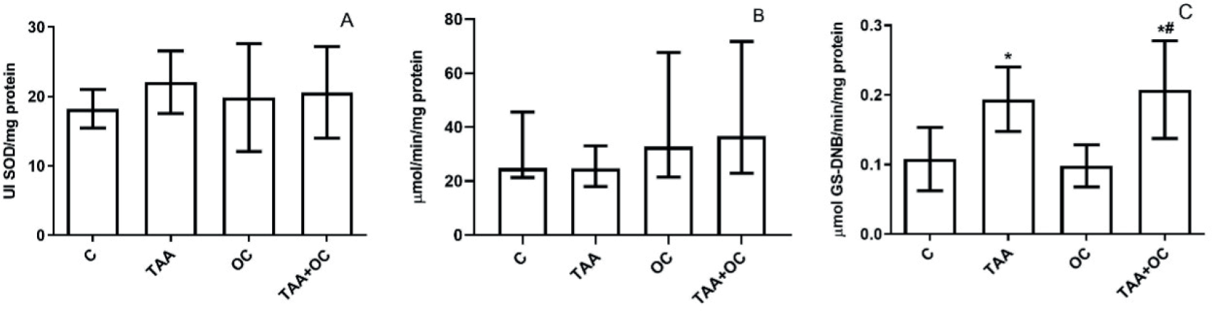
The results demonstrated that there were no significant differences in the activity of SOD and CAT among the groups as shown in Figure 1Aand 1B. However, an increase in GST activity (TAA and TAA+OC groups) can be observed compared to the C group, as indicated in Figure 1C. The TAA+OC group also had higher GST activity compared to the OC group in Figure 1C.
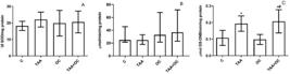
Antioxidant enzymatic activity of Superoxide dismutase (A), Catalase (B) and Glutathione-S-Transferase (C) in the renal tissue of groups C, TAA, OC and TAA+OC. (N = 8). Mean ± standard deviation (SOD, GST). Median and total amplitude (CAT); *P < 0,05 vs C; #P < 0,05 vs OC.
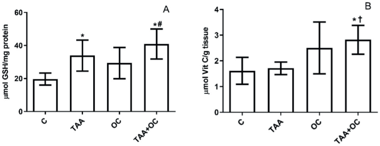
Based on the evaluation of the GSH levels, which is a non-enzymatic antioxidant, it can be observed that there is an increase in both the TAA-treated groups (Figure 2A). On the other hand, the response was similar for vitamin C levels. There was an increase in data for both the OC and TAA+OC groups, but only the TAA+OC group differed from the control and TAA groups, as shown in Figure 2B.
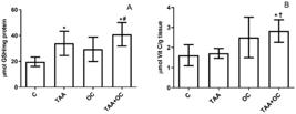
Concentration of non-enzymatic antioxidants GSH (A) and Vit C (B) in the renal tissue of groups C, TAA, OC, and TAA+OC. (N = 8). Mean ± standard deviation. *P < 0,05 vs C; #P < 0,05 vs OC; †P < 0,05 vs TAA.
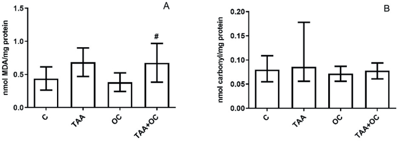
The TBARS analysis showed an increase in this marker, but there was no statistical difference between the groups and the C group. However, in the OC+TAA group, the levels of these substances significantly increased compared to the OC group, as shown in Figure 3A. The analysis of carbonyl proteins did not reveal any significant differences among the analyzed groups, as indicated in Figure 3B.
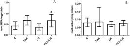
Levels of thiobarbituric acid reactive substances (TBARS) (A) and carbonyl proteins (B) in the renal tissue of groups C, TAA, OC, and TAA+OC. (N = 8). Mean ± standard deviation. Median and total amplitude (Carbonyl); #P < 0,05 vs OC.
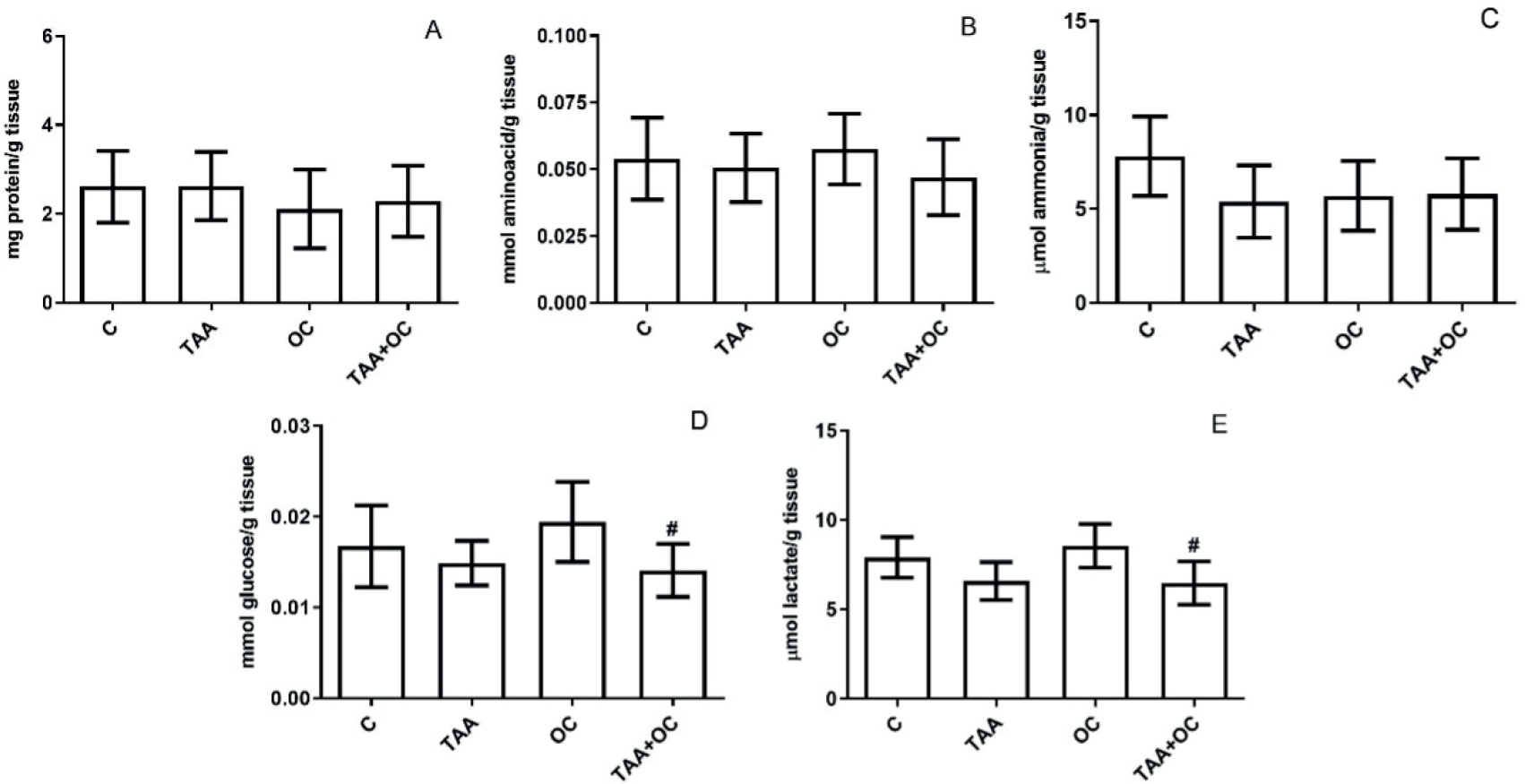
In the same context, for total protein, amino acids, and ammonia there were no statistical differences for the groups when compared to C and TAA (Figure 4A, B and C, respectively). On the other hand, rats treated with TAA and TAA+copaiba oil had reduced levels of glucose and lactate compared to the OC group (Figure 4 D and E, respectively).
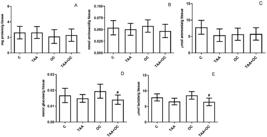
Levels of Total proteins (A); Amino acids (B); Ammonia (C); Glucose (D) and Lactate (E) in the renal tissue of groups C, TAA, OC and TAA+OC. (N = 8). Mean ± standard deviation. #P < 0.05 vs OC.
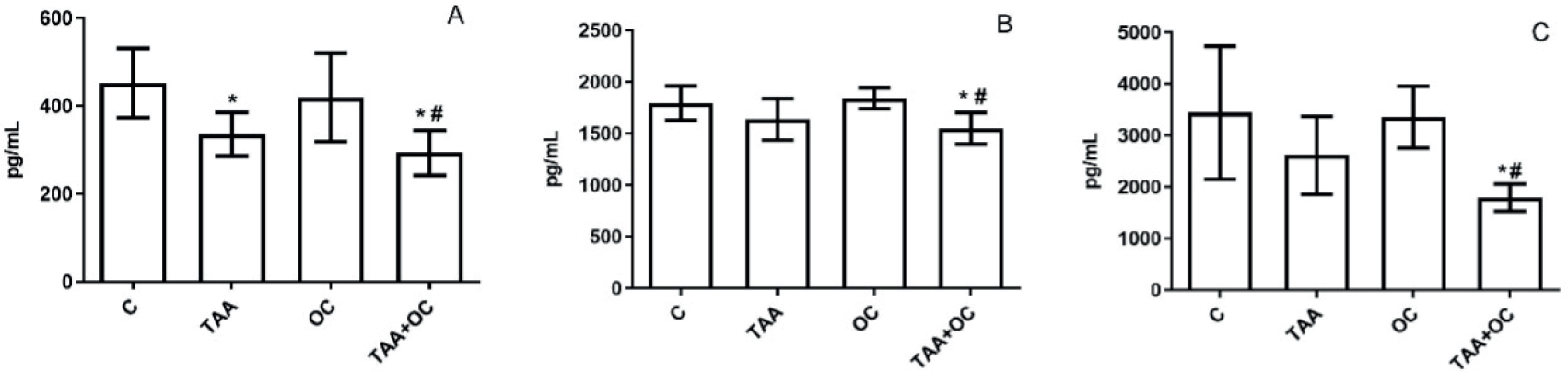
For cytokines analyses in renal tissue, TNF-α level decreased statistically in TAA groups compared with the C group (Figure 5A). However, there was a reduced level of IL-10 and IL-1β in TAA+OC groups when compared to C and OC groups (Figure 5B and C, respectively).
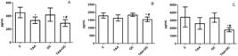
Concentration of cytokines TNF-α (A); IL-10 (B); IL-1β (C) in the renal tissue of groups C, TAA, OC, and TAA+OC. (N = 8). Mean ± standard deviation. *P < 0,05 vs C; #P < 0,05 vs OC.
DISCUSSION
This study aimed to investigate if TAA-induced liver cirrhosis could result in damage to the kidney and if the treatment with copaiba oil-resin could improve the renal injury. Previous studies have shown a connection between cirrhosis and its impact on kidney tissue (Cartaxo et al., 2018), and there are in the literature researches showing the anti-inflammatory effects of copaiba oil (Vásquez, Mendonça, Noda, 2014; Luca et al., 2018). Therefore, we studied the possible effects generated in cirrhotic rats by examining their redox state, metabolic and immunological parameters. Firstly, we investigated the SOD activity, is an important redox state marker that acts as the first line of defense against the superoxide radical by converting it into gaseous oxygen and hydrogen peroxide in the kidneys (Zelko, Mariani, Folz, 2002). In sequence, CAT plays a crucial role in breaking down hydrogen peroxide into water and gaseous oxygen. It is mainly located in peroxisomes and contributes to the beta-oxidation of fatty acids (Brown et al., 1982). However, this study found that there was no significant change in the activity of SOD and CAT due to the inability of thioacetamide to influence the production of ROS in the kidney. Additionally, copaiba oil did not affect the activity of these enzymes. Studies realized by Alqrad et al. (2023) and Alshahrani et al. (2023) showed a decrease in SOD activity and GSH concentration, but the doses administered (200 mg/ kg) were higher than those in this study (100 mg/kg). On the other hand, in studies carried out by Taffarel et al. (2024) using TAA (100 mg/kg), it was possible to observe that there was an increase in SOD, CAT, and GST in the liver of cirrhotic animals and copaiba oil was unable to reverse these enzymatic activities. In this same tissue, GSH levels increased in both groups treated with TAA. However, in that study on hepatic tissue, TAA increased enzymatic antioxidant activity and non-enzymatic antioxidant levels probably as a compensatory mechanism appearing to restore the redox disturbance showed in cirrhotic animals (Nguyen, Nioi, Pickett, 2009). Otherwise, in our study, the results presented in renal tissue suggest that exposure to TAA stimulate GST activity through an additive response, such as an increase observed in TAA groups, thus corroborating the fact that GST is responsible for the conjugation of glutathione with a variety of xenobiotics/ metabolites to form water-soluble conjugates and less toxic (Palodetto et al., 2010). In the same way, Silva et al. (2021) had similar results in liver tissue and indicated the nuclear factorerythroid 2-related factor 2 (Nrf2) antioxidant response signaling pathway as probably responsible for the greater activity of antioxidant enzymes against the damage caused by thioacetamide (Nguyen, Nioi, Pickett, 2009). Furthermore, studies realized by De Paula et al. (2023) demonstrated that copaiba resin oil per se did not influence the activity of SOD, CAT, and GST enzymes in adipose tissue, as in our studies. In the same way, the activity of SOD and GST enzymes did not alter in adipose tissue of rats fed with high sucrose diet (Telles et al., 2022).
For GSH concentration, there was an increase in the groups that had TAA, but administration of OC did not influence this parameter. In this experimental model, with TAA treatment for 8 weeks, the kidney was not so affected, and even the antioxidant biomarkers were stimulated by thioacetamide itself through physiological compensation mechanisms. In the vitamin C analysis, otherwise, it is noticed that the administration of TAA did not significantly alter the data in this group, but OC promoted a good effect increasing this antioxidant when compared to TAA. A very interesting finding was that the OC group showed a tendency to increase GSH and vit C levels in renal tissue, probably due the presence of sesquiterpenes found in OC, differentiating from the findings found for adipose and liver tissues (De Paula et al., 2023; Taffarel et al., 2024 Telles et al., 2022), which demonstrates that depending on the tissue evaluated, the OC may present different effects.
TAA generates a state of oxidative stress due to formation of reactive unstable metabolites (TAA-S-oxide and TAA-S-dioxide), which increase the ROS production and promotes free radical mediated damage to cellular componentes (proteins, lipids, and (DNA, for example) (Hsu et al., 2012; Saad, Oda, Sedeek, 2020). In addition, TAA alters the urea cycle, the profile of circulating amino acids, lipid and lipoprotein metabolism, and fatty acid synthesis (Silvestre, 2015). In our study, the TAA group showed a slight increase in lipoperoxidation in the kidneys, but the increase was not statistically significant. The OC group was unable to reduce this tendency to increase, as evidenced by the TAA+OC group compared to the OC group. When we analyzed the carbonyl data, we observed that TAA and copaiba oil did not cause any change in protein damage, even after 8 weeks of experimental protocol. This indicates that TAA did not cause protein damage, as evidenced by the absence of effects on SOD and CAT, and that copaiba oil did not interfere with carbonyl proteins, showing that its administration is safe, since it maintained the levels of this parameter at control levels. Therefore, it appears that TAA was more harmful to lipids than to proteins present in cell membranes. Although copaiba oil is a lipid, it was not able to have a positive effect on the slight increase in TBARS generated by TAA. It is important to highlight that copaiba oil contains a substance called β-caryophyllene, which is well described in the literature for its antioxidant and anti-inflammatory properties (Fernandes et al., 2007; Ames-Sibin et al., 2018). However, in this study, we used the same oleoresin used in the studies by Telles et al. (2022), which demonstrated a low concentration of this substance, which may justify the fact that we did not find effects on the markers investigated.
Liver cirrhosis generates a catabolic state that culminates in the degradation of glycogen to maintain blood glucose, oxidation of fatty acids, and proteolysis (Islam et al., 2019). In this sense, the kidney also appears to be affected by this condition of lack of energy and may, in part be carrying out the renal gluconeogenesis pathway (Nelson, Cox, 2014) since this organ activates this metabolic pathway and glucose levels appear to be reduced in the TAA group. Therefore, we investigated the metabolic parameters of this tissue, and no significant changes were found in the metabolites for the groups when compared to the control. In the work of Silva et al. (2021), the liver of animals treated with TAA showed a reduction in weight gain, epididymal and retroperitoneal fat, and glucose, a tendency to increase lactate and reduce proteins, and amino acids did not change. In this study, there is a tendency to decrease glucose, lactate, and ammonia in the TAA group, with no changes in proteins and amino acids. It appears that the kidney behaves similarly to the liver regarding the reduction of glucose in these conditions of cirrhosis, however, the kidney does not appear to be carrying out anaerobic glycolysis like the liver, since its levels have not increased in this tissue. Otherwise, as there was also a reduction in ammonia, although not significant, this leads us to suggest that proteolysis did not occur in this tissue. Instead, lipolysis was probably responsible for energy production in this tissue in addition to some glucose consumption.
Regarding the effects of copaiba oil treatment, both glucose and lactate levels were similar in TAA+OC and OC groups, indicating the use of aerobic glycolysis. The presence of TAA in cirrhotic animals likely increased the energetic demand by glucose without the interference of OC.
There is limited understanding of the immune response dynamics in the cirrhosis model induced by TAA. Our group's studies investigated the livers of animals, revealing an intense cellular infiltrate observed histologically. A decrease in IL-10 accompanied this, while pro-inflammatory cytokines IL-6 and TNF-alpha remained unchanged (Taffarel et al., 2024). Conversely, a study published by Kurtoğlu in 2019 found an increase in IFN-gamma, IL-17, and TNF in the livers of mice treated with thioacetamide for 12 weeks. This suggests that there may be a modulation of the Th1 and Th17 type responses and IL-17 producing non-immune cells, such as biliary epithelial cells in these animals' livers.
Nevertheless, there are studies examining the immune response components in the liver of the cirrhosis model induced by TAA, there has been no research, to our knowledge, that evaluates the immune response in the kidneys of these animals. A study conducted by our research group found that treatment with TAA did not alter the histological parameters of the kidneys, and we did not observe any typical cellular infiltrates associated with inflammatory processes, as are commonly found in the liver of this animal model (Da Silva, Aguiar, Bomfim, 2023). Additionally, the TAA-induced cirrhosis model exhibited minimal changes in the cytokine profile assessed in the kidneys, suggesting that, at least in our study, we did not detect significant immunological changes in that organ.
Although the changes in cytokine levels within the kidneys of the TAA-treated animals were relatively minor, treatment with copaiba oil-resin led to a reduction in the concentrations of IL-1β, IL-10, and TNF-α. This observation suggests that copaiba oil may exert a negative modulation of the immune response, specifically in the TAA-treated group. This study on the use of copaiba oil as a supplement in the thioacetamide-induced liver cirrhosis model did not show any interference with redox status parameters on renal tissue as expected. It also did not promote any changes in the metabolic and immunological parameters that were investigated.
ACKNOWLEDGMENT
The authors acknowledge to Conselho Nacional de Desenvolvimento Científico e Tecnológico (CNPq) for granting the scholarships to A.J.L.B.F., G.V.F.S., and J.M.A.F.
-
FUNDING
This study was supported by Fundação de Amparo a Pesquisa do Estado de Mato Grosso - FAPEMAT (process - 571424/2014).
-
ETHICS STATEMENTS
The study was approved by the Ethics Committee on the Use of Animals of Universidade Federal de Mato Grosso (CEUA/UFMT -23108.050625/2019-38).
DATA AVAILABILITY STATEMENT
Data available from the corresponding author upon reasonable request.
REFERENCES
-
Alqrad MAI, El-Agamy DS, Ibrahim SEM, Sirwi A, Abdallah HM, Abdel-Sattar E, et al. SIRT1/Nrf2/ NF-B Signaling Mediates Anti-Inflammatory and Anti-Apoptotic Activities of Oleanolic Acid in a Mouse Model of Acute Hepatorenal Damage. Medicina. 2023;59:1351. https://doi.org/10.3390/medicina59071351
» https://doi.org/10.3390/medicina59071351 -
Alshahrani MY, Ebrahim HA, Alqahtani SM, Bayoumy NM, Kamar SS, ShamsEldeen AM, et al. Metformin Suppresses Thioacetamide-Induced Chronic Kidney Disease in Association with the Upregulation of AMPK and Downregulation of Oxidative Stress and Inflammation as well as Dyslipidemia and Hypertension. Molecules. 2023;28:2756. DOI:10.3390/ molecules28062756
» https://doi.org/10.3390/molecules28062756 -
Ames-Sibin AP, Barizão CL, Castro-Ghizoni CV, Silva FMS, Sá-Nakanishi AB, Bracht L, et al. β-Caryophyllene, the major constituent of copaiba oil, reduces systemic inflammation and oxidative stress in arthritic rats. J Cell Biochem. 2018;119(12):10262-10277. DOI: 10.1002/jcb.27369.
» https://doi.org/10.1002/jcb.27369. -
Bradford MMA. A rapid and sensitive method for the quantification of microgram quantities of protein utilizing the principle of protein-dye binding. Anal Biochem. 1976;72:248-254. DOI: 10.1016/0003-2697(76)90527-3.
» https://doi.org/10.1016/0003-2697(76)90527-3. - Brown FR, Mcandams AJ, Cummins JW, Konkol R, Singh I, Moser HW. Cerebro-hepato-renal (Zellweger) syndrome and neonatal adrenoleukodystrophy: Similarities in phenotype and accumulation of very long chain fatty acids. Johns Hopkins Med J 1982;151(6):344-351. PMID: 7176294.
-
Buege JA, Aust SD. Microsomal lipid peroxidation. Methods Enzymol. 1978;52:302-310. DOI: 10.1016/ S0076-6879(78)52032-6.
» https://doi.org/10.1016/S0076-6879(78)52032-6 - Cartaxo YA, Junior FCO, Silva ML, Feitosa ANA. Diagnóstico e tratamento da síndrome hepatorrenal: Uma Revisão Integrativa. Rev Interdisc Saúde. 2018;5(4):724-742. ISSN: 2358-7490.
-
Colombo G, Clerici M, Garavaglia ME, Giustarini D, Rossi R, Milzani A, Dalle-Donne I. A step-by-step protocol for assaying protein carbonylation in biological samples. J Chromatogr B Analyt Technol Biomed Life Sci. 2016;1019:178-190. DOI:10.1016/j.jchromb.2015.11.052
» https://doi.org/10.1016/j.jchromb.2015.11.052 -
Da Silva KC, Aguiar DH, Bomfim GF. Assessment of the effect of copaiba oil (copaifera sp.) on the kidneys of rats with hepatic dysfunction. Sci Electron Arch. 2023;16(5). DOI:10.36560/16520231722.
» https://doi.org/10.36560/16520231722. -
De Paula M, Rocha LA, Telles LO, Mendonça ST, Sinhorin VDG, Nascimento AF, et al. Copaiba oleoresin attenuates weight gain without alter inflammatory and redox system markers in adipose tissue of healthy animals. Sci Elec Arch. 2023;16(4):53-59. DOI: 10.36560/16420231694.
» https://doi.org/10.36560/16420231694. -
Duboie M, Gilles KA, Hamilton JK, Rebers PT, Smith F. Colorimetric method for determination of sugars and related substances. Anal Biochem. 1956;28(3):350-358. DOI: 10.1021/ac60111a017. DOI: 10.1111/j.1742-7843.2012.00903.x.
» https://doi.org/10.1021/ac60111a017.» https://doi.org/10.1111/j.1742-7843.2012.00903.x. -
Fernandes ES, Passos GF, Medeiros R, Cunha FM, Ferreira J, Campos MM, et al. Anti-inflammatory effects of compounds alpha-humulene and (-) -trans-caryophyllene isolated from the essential oil of Cordia verbenacea. Euro J Pharmacol. 2007;569:228-236. DOI: 10.1016/j.ejphar.2007.04.059.
» https://doi.org/10.1016/j.ejphar.2007.04.059. -
Furtado KS, Prado MG, Silva MA, Dias MC, Rivelle DP, Rodrigues MAM, Barbisan LF. Coffee and Caffeine Protect against Liver Injury Induced by Thioacetamide in Male Wistar Rats. Basic Clinic Pharmacol Toxicol. 2012;111:339-347. DOI: 10.1111/j.1742-7843.2012.00903.x.
» https://doi.org/10.1111/j.1742-7843.2012.00903.x. -
Gentzkow CJ, Masen JM. An accurate method for the determination of blood urea nitrogen by direct nesslerization. J Biol Chem. 1942;143:531-544. DOI: 10.1016/S0021-9258(18)72642-2. DOI: 10.1016/ S0021-9258(18)72642-2.
» https://doi.org/10.1016/S0021-9258(18)72642-2.» https://doi.org/10.1016/S0021-9258(18)72642-2 -
Gonçalves ES, Silva JR, Gomes CL, Nery MBL, Navarro DMAF, Santos GKN, et al. Effects of the oral treatment with Copaifera multijuga oil on reproductive performance of male Wistar rats. Rev Bras Farmacogn. 2014;24:355-362. DOI: 10.3390/plants12051054.
» https://doi.org/10.3390/plants12051054. -
Habig WH, Pabst MJ, Jacoby WB. Glutathione S-transferase, the first enzymatic step in mercapturic acid formation. J Biol Chem. 1974;249:7130-7139. DOI: 10.1016/S0021-9258(19)42083-8.
» https://doi.org/10.1016/S0021-9258(19)42083-8. -
Harrower JR, Brown CH. Blood lactic acid. A micromethod adapted to field collection of microliter samples. J Appl Physiol. 1972;32:709-711. DOI: 10.1152/jappl.1972.32.5.709.
» https://doi.org/10.1152/jappl.1972.32.5.709. - Horácio BO, Geron VL, Fávero MT, Speretta G, Menezes MF. Anti-inflamatory action of copaíba oil: possible contribution in the treatment of Metabolic Syndrome. Rev Cientif FAEMA. 2017.8(1):144-160.
- Hsu DZ, Chu PY, Li YH, Chandrasekaran VRM, Liu MY. Role of flavin-containing-monooxygenase-dependent neutrophil activation in thioacetamide-induced hepatic inflammation in rats. Toxicology. 2012;298:52-58.
- Islam ASMS, Mamun AA, Mahtab MA, Rahman S. Management of nutrition status in liver cirrhosis patients. BEACON Med J. 2019;2(2):32-37. ISSN: 2616-6178.
- Iwakiri Y. The Systemic and Splanchnic Circulation. In: Ginès P, Kamath P, Arroyo V. Chronic Liver Failure: Mechanisms and Management. Humana Press. 2011.
-
Jones DP. Redefining Oxidative Stress. Antioxid Redox Signal. 2006.8(9):1865-1879. DOI: 10.1089/ ars.2006.8.1865.
» https://doi.org/10.1089/ars.2006.8.1865 -
Kurtoğlu EL, Kayhan B, Gül M, Kayhan B, Akdoğan Kayhan M, Karaca ZM, et al. A bioactive product lipoxin A4 attenuates liver fibrosis in an experimental model by regulating immune response and modulating the expression of regeneration genes. Turk J Gastroenterol. 2019 Aug;30(8):745-757. doi: 10.5152/tjg.2019.18276.
» https://doi.org/10.5152/tjg.2019.18276. -
Liguori L, Russo G, Curcio F, Bulli G, Aran L, Della-Morte D, et al. Oxidative stress, aging, and diseases. Clin Interventions in Aging. 2018;13:757-772. DOI: 10.2147/CIA.S158513.
» https://doi.org/10.2147/CIA.S158513. -
Luca LG, Matos SP, Kreutz T, Teixeira HF, Veiga Jr VF, Araújo BV, et al. Antiinflammatory effect from a hydrogel containing nanoemulsified copaiba oil (Copaifera multijuga Hayne). AAPS Pharm Sci Tech. 2018;19:522-530. DOI: 10.1208/s12249-017-0862-6
» https://doi.org/10.1208/s12249-017-0862-6 -
Maciel MA, Pinto AC, Veiga Jr VF, Grynberg NF, Echevarria A. Plantas medicinais: a necessidade de estudos multidisciplinares. Quím Nova. 2002;25(3):429-38. DOI: 10.1590/S0100-40422002000300016.
» https://doi.org/10.1590/S0100-40422002000300016. - Misra HP, Fridovich I. The role of superoxide anion in the auto-oxidation o epinephrine and a simple assay for superoxide dismutase. J Biol Chem. 1972;247:3170-3175. PMID: 4623845.
- Nelson DL, Cox MM. Princípios de bioquímica de Lehninger. Porto Alegre: Artmed, 2014.
-
Nelson DP, Kiesow LA. Enthalpy of decomposition of hydrogen peroxide by catalase at 25°C (with molar extinction coefficients of H2O2 solution in the UV). Anal Biochem. 1972;49:474-478. DOI: 10.1016/0003-2697(72)90451-4.
» https://doi.org/10.1016/0003-2697(72)90451-4. - Nguyen T, Nioi P, Pickett CB. The Nrf2-Antioxidant Response Element Signaling Pathway and Its Activation by Oxidative Stress. J Biol Chem. 2009;284:13291-13295.
- Oliveira ECP, Lameira AO, Zoghbi MGB. Identificação da época de coleta do óleo-resina de copaíba (Copaifera spp.) no município de Moju, PA. Rev Bras Plant Med. 2006;8(3):14-23.
-
Palodetto B, Postal M, Grinoli CRE, Sartorato EL, Oliveira CA. Influence Of Glutathione S-Transferase on the Ototoxicity Caused by Aminoglycosides. Braz J Otorhinolaryngol. 2010;76(3):306-309. DOI: 10.1590/ S1808-86942010000300006.
» https://doi.org/10.1590/S1808-86942010000300006 - Roe JH. Methods of Biochemical Analysis. Glick D (ed). New York: Interscience Publishers. 1954.
- Ramos MFS. Desenvolvimento de microcápsulas contendo a fração volátil de copaíba por spray-drying: estudo de estabilidade e avaliação farmacológica. [Tese de Doutorado]. São Paulo: Universidade de São Paulo (USP). 2006.
- Saad HM, Oda SS, Sedeek EK. Protective effect of Lactéol® forte against thioacetamide-induced hepatic injury in male albino rats. Alex J Vet Sci. 2020;67:92-98.
-
Schoonbroodt S, Piette J. Oxidative stress interference with the nuclear factorκB activation pathways. Biochem Pharmacol. 2000;60(8):1075-1083. DOI: 10.1016/S0006-2952(00)00371-3.
» https://doi.org/10.1016/S0006-2952(00)00371-3. -
Sedlack J, Lindsay RH. Estimation of total, protein bound, and nonprotein sulphydryl groups in tissue with Ellman’s reagent. Anal Biochem. 1968;25:192-205. DOI: 10.1016/0003-2697(68)90092-4.
» https://doi.org/10.1016/0003-2697(68)90092-4. - Sharma B, John S. Hepatic Cirrhosis. In: Stat Pearls (Internet). Treasure Island (FL): StatPearls Publishing. 2021.
-
Silva BS, Paulino AMB, Taffarel M, Borba IG, Telles LO, Lima VV, et al. High sucrose diet attenuates oxidative stress, inflammation, and liver injury in thioacetamide-induced liver cirrhosis. Life Sci. 2021;267:118944. DIO: 10.1016/j.lfs.2020.118944.
» https://doi.org/10.1016/j.lfs.2020.118944 - Silvestre PM. Avaliação do selante de fibrina como arcabouço para células tronco em fígados cirróticos. [Dissertação de Mestrado]. São Paulo: Universidade Estadual Paulista (UNESP). 2015.
-
Simonetto DA, Gines P, Kamath PS. Hepatorenal syndrome: pathophysiology, diagnosis, and management. Br Med J. 2020;370:2687. DOI: 10.1136/ bmj.m2687.
» https://doi.org/10.1136/bmj.m2687 -
Soares ER, Monteiro EB, Silva RC, Batista A, Sobreira F, Mattos T, et al. Compostos biativos em alimentos, estresse oxidativo e inflamação: uma visão molecular da nutrição. Revista HUPE. 2015;14(3):64-72. DOI: 10.12957/rhupe.2015.19942.
» https://doi.org/10.12957/rhupe.2015.19942. -
Spies JR. Colorimetric procedures for amino acids. Met Enzymol. 1957;3:467-477. DOI: 10.1016/S0076-6879(57)03417-5.
» https://doi.org/10.1016/S0076-6879(57)03417-5. -
Taffarel M, Silva BSD, Paulino AMB, Telles LO, Mendonça ST, Santos CVD, et al. Copaiba Oleoresin Improves Weight Gain and IL-10 Concentration, with No Impact on Hepatic Histology, in Liver Cirrhosis. Biology (Basel). 2024 Oct 23;13(11):853. DOI: 10.3390/biology13110853.
» https://doi.org/10.3390/biology13110853. -
Telles LO, Silva BS, Paulino AMB, Trigueiro SM, Sinhorin VDG, Lima MCF, et al. Copaiba oleoresin presents anti-obesogenic effect and mitigates inflammation and redox imbalance in adipose tissue. Acta Amazonica. 2022;52(4):331-338. DOI: 10.1590/1809-4392202201411.
» https://doi.org/10.1590/1809-4392202201411. -
Túnez I, Muñoz MC, Villavicencio MA, Medina FJ, Prado EP, Espejo I, et al. Hepato-and neurotoxicity induced by thiocetamide: protective effects of melatonin and dimethulsulfoxide. Pharm Res. 2005;52:223-228. DOI: 10.1016/j.phrs.2005.03.007.
» https://doi.org/10.1016/j.phrs.2005.03.007. -
Vásquez SPF, Mendonça MS, Noda SN. Etnobotânica de plantas medicinais em comunidades ribeirinhas do Município de Manacapuru, Amazonas, Brasil. Acta Amazon. 2014;44:457-472. DOI: 10.1590/1809-4392201400423.
» https://doi.org/10.1590/1809-4392201400423. -
Veiga Jr VF, Pinto AC. O Gênero Copaifera L. Quím Nova. 2002;25(2):273-286. DOI: 10.1590/S0100-40422002000200016.
» https://doi.org/10.1590/S0100-40422002000200016. -
Wu CC, Yeung LK, Tsai WS, Tseng CF, Chu P, Huang TY, et al. Incidence and factors predictive of acute renal failure in patients with advanced liver cirrhosis. Clin Nephrol. 2006;65:28-33. DOI: 10.5414/cnp65028.
» https://doi.org/10.5414/cnp65028. -
Zelko IN, Mariani TJ, Folz RJ. Superoxide dismutase multigene family: A comparison of the CuZn-SOD (SOD1), Mn-SOD (SOD2), and EC-SOD (SOD3) gene structures, evolution, and expression. Free Radic Biol Med. 2002;33(3):337-349. DOI: 10.1016/s0891-5849(02)00905-x.
» https://doi.org/10.1016/s0891-5849(02)00905-x.
Edited by
-
Associate Editor:
Daniel Demarque