 Antimicrobial peptide cecropins derived from Antheraea pernyi trigger cell apoptosis through mitochondrial caspase-dependent pathway in MDA-MB-231 cells
Abstract
Cecropins are vital innate immune effectors in pathogen defence of animals. Studies have demonstrated that cecropins also possess anticancer capacity against numerous cancerous cells. In this study, the anticancer effects of A. pernyi cecropin D (ApCecD) and cecropin like protein (ApCecL) were investigated in human breast cancer MDA-MB-231 cells. CCK-8 assay indicated that ApCecD and ApCecL suppressed the proliferation of MDA-MB-231 cells in a dose-dependent manner. Wound healing experiments showed that A. pernyi cecropins inhibited the migration of MDA-MB-231 cells. Hoechst staining suggested that treatment with ApCecD and ApCecL induced apoptotic features, including nuclear-like chromatin condensation and increased in nuclear body fragments. Western blotting results showed that ApCecD and ApCecL induced apoptosis via mitochondria-mediated endogenous pathway by upregulating the expression of caspase 3, BNIP3 and Bax. These findings indicated that ApCecD and ApCecL are promising candidates for developing new drugs in breast cancer treatment.
Keywords:
Antheraea pernyi; Cecropins; Breast cancer; Apoptosis.
INTRODUCTION
Nowadays, breast cancer is become the most incidence and mortality cancer among women. Due to cancer heterogeneity, the therapeutic strategy for breast cancer also differs and commonly includes surgery, hormone treatment, chemo/radiotherapy and specific targeting treatment (Geay, 2013). Although many effective advances have been made in cancer treatment in recent years, the spread and metastasis remain major challenges in breast cancer treatment (Bai et al., 2018). Furthermore, chemotherapy, while a conventional and important approach, often leads to side effects and drug resistance with long-term use (Nedeljković, Damjanović, 2019). As a triple negative breast cancer (TNBC) cell line, MDA-MB-231 is negatively to express estrogen receptor, progesterone receptor and human epidermal receptor. Since TNBC is not sensitive for current clinical targeted therapies and ineffective for anti-hormonal treatment, it is more difficult to treat and has a poor prognosis and high recurrence (Shao, Sun, Deng, 2017). Therefore, screening low toxicity and more effective chemical agents will be helpful for breast cancer treatment, particularly for the TNBC treatment.
Antimicrobial peptides (AMPs) are naturally produced peptides that exist in almost all living organisms, including plants, animals and microorganisms. AMPs are effectors of innate immunity and responsible for defending pathogens in host. Without the function in immunity, many studies have demonstrated that AMPs also have the anticancer ability on numerous cancerous cells. Moreover, some AMPs have been proofed with low toxicity on non-cancerous cells (Wang et al., 2018; Lee, Shin, Kim, 2015), and showed minor resistance for long term treatment (Saido-Sakanaka et al., 2004). AMPs are usually short in length with small molecular weights (less than 10 kDa) and diverse structures (Zhang, Sunkara, 2014). Based on the 3D structures, AMPs usually sort into four groups: α-helix, β-sheet, looped peptides, and extended (Aghamiri et al., 2021). AMPs are commonly cationic at neutral pH and have an amphiphilic structure that responsible for interaction with amphiphilic cell membranes (Lee, Hall, Aguilar, 2016). These properties play key roles in AMPs binding to cancerous cell membranes and perform tumoricidal effect. However, since the various structures they contained, the anti-tumor mechanisms of different AMPs may differ. The mechanism of specific AMP remains to be further studied.
Insect lacks adaptive immunity and relies on innate immunity to defending pathogens. During the innate immune responses, AMPs are produced by insect immune organs and transported throughout the body to resist the invasion of bacteria and fungi (Zhou et al., 2024). Based on the structures and characteristics, insect AMPs can be sorted into four types: α-helix (like cecropin), cysteine rich (like defensin), proline rich (like apidaecin) and glycine rich (like attacin) (Bulet, Stocklin, 2005). Cecropins can protect insects by killing pathogens such as bacteria, virus and fungi. In addition to function in innate immunity, several studies have revealed that cecropins also exhibit the anti-tumor activity. Suttmann et al. (2008) demonstrated that cecropin A and B disrupted cell membrane and induced cytolysis in bladder cancer cells. Bombyx mori Cecropin XJ has been shown to inhibit human gastric cancer cells proliferation by inducing cell apoptosis (Wu et al., 2015). Similarly, B. mori cecropin A and cecropin D suppressed the viability of esophageal cancer cells by activating mitochondria-mediated caspase pathway (Xu et al., 2020). Antheraea pernyi, also known as the oak silkworm, is usually reared in the wild and exhibits high active innate immune responses.
Our group’s previous study has shown that a novel cecropin-like peptide (ApCecL) from A. pernyi has anti-bacteria activity and possesses a low cytotoxicity to mouse blood cells (Fang et al., 2017). In this study, the anticancer effects of A. pernyi ApCecD and ApCecL against MDA-MB-231 cells were investigated. ApCecD and ApCecL were found to inhibit cell proliferation and migration, and trigger apoptosis of MDA-MB-231 cells. Furthermore, the cecropins affected proteins expression in mitochondrion-dependent apoptosis pathway. These results indicated that ApCecD and ApCecL are promising candidates to develop new anticancer drugs.
MATERIAL AND METHODS
A. pernyi ApCecD and ApCecL peptide synthesis
A. pernyi ApCecD and ApCecL were synthesized using the solid-phase synthetic method (Sangon, China). The sequence information of ApCecD (GenBank: ACB45561.1) and ApCecL (unsubmitted) were WNPFKELERAGQRVRD AIISAGPAVATVAQATALAKGK and K W K F F K K I E R V G Q N I R D G I I K A G PAVAVVGQATNIAKG, respectively. Both cecropins were chemically modified by amidation in C-terminus. Mass spectrometry was carried out to confirm that the sequences were correct (Figure 1). High performance liquid chromatography was carried out to detect the purity of the peptides. The purity was 94.72% for ApCecD and 93.94% for ApCecL (Figure 1). The physicochemical properties of ApCecD and ApCecL were detected via antimicrobial peptide database server APD3 (http://aps.unmc.edu). The 3D structures of ApCecD and ApCecL were predicted by SWISS-MODEL server (https://swissmodel.expasy.org/) and visualized by SPDBV 4.10 software.
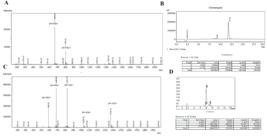
Purity and molecular mass of ApCecD and ApCecL. (A) Liquid chromatographymass spectrometry (LC-MS) of purified ApCecD. (B) High performance liquid chromatography (HPLC) of purified ApCecD. (C) LC-MS of purified ApCecL. (D) HPLC of purified ApCecL.
Cell culture and AMPs administration
MDA-MB-231 cells were cultured in DMEM/F-12(1:1) culture medium supplemented with 10% FBS and 100 U/mL penicillin. The cells were maintained in 5% CO2 at 37°C with saturated humidity. In experimental group, the cells were incubated with varying concentrations of ApCecD and ApCecL, while the negative control group was treated with an equivalent volume of PBS in all experiments. 5-FU (Beyotime, China) was administrated as positive control.
Cell viability assay
The inhibitory effect of ApCecD and ApCecL on MDA-MB-231 cells was assessed by Cell Counting Kit-8 (CCK-8). MDA-MB-231 cells were digested and diluted to 8×10⁴ cells/ml, then seeded in 96-well plate and cultured overnight. Different concentrations of ApCecD and ApCecL (0, 50, 100, 200, 300 and 400 μg/ml) were added to the medium for 12, 24 and 48 h, respectively. Positive control group was treated with 5-FU (1 μg/ml). After treatment, 10 μl CCK-8 (Sangon, China) was added, then incubated in 37°C for 2 h. After incubation, the absorbance at 450 nm was measured by using SuPerMax 3100 (Shanghai Flash Spectrum, China). The growth inhibition rate (%) was calculated as: (AbC - AbT)/ AbC × 100%. AbC and AbT indicated the absorbance in negative control group and treatment group, respectively.
Wound healing assay
MDA-MB-231 cells (2×10⁶ cells/well) grown logarithmically were seeded into 6-well plate, and cultured for stably adherence. A sterilized 100 μl pipette tip was used to scratch three straight lines in the plate to create a wound gap. After washing 3 times with PBS, 0, 100 and 200 μg/ml ApCecD and ApCecL were administrated. 5-FU (1 μg/ml) treatment is positive control. Following treatment for 24 and 48 h, the cell scratches were observed and recorded by inverted microscope (Olympus IX71, Olympus, Japan). Wound areas were analyzed by Image J software. The migration rate was calculated as previously described (Ding et al., 2022).
Hoechst stain
After treatment with different concentration of ApCecD and ApCecL, MDA-MB-231 cells were fixed with 4% paraformaldehyde for 0.5 h. After washing with PBS, the cells were stained with Hoechst 33342 (1 μg/ml; MCE, USA) for 10 min. Then, the cells were washed 3 times with PBS and observed under inverted fluorescence microscope (Olympus IX71, Olympus, Japan).
Western blotting
After treatment with ApCecD and ApCecL (50 and 100 μg/ml), MDA-MB-231 cells were collected and lysed in RIPA lysis buffer (Beyotime, China). The concentration of extracted proteins was detected by BCA protein concentration assay kit (Beyotime, China). Protein samples were separated using 12% SDS-PAGE and transferred to polyvinylidene fluoride (PVDF) membrane. The membrane was blocked with 5% skim milk in TBST buffer for 1 h, and later incubated with primary antibody at 4°C for overnight. After washing with TBST, the membrane was incubated with the secondary antibody for 3 h and then visualized using chemiluminescence detection system (Tanon, China).
The antibodies against β-Tubulin, BNIP3 were purchased from Sangon (Sangon, China). The antibodies against p53 and Bax were obtained from Transgen (Transgen, China). The antibody against caspase 3 was obtained from ABclonal (ABclonal, China).
Statistical analysis
All data were analyzed using SPSS software by performing one-way analysis of variance. The data were presented as mean ± SD. P<0.05 were considered statistically significant, and indicated in the figures with *.
RESULTS
ApCecD and ApCecL inhibit proliferation of MDA-MB-231 cells
The inhibitory effects of ApCecD and ApCecL were determined by CCK-8. Compared with the negative control, the OD450 values of MDA-MB-231 cells were significantly decreased after ApCecD treatment for 24 and 48 h (Figure 2A). The inhibition ratio did not change obviously after 12 h treatment, but it increased significantly with the ApCecD concentration increase after treatment 24 and 48 h (Figure 2B). Besides, the inhibitory effects were varied after treatment with different doses of ApCecD. The cell growth was not obviously suppressed until treatment with 50 μg/ml ApCecD for 48 h. ApCecD treatment for 24 and 48 h both had certain suppressive effects in 100-400 μg/ml dose groups (Figure 2B). However, since the theoretical IC50 value of ApCecD was at a higher concentration of 1335±336.5 μg/ml after 48 h treatment, the growth inhibition effect of ApCecD remained unsatisfactory. ApCecL obviously decreased the OD450 values after 24 and 48 h of treatment, particularly in high dose treatment groups (Figure 2C). The inhibition ratio results showed that 50 μg/ml ApCecL suppressed cell viability after 24 h treatment. However, the inhibitory rates did not show a significant difference in 100 μg/ml ApCecL treatment in all tested time points. The inhibition ratio increased with the ApCecL concentration in 200-400 μg/ml dose groups (Figure 2D). The theoretical IC50 values of ApCecL were 295.3±10.4 μg/ml after 48 h of treatment. In positive control group, 5-Fu treatment exhibited high inhibition ratios at all tested time points. These results imply that ApCecD and ApCecL have growth suppression effects on MDA-MB-231 cells. Furthermore, compared to ApCecD, ApCecL treatment demonstrated a superior inhibitory effect on the proliferation of MDA-MB-231 cells.

Inhibitory effect of ApCecD and ApCecL on the proliferation of breast cancer MDA-MB-231 cells. (A) CCK-8 assay of MDA-MB-231 cells after treated with different concentrations ApCecD 12, 24 and 48 h. (B) Inhibitory rate of MDA-MB-231 cells treated with ApCecD. (C) CCK-8 assay of MDA-MB-231 cells after treated with different concentrations ApCecL 12, 24 and 48 h. (D) Inhibitory rate of MDA-MB-231 cells treated with ApCecL. * indicates 5-Fu positive control. * above the error bars represent P < 0.05 and ** represent P < 0.01 in each group.
ApCecD and ApCecL inhibit migration of MDA-MB-231 cells
Compared with the control, MDA-MB-231 cells displayed larger wound areas after ApCecD treatment 24 and 48 h (Figure 3A). This result was consistent with the positive control group, and there was an obvious gap after 5-Fu treatment. The migration ratio results also showed that MDA-MB-231 cells was significantly suppressed after ApCecD or 5-Fu treatment 24 and 48 h (Figure 3B). Similarly, the ApCecL treatment groups also had larger wound areas, particularly the 200 μg/ ml treatment group (Figure 3C). ApCecL-treated cells were very significantly inhibited in their migration. The relative migration ratios were significantly decreased after ApCecL or 5-Fu treatment 24 and 48 h (Figure 3D). These results suggest that ApCecD and ApCecL can suppress the migration of MDA-MB-231 cells.

ApCecD and ApCecL inhibited cell migration in MDA-MB-231 cells. (A) Wound healing of MDA-MB-231 cells after ApCecD treated 24 and 48 h. Scale bar 500 μm. (B) The relative migration ratio after ApCecD treated. (C) Wound healing of MDA-MB-231 cells after ApCecL treated 24 and 48 h. Scale bar 500 μm. (D) The relative migration ratio after ApCecL treated. Bars represent mean ± SD (n = 3).* represent P < 0.05, ** represent P < 0.01, *** represent P < 0.001 in each time point.
ApCecD and ApCecL induce apoptosis via mitochondrial apoptotic pathway
To explore the mechanism of A. pernyi cecropins in inhibiting of MDA-MB-231 cells, hoechst staining was performed to detect cell apoptosis after cecropins treatment. MDA-MB-231 cells were stained evenly without bright fluorescence in the control group. However, the fluorescence intensity and refraction were obviously increased after ApCecD treatment in 100 and 200 μg/ml dose groups (Figure 4A). Similarly, compared with the control group, the number of cells exhibiting bright fluorescence was increased after ApCecL treatment (Figure 4B). Further results showed that A. pernyi cecropins-treated cells displayed apoptosis features such as nuclear-like chromatin condensation and increased nuclear body fragments (Figure 4C). Based on these results, ApCecD and ApCecL can induce MDA-MB-231 cells apoptosis to exert suppressive effect.
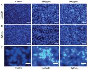
ApCecD and ApCecL induced apoptotic morphological changes in MDA-MB-231 cells. Hoechst staining of MDA-MB-231 cells after (A) ApCecD and (B) ApCecL treated 48 h and observed by fluorescence microscopy. Scale bar 100 μm. (C) The observation of nuclear body fragments in MDA-MB-231 cells after ApCecD and ApCecL treated 48 h. Scale bar 25 μm.
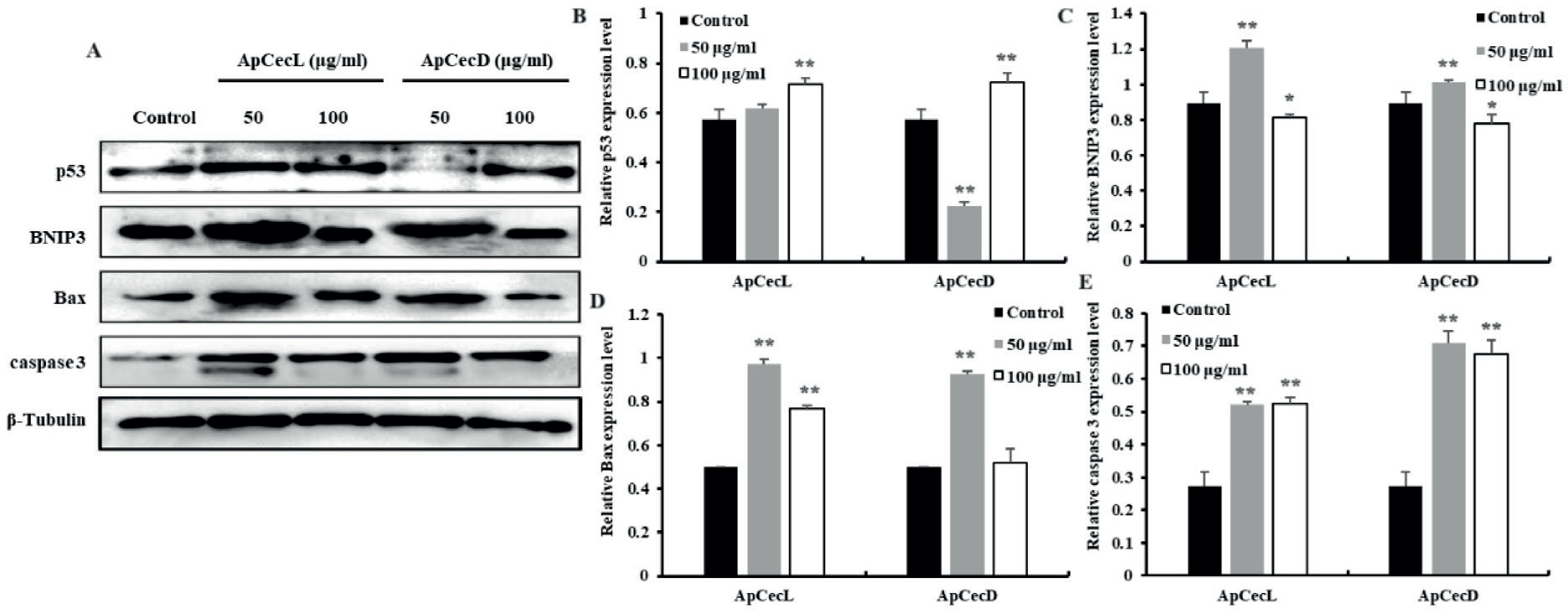
To further confirm the mechanism of A. pernyi cecropins, western blot was performed. The results showed that 50 μg/ml of ApCecL treatment did not significantly change p53 expression, whereas its expression was notably down-regulated after treatment with 50 μg/ml of ApCecD (Figure 5A and 5B). However, both cecropins significantly up-regulated p53 expression at a concentration of 100 μg/ml. BNIP3 expression was significantly increased after treatment with the cecropins at a dose of 50 μg/ml. Conversely, the expression was decreased after treatment with 100 μg/ml of the cecropins (Figure 5A and 5C). Bax, an important molecular of mitochondrial apoptotic pathway, also significantly increased after ApCecD and ApCecL treatment (Figure 5A and 5D). Besides, caspase 3 protein level also significantly increased, and the cleaved caspase 3 was also increased and observed clearly after ApCecD and ApCecL treatment (Figure 5A and 5E). The above results suggest that ApCecD and ApCecL trigger caspase-dependent apoptosis via the mitochondrial apoptotic pathway.

ApCecD and ApCecL induced cell apoptosis in MDA-MB-231 cells. (A) Western blot analysis of MDA-MB-231 cells after ApCecD and ApCecL treatment. β-Tubulin was used as an internal control. (B), (C), (D) and (E) Represent relative protein levels of p53, BNIP3, Bax and caspase 3, respectively. The relative protein level was quantified by normalized with β-Tubulin. * represent P < 0.05 and ** represent P < 0.01 in each treatment group.
ApCecD and ApCecL physicochemical properties and structures
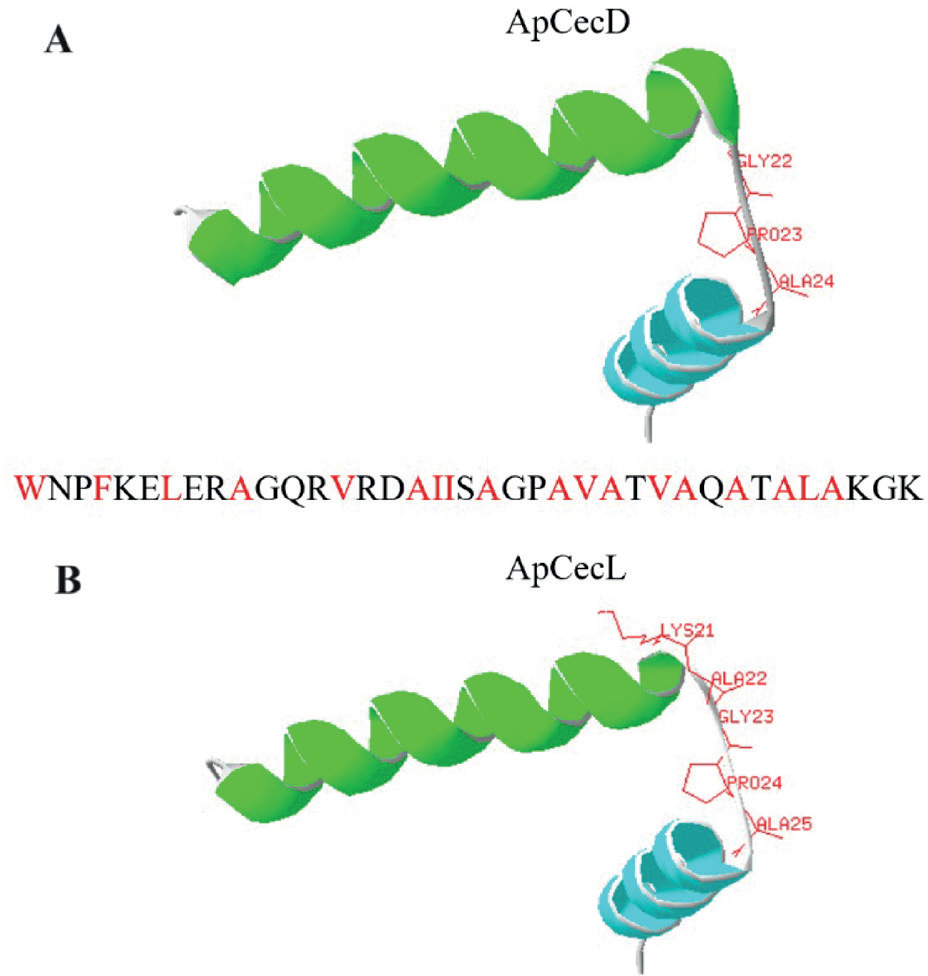
The antimicrobial function of cecropin is related to its amino acid composition and 3 D structure. ApCecD consists of 38 amino acids, out of which 17 are hydrophobic (Figure 6A). The total net charge of ApCecD is +3, and the 3 D structure shows that ApCecD has two α-helix structures connected by 3 amino acids (G22P23A24). In addition, the α-helix is amphipathic in NH2-terminal, while the another is hydrophobic (Figure 6A). ApCecL is also composed of 38 amino acids, and 17 of them are hydrophobic amino acids (Figure 6B). The total net charge of ApCecL is +6. The 3 D structure of ApCecL has two α-helix structures connected by 5 amino acids (K21A22G23P24A25). ApCecL also exhibits amphipathic properties, the NH2-terminal α-helix is amphipathic and another is hydrophobic (Figure 6B).
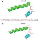
Structure of ApCecD and ApCecL. (A) Primary structure and 3 D structure of ApCecD. (B) Primary structure and 3 D structure of ApCecL. Hydrophobic amino acids are shown in red text in primary structures.
DISCUSSION
Breast cancer is the most common and malignant cancer among women. Among breast cancer treatment strategies, chemotherapy is still the most common non-surgical option (Maughan, Lutterbie, Ham, 2010). However, cancer heterogeneity and chemotherapy induced side effects and drug resistance are still barriers in breast cancer treatment. Therefore, to explore safe and non-resistant drugs are needed in breast cancer treatment, especially to TNBCs. As a novel and promising candidate, AMPs have been proven to have anticancer properties (Teng et al., 2020; Zandsalimi et al., 2020). Apart from the anti-pathogen capacity in innate immunity, AMPs also have the anti-cancer abilities and have been evaluated on many cancer cells, such as hepatocellular cancer, gastric cancer, bladder cancer, and ovarian cancer (Ziaja et al., 2020). Cecropins belong to arthropod AMPs. Several studies indicated that cecropins can kill cancer cells, but not toxic to noncancerous cells. In the present study, two cecropins derived from A. pernyi against a TNBC cell line (MDA-MB-231) was investigated. Cecropins are α-helix peptides, displaying amphipathic properties and including A, B, C, D, P1, and other types. Without exception, ApCecD and ApCecL are also amphipathic peptides and contain two α-helices. Additionally, the NH2-terminal α-helices of ApCecD and ApCecL are amphipathic, while the COOH-terminal α-helices are hydrophobic. ApCecL has higher sequence similarity with Hyalophora cecropia cecropin A and can be considered as cecropin A type AMP (Sato, Feix, 2006). This amphipathic structure could contribute to binding to the cell membrane or organelle membrane and lead to cell depolarization, cell destruction, and cell death (Boman, 2003).
In a previous study, Zhang et al. (2003) proved that A. pernyi cecropins selectively inhibited human colon cancer cells LS-174T growth in vitro, and had no cytotoxic effect on human gastric epithelial cells GES-1. However, the study was performed using a mixture of A. pernyi cecropins and did not mention the precise cecropin types and the related sequences. In our previous study, we identified a new cecropin-like peptide (ApCecL) that had anti-bacteria properties by destroying bacterial cell membranes but had low toxicity to mouse blood cells (Fang et al., 2017). Therefore, we selected ApCecD and ApCecL to determine the anti-proliferation and anti-migration capacities in a TNBC cell line. Our results indicated that ApCecD and ApCecL both strongly inhibit the proliferation and migration of MDA-MB-231 cells. Similarly, B. mori cecropin XJ, a cecropin B family peptide, has been verified to inhibit the proliferation of human hepatocellular carcinoma cells by arresting cell cycle and promoting cell apoptosis (Xia et al., 2016). This inhibitory effect may be associated with the cationic sequence of cecropin interacting with the non-polar anionic cytomembrane (Harris et al., 2013). Further study also demonstrated that B. mori cecropin D recognizes the phosphatidylserine headgroup in the surface of cancer cells or interacts with mitochondrial cardiolipin to induce cell apoptosis (Ramos-Martín, Herrera-León, D’Amelio, 2022).
Although cecropins are all similar in second structures, they differ in the composition of hydrophilic and hydrophobic amino acids. Therefore, the mechanism of cecropin in cancer treatment is still controversial. One theory assumes that cecropin may accumulate on the cytomembrane, change the membrane conformation, and finally cause cytomembrane permeabilization and disruption (Ziaja et al., 2020). Another theory proposes that cecropin destroys cancer cells by affecting cellular signal pathways, including mitochondrial membrane disruption, cell cycle arrest, and cell apoptosis induction (Ceron et al., 2010). Our results revealed that ApCecD and ApCecL induced apoptosis with the nuclear structure disruption in breast cancer cells. Consistent with our results, Musca domestica cecropin also induced apoptotic morphology changes, such as folded nuclear membrane and condensed chromatin in hepatocellular carcinoma cells (Jin et al., 2010). Except for morphology changes, cecropin also affects molecules in apoptosis pathway. Our results showed that the pro-apoptotic proteins, including Bax and BNIP3, were significantly up-regulated after treatment with ApCecD and ApCecL at a concentration of 50 μg/ml. But, the expression of these two proteins was decreased with 100 μg/ml of ApCecD and ApCecL treatment. This difference in expression may be related to the concentration of treatment. Furthermore, caspase 3, the crucial effector of apoptosis, was also increased and activated after treatment. These results suggest that ApCecD and ApCecL induce caspase-dependent apoptosis through the mitochondrial apoptotic pathway in MDA-MB-231 cells. B. mor.i cecropin A also triggered mitochondria-related apoptosis by activating caspase 3, increasing Bax expression, and decreasing B-cell lymphoma 2 expression in esophageal cancer cells (Xu et al., 2020). Interestingly, p53, the key transcription factor involved in apoptosis regulation, was not up-regulated after treatment with 50 μg/ml ApCecD and ApCecL. However, its expression was increased significantly after 100 μg/ml of cecropins treatment. Actually, p53 was highly mutated at Arg280 in MDA-MB-231 cells (Gasco, Shami, Crook, 2002). This mutant site is located in the DNA-binding domain and influences the transcriptional activity of p53. Therefore, p53 may be induced by A. pernyi cecropins, but may not activate p53-dependent and Fas mediated exogenous apoptosis pathway as the wild-type p53 does (Muller et al., 1998). Thus, ApCecD and ApCecL probably induce MDA-MB-231 cell apoptosis via the mitochondria-mediated endogenous pathway. Our findings highlight the potential of ApCecD and ApCecL as natural agents in human breast cancer treatment. Further study is also needed and is underway to evaluate the anti-cancer ability in vivo.
ACKNOWLEDGMENTS
The present work was supported by the General Program of Natural Science Foundation of Anhui Province (grant no. 2308085MC79); and the Scientific Research Project of Universities in Anhui Province (grant no. 2023AH040055).
DATA AVAILABILITY STATEMENT
Data available from the corresponding author upon reasonable request.
REFERENCES
- Aghamiri S, Zandsalimi F, Raee P, Abdollahifar MA, Tan SC, Low TY, et al. Antimicrobial peptides as potential therapeutics for breast cancer. Pharmacol Res. 2021;171:105777.
- Bai X, Ni J, Beretov J, Graham P, Li Y. Cancer stem cell in breast cancer therapeutic resistance. Cancer Treat Rev. 2018;69:152-163.
- Boman HG. Antibacterial peptides: basic facts and emerging concepts. J Intern Med. 2003;254(3):197-215.
- Bulet P, Stocklin R. Insect antimicrobial peptides: structures, properties and gene regulation. Protein Pept Lett. 2005;12(1):3-11.
- Ceron JM, Contreras-Moreno J, Puertollano E, De Cienfuegos GA, Puertollano MA, De Pablo MA. The antimicrobial peptide cecropin A induces caspase-independent cell death in human promyelocytic leukemia cells. Peptides. 2010;31(8):1494-1503.
- Ding W, Chen C, Li J, Geng X, Zhang H, Sun Y. Quercus acutissima Carruth. root extract triggers apoptosis, autophagy and inhibits cell viability in breast cancer cells. J Ethnopharmacol. 2022;289:115039.
- Fang SL, Wang L, Fang Q, Chen C, Zhao XS, Qian C, et al. Characterization and functional study of a Cecropin-like peptide from the Chinese oak silkworm, Antheraea pernyi Arch InsectBiochem Physiol. 2017;94(1):e21368.
- Gasco M, Shami S, Crook T. The p53 pathway in breast cancer. Breast Cancer Res. 2002;4(2):70-6.
- Geay JF. Physiopathologie, diagnostic et thérapeutique du cancer du sein [Physiopathology, diagnosis and treatment of breast cancer]. Soins. 2013;776:25-29.
- Harris F, Dennison SR Singh J, Phoenix DA. On the selectivity and efficacy of defense peptides with respect to cancer cells. Med Res Rev. 2013;33(1):190-234.
- Jin X, Mei H, Li X, Ma Y, Zeng AH, Wang Y, et al. Apoptosis-inducing activity of the antimicrobial peptide cecropin of Musca domestica in human hepatocellular carcinoma cell line BEL-7402 and the possible mechanism. Acta Biochim Biophys Sin(Shanghai). 2010;42(4):259-265.
- Lee E, Shin A, Kim Y. Anti-inflammatory activities of cecropin A and its mechanism of action. Arch. Insect Biochem. Physiol. 2015;88:31-44.
- Lee TH, Hall KN, Aguilar MI. Antimicrobial peptide structure and mechanism of action: a focus on the role of membrane structure. Curr TopMed Chem. 2016;16(1):25-39.
- Maughan KL, Lutterbie MA, Ham PS. Treatment of breast cancer. Am Fam Physician. 2010;81(11):1339-1346.
- Muller M, Wilder S, Bannasch D, Israeli D, Lehlbach K, Li-Weber M, et al. p53 activates the CD95 (APO-1/ Fas) gene in response to DNA damage by anticancer drugs. J Exp Med. 1998;188(11):2033-2045.
- Nedeljković M, Damjanović A. Mechanisms of Chemotherapy Resistance in Triple-Negative Breast Cancer-How We Can Rise to the Challenge. Cells. 2019;8(9):957.
- Ramos-Martín F, Herrera-León C, D’Amelio N. Bombyx mori Cecropin D could trigger cancer cell apoptosis by interacting with mitochondrial cardiolipin. Biochim Biophys Acta Biomembr. 2022;1864(10):184003.
- Saido-Sakanaka H, Ishibashi J, Momotani E, Amano F, Yamakawa M. In vitro and in vivo activity of antimicrobial peptides synthesized based on the insect defensin. Peptides. 2004;25(1):19-27.
- Sato H, Feix JB. Peptide-membrane interactions and mechanisms of membrane destruction by amphipathic alpha-helical antimicrobial peptides. Biochim Biophys Acta. 2006;1758(9):1245-1256.
- Shao F, Sun H, Deng CX. Potential therapeutic targets of triple-negative breast cancer based on its intrinsic subtype. Oncotarget. 2017;8(42):73329-73344.
- Suttmann H, Retz M, Paulsen F, Harder J, Zwergel U, Kamradt J, et al. Antimicrobial peptides of the Cecropin-family show potent antitumor activity against bladdercancer cells. BMC Urol. 2008;8:5.
- Teng QX, Luo X, Lei ZN, Wang JQ, Wurpel J, Qin Z, Yang DH. The multidrug resistance-reversing activity of a novel antimicrobial peptide. Cancers. 2020;12(7):1963.
- Wang J, Ma K, Ruan M, Wang Y, Li Y, Fu YV, Song Y, Sun H, Wang J. A novel cecropin B-derived peptide with antibacterial and potential anti-inflammatory properties. PeerJ. 2018;6:e5369.
- Wu YL, Xia LJ, Li JY, Zhang FC. CecropinXJ inhibits the proliferation of human gastric cancer BGC823 cells and induces cell death in vitro and in vivo. Int J Oncol. 2015;46(5): 2181-2193.
- Xia L, Wu Y, Ma JI, Yang J, Zhang F. The antibacterial peptide from Bombyx mori cecropinXJ induced growth arrest and apoptosis in human hepatocellular carcinoma cells. Oncol Lett. 2016;12(1):57-62.
- Xu, P, Lv D, Wang X, Wang Y, Hou C, Gao K, Guo X. Inhibitory effects of Bombyx mori antimicrobial peptide cecropins on esophageal cancer cells. Eur J Pharmacol. 2020;887: 173434.
- Zandsalimi F, Talaei S, Noormohammad Ahari M, Aghamiri S, Raee P, Roshanzamiri S, et al. Antimicrobial peptides: a promising strategy for lung cancer drug discovery? Expert Opin Drug Discov. 2020;15(11):1343-1354.
- Zhang G, Sunkara LT. Avian antimicrobial host defense peptides: from biology to therapeutic applications. Pharmaceuticals. 2014;7(3):220-247.
- Zhang WM, Lai ZS, He MR, Xu G, Huang W, Zhou DY. [Effects of the antibacterial peptide cecropins from Chinese oak silkworm, Antheraea pernyi on 1, 2-dimethylhydrazine-induced colon carcinogenesis in rats]. Di Yi Jun Yi Da Xue Xue Bao. 2003; 23(10):1066-1068.
- Zhou L, Meng G, Zhu L, Ma L, Chen K. Insect antimicrobial peptides as guardians of immunity and beyond: a review. Int J Mol Sci. 2024;25(7):3835.
- Ziaja M, Dziedzic A, Szafraniec K, Piastowska-Ciesielska A. Cecropins in cancer therapies-where we have been?. Eur J Pharmacol. 2020;882:173317.
Edited by
-
Associate Editor:
Márcia Consolaro