
 Caralluma dalzielii as a potential anti-venom agent against Naja nigricollis envenomation in Wistar rats
Abstract
The World Health Organization has listed Snakebite Envenomation as a significantly ignored tropical disease with a worldwide annual snakebite affecting millions of people, especially in Africa. This study evaluated the in vivo and in vitro neutralising effects of the aqueous extract from the aerial parts of Caralluma dalzielii against snake venom. In the in vivo study, 35 Wistar rats were divided into seven groups, receiving snake venom and treatments with plant extract or standard anti-venom. Mortality rates, haematological, and biochemical parameters were recorded. Whereas, in the in vitro study, different concentrations of plant extract were tested for enzyme inhibition against Naja nigricollis venom. The results showed that the plant extract prevented mortality in all treated groups, comparable to standard anti-venom, and exhibited immunostimulatory effects, increasing WBC levels. It also prevented biochemical disturbances caused by venom, with the highest dose (500 mg/kg) maintaining normal liver enzyme levels. The extract at 100 mg/ml achieved 64.29% enzyme inhibition of PLA2, while the standard anti-venom reached 82.63%, showing concentration-dependent inhibition. In conclusion, the aqueous extract of C. dalzielii exhibits significant anti-venom activity both in vivo and in vitro, demonstrating its potential as a therapeutic option for treating snake bites.
Keywords:
Caralluma dalzielii; Anti-Venom; Naja nigricollis; PLA2 enzyme; Snakebite.
INTRODUCTION
Snakebite Envenomation (SBE) has been listed by the World Health Organization as a significantly ignored tropical disease, with a worldwide annual snakebite affecting 5.4 million people and injuring 2.7 million lives (Bala et al., 2023). The effects of snakebite envenomation are usually species-specific (Bhaumik et al., 2020; Malik, Ada, Udeh, 2021). Snake venoms are generally classified as cytotoxic, hemotoxic, or neurotoxic, and they can all act synergistically involving multiple tissues and organs (Kalita, Saviola, Mukherjee, 2021; Adrião et al., 2022; Osipov, Utkin, 2023). It constitutes a significant public health challenge, especially for people living in rural tropical areas of developing countries, owing to delayed access to healthcare facilities and the conventional anti-snake venom (ASV); which is the most effective countermeasure for treating snake bites (Dossou et al., 2014; Afroz et al., 2024).
The use of traditional remedies, such as medicinal plants rich in bioactive compounds with therapeutic activities, and reported to exhibit anti-venom properties has much been recognised as the immediate means of the effects of snakebite management in tropical rural areas due to the challenges mentioned earlier (Michael et al., 2018; Babangida et al., 2020). Several scientific studies have explored various plant families; not limited to Apocynaceae, Asteraceae, Euphorbiaceae, Fabaceae, Lamiaceae, and Rubiaceae, for their anti-venom properties against different snake venoms with significant efficacies (Liaqat et al., 2022; Patel et al., 2023). Phytoconstituents analysis of plant extracts has identified several metabolites, including alkaloids, flavonoids, saponins, tannins, and terpenoids, that may be responsible for the venom-neutralising effects (Aly, El-Shazly, Eldahshan, 2024; Kusar et al., 2024) sequel to snakebite envenomation.
Previously, researchers have identified a plethora of medicinal plant extracts, including Caralluma dalzielii N. E. Brown (Asclepiadaceae) with safety and toxicity concerns (Mugale et al., 2024). Caralluma dalzielii N. E. Brown (Asclepiadaceae) is a cactus-like shrub widely used in traditional medicine for the treatment of rheumatoid arthritis, diabetes, infertility and impotence (Ugwah-Oguejiofor et al., 2013; Ugwah-Oguejiofor et al., 2019). Though reported to show low hepatotoxicity; manifested by mild hepato-cellular distortion with infiltration of chronic inflammatory cells in the liver, Caralluma dalzielii N. E. Brown has an anti-ulcerogenic effect (Ugwah-Oguejiofor et al., 2019). GC-MS profile of this plant has been previously assessed and shown to contain constituents (Ugwah-Oguejiofor et al., 2024) some of which may be responsible for antisnake activity. Information on the antisnake venom activity of Caralluma dalzielii N. E. Brown (Asclepiadaceae), following snake envenomation is ongoing in Nigeria.
This study evaluated the in vivo and in vitro neutralising effects of the aqueous extract from the aerial parts of Caralluma dalzielii (CDAP) against snake venom.
MATERIAL AND METHODS
Chemicals and Reagents
Snake venom, Anti-snake venom (Polyvalent, Enzyme refined, Equine anti-venom fragment, B.NO:07AS20001, MFG: 01/2020, EXP:12/2023), calcium chloride, sodium deoxycholate, NaOH solution, Normal saline.
Collection of snake venom and Anti-snake venom
The Lyophilized venom of Naja nigricollis as well as the polyvalent, enzyme-refined, Equine anti-venom fragment (ordered from the manufacturer in India) was obtained from the Department of Pharmaceutical and Medicinal Chemistry, Faculty of Pharmaceutical Sciences, Usmanu Danfodiyo University, Sokoto, Nigeria. They were both preserved at 4 oC before the experiment.
Experimental Animal
Seventy albino Wistar rats of both sexes, weighing between 150-180 g were obtained from the animal house of the Department of Pharmacology and Toxicology, Ahmadu Bello University, Zaria. They were housed in well-ventilated animal cages and acclimatized in the animal house of the Department of Pharmacology and Toxicology, Faculty of Pharmaceutical Sciences, Usmanu Danfodiyo University, Sokoto. The animals were properly fed with viable grower feeds, and drinking water was provided ad libitum before being transferred to the laboratory for the experiment. All experimental procedures were approved by the Animal Rights and Ethics Committee of the University (NHREC/UDU-HREC/25/06/2023).
Collection of plant material
The aerial parts of Caralluma dalzielii were obtained from the Wamako Local government area in Sokoto, and the authentication and taxonomic identification were carried out in the Department of Pharmacognosy and Ethnomedicine where a voucher number (Pcg/UDUS/Asdy/001) was received.
Preparation of the plant extract
The fresh aerial parts of the plant were collected and properly air-dried in the laboratory until a constant weight was obtained. The dried aerial parts were finely pulverised to a homogeneous size, yielding 90 g. The 90 g pulverized plant was soaked with 900 mL of distilled water. After 24 h, it was filtered with Whatman No. 1 filter paper. The filtrate obtained was evaporated to dryness on water bath at 45-55 OC. This dried extract was denoted as CDAP.
Phytochemical analysis
The CDAP underwent qualitative analysis following the procedures outlined by Trease and Evans (1983). Tests were conducted to detect the presence of alkaloids, glycosides, tannins, saponins, terpenoids, flavonoids, cardiac glycosides, anthraquinones, volatile oils, and steroids.
In vivo experiments
Venom lethal dose determination
Thirty adult rats of both sexes were randomly selected from the pool of seventy (70) rats and these rats were divided into six groups of five rats in a group (n=5). Each group was housed separately in well-ventilated animal cages. Group 1, served as the control group and was administered with normal saline via intraperitoneal route (i.p.). Groups 2 - 6 received different doses of the snake venom at 0.6, 1.2, 2.4, 4.8 and 9.6 mg/kg body weight of the animals, i.p., respectively. The animals were observed for 24 h for possible signs of toxicity and mortality. Thereafter, the results obtained were recorded. Probit analysis was used to analyse the results that gave rise to the obtained LD50 and LD75 of the venom according to Turner’s method (Turner, 1965).
Anti-venom experiment
Thirty-five (35) Wistar rats of both sexes were divided into seven different groups, of five animals each. Group 1 received 0.2 mL of normal saline intraperitoneally and this served as the normal control group. Groups 2, 3, 4, 5 and 6 received 1.26 mg/kg (LD75) of snake venom intraperitoneally. After thirty minutes, groups 2, 3 and 4 were treated orally with 150, 250 and 500 mg/kg of aqueous extract of Caralluma dalzielii, respectively while group 5 received 0.6 mg/ mL of standard polyvalent anti-snake venom (ASV) through intraperitoneal route. The negative control (group 6) which had received 1.26 mg/kg of snake venom was left untreated while in Group 7, the extract-treated group was administered orally with 500 mg/kg of plant extract only. The animals were observed for 24 h. The survivors of the envenomed treated groups (Group 2, 3 & 4) and extract control group (Group 7) were further treated with plant extract for seven days. The average weights of all the animals before envenomation and after treatment were obtained.
Blood sample collection and dissection
At the end of the experiment, the animals were euthanised via cardiac puncture under anaesthesia (ketamine 80 mg/kg and xylazine 10 mg/kg combination), and blood samples were collected for haematological analysis in EDTA bottles and for biochemical analysis in plain bottles.
Evaluation of liver enzymes and creatinine
Aspartate Aminotransferase (AST), Alanine Aminotransferase (ALT), Alkaline Phosphatase (AL) and creatinine were determined using a biochemical auto-analyser.
Haematological analysis
The values of the white blood cell (WBC), haematocrit (HCT), erythrocyte (RBC), haemoglobin (HGB), platelet count (PLT), mean corpuscular volume (MCV), mean corpuscular haemoglobin (MCH), mean corpuscular haemoglobin concentration (MCHC), red distribution width (RDW), platelet distribution width (PDW), and mean platelet volume (MPV) were all analysed using haematology auto-analyser and the results recorded.
In vitro experiment
Phospholipase A2 Enzyme Assay
The acidimetric assay for PLA2 enzymes was performed following the method outlined by Tan and Tan (1988). A constant volume of substrate, consisting of calcium chloride (18 mM) and sodium hydroxide (1 M), was prepared, and the pH of the solution was adjusted to 8.0. Snake venom (0.1-8 mg/0.1 mL) was added to the 15 mL mixture to initiate hydrolysis, with saline serving as the control. The pH decrease was recorded after 2 minutes using a pH meter. A 1.0 unit drop in pH corresponded to the release of 133 µmols of fatty acids in the egg yolk mixture. Phospholipase A2 activity was then determined as the micromoles of fatty acid released per minute. To assess the anti-venom potential of CDAP, the venom (0.1 mg) was pre-incubated with varying concentrations of the extract to neutralise PLA2 hydrolytic activity. The protective effect of the plant extract against phospholipase A2 was quantified and expressed as a percentage.
Data Analysis
Data are presented as mean ± Standard Error of Mean. One-way Analysis of Variance (ANOVA) was used to compare significant (p<0.05) differences among groups. While Dunnett’s post hoc test was used to compare treated groups with control. Chi-square was used to compare proportions. All statistical analyses were done using GraphPad Prism version 8.
RESULTS
Phytochemical analysis
Flavonoids, saponins and saponin glycosides, steroids, alkaloids, terpenoids, glycosides and cardiac glycosides were identified in CDAP.
Determination of lethal dose LD50 and LD75 of N. nigricollis venom
The results of lethal doses of Naja nigricollis venom LD50 and LD75 are shown in Table I and Figure 1. The LD50 and LD75 values of the venom were determined to be 0.25 and 1.26 mg/kg respectively.
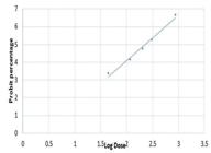
The Probit Graph of Venom Lethality Dose. At probit 50%, the log is 2.4 Antilog 2.4 is 0.25, thus the LD50 of the N. nigricollis venom equals 0.25 mg/kg. At probit 75%, log dose is 3.1 Antilog of 3.1 is 1.26 thus; LD75 of N. nigricollis venom equals 1.26 mg/kg LD50 and LD75 of N. nigricollis venom were found to be 0.25 mg/kg and 1.26 mg/kg respectively.
In vivo anti-venom activity of CDAP
CDAP at doses of 150, 250 and 500 mg/kg was able to prevent mortality in all Wistar rats envenomed with 1.26 mg/kg (LD75) venom of N. nigricollis. Mortality of 42.86% was recorded only in group 6 which served as negative control (Table II).
Body weight of the envenomed treated within the experimental groups
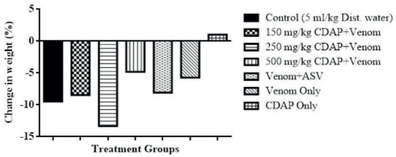
There was a reduction in the average weight of all envenomed treated groups as shown in Figure 2. The highest weight reduction was at 250 mg/kg while the lowest was at 500 mg/kg. Only the extract group showed an increase in average weight by 1.03% (Figure 2).
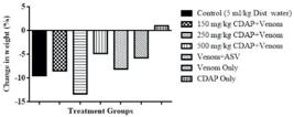
Effects of CDAP on liver enzymes and creatinine in Wistar rats envenomed with venom of N. nigricollis
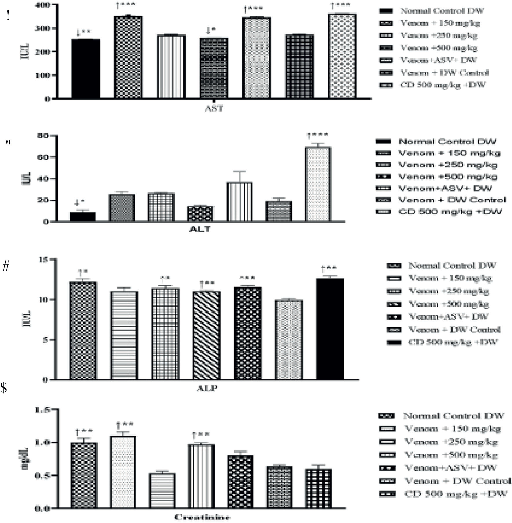
There was a significant (p<0.001) rise in serum AST and ALT levels of the envenomed groups, with a non-significant (p<0.01) increase in ALP when compared with the normal control group. AST and ALT values were, however, significantly (p<0.05) reduced only in group 4, when compared to the venom control group. The serum values of the liver enzymes; AST, ALT, and ALP were significantly (p<0.001) increased, considerably in group 7. There was no significant increase in serum creatinine in treated groups when compared with control groups (Figure 3a-d).
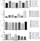
Effect of Caralluma dalzielii on biochemical parameters in rats envenomed with Naja nigricollis. Data presented as Mean±SEM
Effects of CDAP on haematological parameters
Table III shows a significant (p<0.05) increase in WBC and LYM (p<0.01 for 150 mg/kg) values of all the venom-extract exposed groups, as well as in the extract-DW exposed group. Values of MID and NEU have significantly (p<0.01) increased in venom-extract groups at 150 mg/kg and 250 mg/kg when compared to the venom control group. The values of RBC and HBG were significantly (p<0.05) reduced only in the venom-ASV exposed group. MCV values were significantly decreased in 250 mg/kg (p<0.05), 500 mg/kg (p<0.01), ASV (p<0.01), and CDAP-only groups (p<0.01) when compared to the negative control group (Table III).
Effect of C. dalzielii on haematological parameters of rats envenomed with venom of N. nigricollis
In vitro anti-venom activity of CDAP
Effect of CDAP against N. nigricollis PLA2 enzyme
CDAP at a concentration of 100 mg/ml showed a percentage enzyme inhibition of 64.29% against PLA2 while standard anti-snake venom showed 82.65% after 10 mins incubation. All enzyme inhibition shown by the plant extract is concentration-dependent (Table IV).
Effect of CDAP against N. nigricollis PLA2 enzyme after 30 minutes incubation
Enzyme inhibition of CDAP at different concentrations after 30 mins was lower than what was obtained after 10 mins. At a concentration of 100 mg/ ml, the plant extract showed a percentage enzyme inhibition of 42.25%. Inhibitions were shown to be concentration-dependent (Table V).
DISCUSSION
The findings that the LD50 and LD75 of Naja nigricollis venom in Wistar rats are 0.25 mg/kg and 1.26 mg/kg, respectively, indicate that the venom is highly toxic, with 75% of the test population succumbing at a dose greater than the LD50. The negative control group had a mortality rate of 42.86%. In contrast, treatment of the Naja nigricollis envenomed Wistar rats with the CDAP showed that the extract was able to prevent mortality in all the rats at these doses, indicating its potential as a treatment for venomous snakebites. The reduction in the average weight of all envenomed treated groups of rats conforms with the previously reported findings due to snake envenomation, perhaps as a result of several factors; such as toxicity and inflammation, stress response, loss of appetite, increased water consumption, and metabolic changes (Bhattacharya et al., 2020; Ajisebiola et al., 2022).
Naja nigricollis envenomation caused reduced levels of creatinine, AST, ALT, and ALP in our study. This could be due to organ dysfunction, particularly in the liver and kidneys, which impairs their release into the bloodstream (Da Silva et al., 2022). The venom could also have disrupted normal metabolic processes, lowering the production and release of these markers. Furthermore, the venom’s immune-suppressive and systemic effects may have reduced the typical enzyme response to tissue damage. The significant rise in these parameters in the treated groups might be a result of the toxins-neutralising effects of CDAP, which agrees with the previous report (Sani et al., 2020), thereby reducing the damage to liver cells and normalising their levels. AST and ALT values were, however, significantly reduced only in the CDAP 500 mg/kg group suggesting that the extract might have enhanced liver function, leading to a more efficient clearance of AST and ALT from the serum, resulting in lower levels as previously reported (Ukpabi-Ugo, Ndukwe, Iwuoha, 2019; Khalid et al., 2023) or perhaps may have exhibited antioxidant properties, which could have mitigated the oxidative stress caused by the venom thus reducing the rise in liver enzymes (Adeyi et al., 2021). The serum values of the liver enzymes; AST, ALT, and ALP were significantly increased, considerably in the CDAP-only group at 500 mg/kg. The non-significant increase in serum creatinine in treated groups may indicate that there is no substantial risk of antagonistic outcomes, following treatment with CDAP such as mortality, associated with these changes.
The harmful effects of Naja nigricollis have been linked to disruptions in blood-related functions (Dobson et al., 2024). A decrease in WBC was observed in the venom-only group in our study. The decrease in WBC in Naja nigricollis envenomed Wistar rats could be due to the venom’s immunosuppressive and cytotoxic effects, which damage immune cells directly. The venom triggers an inflammatory response, drawing white blood cells to the site of injury, and reducing their presence in circulation. Additionally, venom components can suppress bone marrow activity, hindering new white blood cell production. Systemic toxicity and organ damage, particularly in the spleen and liver, further contribute to the decline in WBC levels. As the rats were treated, an increase in WBC counts was observed, which may indicate an adaptive response or compensatory mechanism that helps cells counteract the damage caused by the venom. WBC values of all the venom-extract exposed groups, as well as in the extract-DW exposed group were increased, suggesting that the CDAP may have immunoprotective properties (Menaldo et al., 2019; Maheshwari et al., 2022).
Again, in Naja nigricollis envenomed Wistar rats, lymphocytes decrease due to the venom’s cytotoxic effects, immune suppression, and bone marrow inhibition just like in the WBC. Lymphocytes may migrate to tissues during the body’s inflammatory response, reducing their levels in the bloodstream (Minutti-Zanella, Gil-Levya, Vergara, 2021). CDPA improved this count just like the WBC probably by neutralising the venom’s toxins, preventing further damage to lymphocytes and other immune cells. This process could help restore normal immune function, allowing the animals to recover from the venom’s cytotoxic effects, reduce inflammation, and support the production and replenishment of WBC, including lymphocytes, by the bone marrow.
In envenomed Wistar rats, a decrease in MID (monocytes, eosinophils, basophils) indicates suppression of the immune cells involved in inflammation and allergic responses. Neutrophils (NEU) may initially increase as the body mounts a defence against the venom, but severe toxicity can lead to their depletion, compromising the immune response. In our study, the venom-only group showed marked depression of MID and NEU. These changes in MID and NEU reflect the degree of immune system disruption caused by the venom (Andrés et al., 2022). However, treatment with the extract, neutralized the venom’s toxic effects, helping to restore immune balance by preventing further suppression or destruction of immune cells. It also stabilized MID levels by protecting monocytes, eosinophils, and basophils from venom-induced damage, allowing them to function properly in the immune response. For neutrophils (NEU), CDAP may have reduced excessive inflammation, helping the maintenance of a normal count and ensuring these cells can continue to protect the body without being overwhelmed or depleted.
RBC and HGB levels were not significantly affected by the envenomation, this could indicate minimal haemolysis and effective compensatory mechanisms by the rats (Williams et al., 2018). It may also suggest that the venom’s primary impact may be on other immune components rather than directly affecting RBC and HGB. The values of RBC and HBG were significantly reduced only in the venom-ASV-exposed group. This may result from an immune reaction to the anti-venom or could reflect the severe underlying effects of the venom being addressed by the anti-venom.
Envenomation can increase MCV due to the release of larger red blood cells from the bone marrow following haemolysis (Williams et al., 2018). Again, the body may produce larger red blood cells as a compensatory response to the venom’s effects. This was evident from our study. However, CDAP at all doses reduced MCV counts. This could probably be by neutralising venom effects that caused increased red blood cell size. This stabilization leads to a reduction in the production of larger, immature red blood cells, normalizing the MCV as the body recovers from venom-induced damage (Ajisebiola, Oladele, Adeyi, 2023).
Inhibiting PLA2 prevents cell membrane damage and reduces tissue destruction, protecting cells from venom-induced lysis. It also decreases inflammation, minimizing swelling and pain, and leading to quicker recovery from venom toxicity. Our extract dose-dependently inhibited PLA2 enzyme indicating that the CDAP has significant inhibitory activity, and may serve as a potential natural alternative for managing snakebite envenomation (Ugwah-Oguejiofor et al., 2018). Fascinatingly, the inhibitory activity observed after 30 mins though lower compared to the inhibition reported after 10 mins suggests that the inhibitory effect of the extract on PLA2 enzyme activity remained concentration-dependent and may decrease over time. The ability of the CDAP to inhibit PLA2 enzyme activity from N. nigricollis venom highlights its potential as a natural therapeutic agent against snakebite envenomation.
As was identified in CDAP, flavonoids, saponins, and terpenoids may contribute to anti-venom activity through their anti-inflammatory, antioxidant, and immunomodulatory effects, helping to reduce tissue damage and support recovery. Alkaloids and steroids are known to provide additional benefits by reducing pain, and inflammation, and stabilizing cell membranes, while glycosides and saponin glycosides may further enhance immune response and recovery processes. Cardiac glycosides, though primarily affecting heart function, could help manage venom-induced cardiovascular symptoms. Further research into these compounds may lead to the development of effective treatments for snakebite envenomation.
CONCLUSION
The study has shown that CDAP possesses both in vitro and in vivo anti-snake venom activity by inhibiting the pathologic effect of the venom of N. nigricollis. This further supports its traditional use in the treatment of snakebite. Further research is needed to fully understand the mechanisms behind the inhibitory activity of the CDAP and to optimize the dosage and duration of treatment. Additionally, isolation and characterization of the bioactive compounds are also needed.
ACKNOWLEDGEMENT
The authors thank Mal Usman Arzika for assisting with the in vivo studies and Mr Mustapha Salihu for the in vitro studies.
DATA AVAILABILITY STATEMENT
The data supporting the findings of this study are available within the article and its supplementary materials.
REFERENCES
- Adeyi AO, Ajisebiola BS, Adeyi OE, Adekunle O, Akande OB, James AS, et al. Moringa oleifera leaf fractions attenuated Naje haje venom-induced cellular dysfunctions via modulation of Nrf2 and inflammatory signalling pathways in rats. Biochem Biophys Rep. 2021 Mar 1;25:100890.
- Adrião AA, Dos Santos AO, de Lima EJ, Maciel JB, Paz WH, da Silva FM, et al. Plant-derived toxin inhibitors as potential candidates to complement antivenom treatment in snakebite envenomations. Front Immunol. 2022 May 9;13:842576.
- Afroz A, Siddiquea BN, Chowdhury HA, Jackson TN, Watt AD. Snakebite envenoming: A systematic review and meta-analysis of global morbidity and mortality. PLOS NTDs. 2024 Apr 4;18(4):e0012080.
- Ajisebiola BS, Adeniji OB, James AS, Ajayi BO, Adeyi AO. Naja nigricollis venom altered reproductive and neurological functions via modulation of pro-inflammatory cytokines and oxidative damage in male rats. Metabolism Open. 2022 Jun 1;14:100188.
- Ajisebiola BS, Oladele JO, Adeyi AO. Kaempferol from Moringa oleifera demonstrated potent antivenom activities via inhibition of metalloproteinase and attenuation of Bitis arietans venom-induced toxicities. Toxicon. 2023 Sep 1;233:107242.
- Aly SH, El-Shazly M, Eldahshan OA. Antidotes to reptile toxins chemical composition of snake venoms; toxic effects of snake venom. In: Antidotes to Toxins and Drugs. Elsevier. 2024 Jan 1 (pp. 71-97).
- Andrés CM, Pérez de la Lastra JM, Juan CA, Plou FJ, Pérez-Lebeña E. The role of reactive species on innate immunity. Vaccines. 2022 Oct 17;10(10):1735.
- Babangida S, Erhabor O, Erhabor T, Adias TC. A review on Snake Bite and Management in Nigeria-The Need for The Development of a Snake Bite Strategy. Sokoto J Med Lab Sci. 2020 Dec 31;5(4):41-54.
- Bala AA, Mohammed M, Umar S, Ungogo MA, Hassan MA, Abdussalam US, Ahmad MH, Ishaq DU, Mana D, Sha’aban A, Jatau AI. Pre-clinical efficacy of African medicinal plants used in the treatment of snakebite envenoming: A systematic review. Toxicon. 2023 Mar 1; 224:107035.
- Bhattacharya S, Krishnamurthy A, Gopalakrishnan M, Kalra S, Kantroo V, Aggarwal S, Surana V. Endocrine and metabolic manifestations of snakebite envenoming. Am J Trop Med Hyg. 2020 Oct;103(4):1388.
- Bhaumik S, Beri D, Lassi ZS, Jagnoor J. Interventions for the management of snakebite envenoming: An overview of systematic reviews. PLoS NTDs. 2020 Oct 13;14(10):e0008727.
- Da Silva DP, de Sousa Ferreira S, Torres-Rego M, Furtado AA, de Oliveira Yamashita F, da Silva Diniz EA, et al. Antiophidic potential of chlorogenic acid and rosmarinic acid against Bothrops leucurus snake venom. Biomed Pharmacother. 2022 Apr 1;148:112766.
- Dobson J, Chowdhury A, Tai-A-Pin J, van der Ploeg H, Gillett A, Fry BG. The Clot Thickens: Differential Coagulotoxic and Cardiotoxic Activities of Anguimorpha Lizard Venoms. Toxins. 2024 Jun 20;16(6):283.
- Dossou AJ, Fandohan AB, Omara T, Chippaux JP. Comprehensive Review of Epidemiology and Treatment of Snakebite Envenomation in West Africa: Case of Benin. J Trop Med. 2024;2024:1-10.
- Kalita B, Saviola AJ, Mukherjee AK. From venom to drugs: a review and critical analysis of Indian snake venom toxins envisaged as anticancer drug prototypes. Drug Discov Today. 2021 Apr 1;26(4):993-1005.
- Khalid S, Arshad M, Raza K, Mahmood S, Siddique F, Aziz N, et al. Assessment of hepatoprotective, nephroprotective efficacy, and antioxidative potential of Moringa oleifera leaf powder and ethanolic extract against PCOS-induced female albino mice (Mus Musculus). Food Sci Nutr. 2023 Nov;11(11):7206-17.
- Kusar S, Saddiqe Z, Asad MH, Ali F, Kirmani F. Phytochemical characterization and phospholipase A2 inhibitory effect of Vitex negundo L. root extracts. J Ethnopharmacol. 2024 Apr 6;323:117671.
- Liaqat A, Mallhi TH, Khan YH, Khokhar A, Chaman S, Ali M. Anti-snake venom properties of medicinal plants: a comprehensive systematic review of literature. Braz J Pharm Sci. 2022 Sep 2;58:e191124.
- Maheshwari S, Kumar V, Bhadauria G, Mishra A. Immunomodulatory potential of phytochemicals and other bioactive compounds of fruits: A review. Food Front. 2022 Jun;3(2):221-38.
- Malik R, Ada G, Udeh CA. Snakebite envenomation in Benue state: A study of prevalence and treatment in Agatu local government area, Benue state-Nigeria. J Res For Wildl Environ. 2021 May 13;13(1):94-107.
- Menaldo DL, Costa TR, Ribeiro DL, Zambuzi FA, Antunes LM, Castro FA, et al. Immunomodulatory actions and epigenetic alterations induced by proteases from Bothrops snake venoms in human immune cells. Toxicol in Vitro. 2019 Dec 1;61:104586.
- Michael GC, Grema BA, Aliyu I, Alhaji MA, Lawal TO, Ibrahim H, et al. Knowledge of venomous snakes, snakebite first aid, treatment, and prevention among clinicians in northern Nigeria: a cross-sectional multicentre study. Trans R Soc Trop Med Hyg. 2018 Feb 1;112(2):47-56.
- Minutti-Zanella C, Gil-Leyva EJ, Vergara I. Immunomodulatory properties of molecules from animal venoms. Toxicon. 2021 Feb 1;191:54-68.
- Mugale MN, Dev K, More BS, Mishra VS, Washimkar KR, Singh K, et al. A Comprehensive Review on Preclinical Safety and Toxicity of Medicinal Plants. Clin Complement Med Pharmacol. 2024 Jan 13:100129.
- Osipov A, Utkin Y. What are the neurotoxins in hemotoxic snake venoms? Int J Mol Sci. 2023 Feb 2;24(3):2919.
- Patel N, Patel S, Adeyemi SB, Rahaman AA, Olorunmaiye KS, Krishnamurthy R. Sustainable Agricultural Practices of Industrially Utilized Tropical Medicinal Plants. In Sustainable Uses and Prospects of Medicinal Plants 2023 May 5 (pp. 189-225). CRC Press.
- Sani I, Umar RA, Hassan SW, Faruq UZ, Bello F, Aminu H, Sulaiman A. Hepatoprotective effect of Azadirachta indica leaf fractionated extracts against snake venom toxicity on albino rats. Saudi J Biomed Res. 2020;5(6):112-7.
- Turner RA. Screening methods in Pharmacology. New York: Academic Press; 1965.
- Ugwah-Oguejiofor CJ, Abubakar K, Ugwah MO, Njan AA. Evaluation of the antinociceptive and anti-inflammatory effect of Caralluma dalzielii J Ethnopharmacol. 2013;150(3):967-72.
- Ugwah-Oguejiofor CJ, Alkali YI, Inuwa AM, Pender GC, Chindo BA. Studies on neurobehavioural properties of Caralluma dalzielii NE Br. aqueous aerial parts extract in mice. J Ethnopharmacol. 2024 Apr 24;324:117774.
- Ugwah-Oguejiofor CJ, Ogbulie CS, Ugwah MO, Umaru ML, Adebisi IM, Abubakar K, Alkali Y. Effect of aqueous extract of the aerial parts of Caralluma dalzielii N. E Brown on the testosterone levels of male Wistar rats. J Pharm Bioresour. 2018 Dec 5;15(2):175-9.
-
Ugwah-Oguejiofor CJ, Okoli CO, Ugwah MO, Umaru ML, Ogbulie CS, Mshelia HE, et al. Acute and sub-acute toxicity of aqueous extract of aerial parts of Caralluma dalzielii NE Brown in mice and rats. Heliyon. 2019 Jan 1;5(1): e01179 https://doi.org/10.1016/j.heliyon.2019.e01179
» https://doi.org/10.1016/j.heliyon.2019.e01179 - Ukpabi-Ugo JC, Ndukwe PA, Iwuoha AG. Hepatoprotective effect of methanol extract of Justicia carnea leaves on carbon tetrachloride-intoxicated albino rats. Biochem Anal Biochem. 2019;8(381):2161-1009.
- Williams HF, Hayter P, Ravishankar D, Baines A, Layfield HJ, Croucher L, et al. Impact of Naja nigricollis venom on the production of methaemoglobin. Toxins. 2018; 10: 539.
Edited by
-
Associate Editor:
Skylar Carlson

 *p<0.05; **p<0.01; ***p<0.001, ↑= increase; ↓= decrease compared to the negative control group CD= Caralluma dalzielii; AST= Aspartate Aminotransferase, ALT= Alanine Aminotransferase, ALP= Alkaline phosphatase. (continue)