 Evaluation of the antihyperlipidemic activity of Dracaena trifasciata (Prain) Mabb. ethanolic extract in high-fat diet-induced hyperlipidemia in rats
Hyperlipidemia, characterized by elevated cholesterol or triglyceride levels, is a key risk factor for cardiovascular diseases. This study explores the antihyperlipidemic potential of Dracaena trifasciata (Prain) Mabb., a perennial herb with diverse pharmacological properties. The ethanolic extract of D. trifasciata leaves (EEDT) was prepared and analyzed for phytochemicals. Wistar rats fed a high-fat diet (HFD) were treated with EEDT (200 mg/kg and 400 mg/kg), with statin as a positive control. Parameters assessed included lipid profiles, atherogenic indices (AIP, CRR, CPI, AC), lipid peroxidation, antioxidant levels, liver enzymes, and histopathology. Phytochemical analysis revealed flavonoids, phenols, alkaloids, and saponins, with high phenolic (776.33 mg GAE/g) and flavonoid (284 mg RT/g) content. EEDT significantly (p < 0.001) reduced total cholesterol, triglycerides, LDL, and VLDL while increasing HDL and CPI. Atherogenic indices (AIP, CRR, AC) were also significantly lowered (p < 0.001). Antioxidant markers improved (p < 0.05), and liver enzyme levels (AST, ALT, ALP) decreased (p < 0.01). Histopathological findings supported these results. EEDT demonstrated potent antihyperlipidemic and antioxidative effects, suggesting its potential as a natural therapeutic agent for hyperlipidemia and oxidative stress. However, further clinical studies are needed to confirm its efficacy and safety for therapeutic use.
Keywords:
Antioxidant activity; Atherogenic indices; Dracaena trifasciata (Prain) Mabb; Hyperlipidemia; Lipid profile.

INTRODUCTION
Hyperlipidemia is a condition marked by elevated levels of plasma lipids, including cholesterol, triglycerides, cholesterol esters, phospholipids, and in some cases, very low-density lipoprotein (VLDL) and low-density lipoprotein (LDL). This condition is often associated with reduced levels of high-density lipoprotein (HDL). As a widespread metabolic disorder, hyperlipidemia is a significant risk factor for various cardiovascular and metabolic diseases, such as heart disease, atherosclerosis, hypertension, diabetes mellitus, hypercholesterolemia, and obesity (Mishra et al., 2011; Jeyabalan, Palayan, 2009; Duraipandiyan et al., 2016). Its prevalence has become a global concern, contributing to over four million deaths annually due to its complications and associated health risks. These factors increase morbidity and mortality rates worldwide (Shattat, 2015; Kumar et al., 2012).
The management of hyperlipidemia primarily focuses on lifestyle modifications. Key recommendations for patients include reducing the intake of fatty foods, quitting smoking, engaging in regular aerobic exercise, and adopting a healthy diet aimed at improving overall lipid profiles (Mannu et al., 2013). These lifestyle changes are critical in preventing the onset of complications. Along with lifestyle interventions, pharmacological treatments play a vital role in managing the condition.
Current antihyperlipidemic drugs, such as statins (e.g., atorvastatin), fibrates (e.g., fenofibrate), bile acid sequestrants (e.g., cholestyramine), PCSK9 inhibitors (e.g., evolocumab), niacin, and ezetimibe, are effective in reducing lipid levels and preventing the progression of cardiovascular diseases. However, they are also associated with adverse effects, including gastrointestinal discomfort, muscle-related issues such as rhabdomyolysis and myopathy, dizziness, kidney damage, and an increased risk of developing type 2 diabetes, particularly at higher dosages (Shattat, 2015; Gupta et al., 2010; Nelson, 2013; Anees et al., 2024).
Due to the adverse effects associated with conventional lipid-lowering medications, there is growing interest in exploring alternative therapeutic approaches, particularly herbal medicines with hypolipidemic properties. Several plant-based compounds, including those derived from garlic, green tea, and bergamot, have demonstrated cholesterol-lowering potential with fewer side effects, making them viable alternatives for patients who are intolerant to statins (Hasani-Ranjbar et al., 2010; Bahmani et al., 2015). Given the multifactorial nature of hyperlipidemia and its association with chronic conditions such as diabetes, hypertension, and stroke, it is imperative to investigate novel therapeutic targets beyond traditional statins. This necessitates evaluating the efficacy and safety of alternative drug classes and non-pharmacological interventions. Natural remedies, such as herbal medicines, may complement existing treatments or offer standalone benefits. However, more research is required to fully elucidate their efficacy, mechanisms of action, and long-term safety profiles. Investigating these alternatives is particularly valuable, as they may provide effective lipid-lowering benefits with a reduced risk of adverse effects compared to synthetic medications, thereby improving overall health outcomes.
Dracaena trifasciata (Prain) Mabb., formerly known as Sansevieria trifasciata Prain, is a perennial herb that belongs to the Asparagaceae family. Native to tropical West Africa, this plant is commonly grown as an ornamental plant worldwide, and it is known by various names, including mother-in-law’s tongue, snake plant, viper’s bowstring hemp, and Saint George’s sword. Traditionally, D. trifasciata has been valued for its therapeutic properties, exhibiting potential in the treatment of various ailments, including acne, fungal infections, skin pruritus, ulcers, earaches, allergies, helminth infestations, jaundice, pharyngitis, and urinary disorders. Beyond its analgesic and antipyretic effects, the plant holds cultural significance in Africa, where it is traditionally believed to offer protection against malevolent forces and bewitchment. It is also cultivated for its durable fibre in several tropical countries (Stafford et al., 2008; Qomariyah, 2012; Rwawiire, Tomkova, 2015). The leaves of D. trifasciata contain several bioactive phytoconstituents, which are responsible for its diverse pharmacological activities. These phytoconstituents contribute to its anti-inflammatory, analgesic, antipyretic, antiulcerative, antibacterial, antidiabetic, hepatoprotective, wound healing, antioxidant, and anthelmintic properties (Babu, Prabhu, 2024). Despite its wide range of medicinal uses, there is limited research on its antihyperlipidemic potential. Given the rising interest in herbal alternatives, the present study aims to evaluate the antihyperlipidemic activity of the ethanolic extract of D. trifasciata in a high-fat-diet-induced hyperlipidemia model in Wistar rats.
MATERIAL AND METHODS
Plant material
The leaves of Dracaena trifasciata (Prain) Mabb. were collected during the winter season in January 2024 from a local nursery in Hyderabad. The leaves of D. trifasciata were identified and authenticated by Dr. L. Rasingam (Scientist ‘E’ & HoO), Botanical Survey of India (BSI) Koti, Hyderabad, India. Authentication number: BSI/DRC/2023-2024/642.
Preparation of plant extract
The leaves were washed thoroughly under running water to remove impurities and then cut into smaller pieces. These pieces were air-dried under indirectsunlight while being covered with a black cloth to prevent direct exposure until completely dry. Once dried, the samples were ground into a fine powder using a mechanical grinder and sieved through a 22-mesh sieve to ensure uniform particle size. A 200 g portion of this powdered material was then subjected to maceration with ethanol in a 1:4 ratio for three days. Ethanol was selected as the extraction solvent due to its ability to dissolve a broad spectrum of polar and non-polar bioactive compounds, making it highly effective for extracting phytochemicals with potential pharmacological properties. Compared to other solvents, ethanol is preferred for its relatively low toxicity, ease of evaporation, and compatibility with both hydrophilic and lipophilic compounds. After maceration, the mixture was filtered, and the filtrate was concentrated using a rotary evaporator to obtain a thick extract. The final extract was stored at 4°C for further experimental procedures (Azwanida, 2015).
Preliminary phytochemical screening
A qualitative chemical analysis of EEDT was conducted to identify the presence of various phytoconstituents, following established protocols (Trease, Evans, 1989; Trease, Evans, 1996).
Quantitative analysis
Quantitative analysis was done to determine the total phenolic and total flavonoid contents of EEDT.
Total phenolic content (TPC)
The Folin-Ciocalteu method was used to estimate total phenolic content (TPC), with gallic acid as the standard. A 1 mg/ml extract solution was prepared, and 1 ml of this was mixed with 0.5 ml of 2N Folin-Ciocalteu reagent. After 5 min, 1.5 ml of 20% sodium bicarbonate was added, and the volume was adjusted to 8 ml with distilled water. Following a 2 h incubation at room temperature, absorbance was measured at 765 nm. TPC was calculated as mg gallic acid equivalents (GAE)/g extract (Naaz et al., 2024; Banu, Samreen, Mujeeb, 2025).
Total flavonoid content (TFC)
Using rutin as the standard, the aluminium chloride colorimetric method measured total flavonoid content (TFC). A 0.5 ml sample was combined with 3 ml of methanol, 0.2 ml of 10% aluminium chloride, 0.2 ml of 1M potassium acetate, and 5 ml of water. After a 30-min incubation at room temperature, absorbance was recorded at 415 nm. TFC was expressed as mg rutin equivalents (RE)/g extract (Naaz et al., 2024; Banu, Samreen, Mujeeb, 2025).
Experimental animals
Male Wistar rats (150-200 g) obtained from Jeeva Life Sciences (An ISO 9001:2015 Certified Company), Hyderabad were used in the study. The animals were acclimatised to the laboratory for 5 days before the experiment. All procedures adhered to institutional guidelines and CCSEA regulations. Rats were housed in polypropylene cages with controlled temperature (65% ± 2%) and a 12-hour light/dark cycle. The study was approved by the Institutional Animal Ethics Committee (RBVRR 1328/05/2024).
Sample size determination
The sample size for this study was calculated using power analysis (G*Power, version 3.1) with 80% power (α = 0.05) and an expected medium effect size (Cohen’s f = 0.25). Based on this analysis, each group required 6 animals to detect statistically significant differences in lipid parameters among the experimental groups. This resulted in a total of 30 animals across 5 groups for the main study (6 animals per group × 5 groups).
For the acute toxicity study, the sample size was determined following OECD Guideline 420 (Acute oral toxicity - Fixed Dose Procedure), which recommends a minimum of 5 animals per dose group. Accordingly, 2 groups of 5 animals each were used. This sample size is sufficient to identify toxic effects, determine the NOAEL, and assess the safety profile of D. trifasciata extract while adhering to ethical principles of animal use reduction.
Acute toxicity study
Following the fixed dose procedure (OECD 420), a single oral dose of 2000 mg/kg body weight of the EEDT was administered to a group of rats to assess potential toxicity. Ten rats were randomly divided into two groups: A control group and a treated group (5 rats each). Before dosing, all animals were fasted for 3-4 h and weighed. The treated group received the extract via oral gavage, while the control group received normal saline. Following treatment, animals were monitored for signs of toxicity, such as changes in skin and fur, eye health, respiratory patterns, and behaviour, with observations at intervals during the first 24 h (0.5, 1, 6, and 24 h) and once daily for 14 days thereafter. On day 15, all animals were sacrificed, and a thorough necropsy was conducted to examine any gross pathological changes (Banu et al., 2016; Naaz, Banu, 2024).
Evaluation of the antihyperlipidemic activity of EEDT
Preparation of a high-fat diet
A high-fat diet (HFD) was formulated to induce hyperlipidemia in rats, and the composition of this diet is detailed in Table I. To prepare the HFD, cow fat and dalda/ghee are melted and gradually incorporated into crushed rat pellets in a mixing container. Once a semi-solid consistency is achieved, milk powder and coconut oil are added for uniform blending. The mixture is kneaded thoroughly, shaped into pellets, and dried in a hot air oven. The final HFD pellets are stored in an airtight container to prevent oxidation and are used to replace the standard diet in experimental models for hyperlipidemia studies (Jing et al., 2022).
Induction of hyperlipidemia
After a one-week acclimatization period, Wistar rats were fed a high-fat diet (HFD) designed to induce hyperlipidemia over a 30-day period. The HFD composition, expressed on a weight/weight (w/w) basis, included 6% cow’s fat, 6% Dalda, 6% coconut oil, 14% milk powder, and 68% standard rat pellet, as outlined in Table I. Following the initial 30-day hyperlipidemia induction phase, the rats continued to receive the HFD while being administered varying doses of EEDT for an additional 15 days (Table II). The EEDT doses were selected based on acute toxicity studies, which revealed an LD50 value greater than 2000 mg/kg. Accordingly, doses equivalent to 1/5th and 1/10th of the LD50 were utilized in this study. Blood samples were collected at 7, 14, 30, and 45 days via retro-orbital puncture for subsequent determination of lipid profile parameters.
At the end of the experimental period, overnight-fasted rats were sacrificed and the liver was carefully excised for subsequent biochemical analysis and histopathological examination (Jing et al., 2022).
Effect on Body weight, BMI, and Lipid profile
Effect on weight
The rats’ weights were recorded immediately after grouping and then every three days over a 30-day period, which included both the high-fat diet and treatment phases. Weight changes were compared on day one, after hyperlipidemia induction, and at the end of the treatment period to assess the effects of the diet and treatment.
Determination of Body mass index (BMI)
BMI was calculated as the ratio of body weight (in grams) to the square of body length (in cm). The length of the rats was measured from the nose to the base of the tail. The formula used was:
Lipid profiles
Blood samples were obtained from the retro-orbital plexus and left to sit for 10 min. They were then centrifuged at 3000 rpm for 10 min to separate the serum. The lipid profile, which included total cholesterol (TC), triglycerides (TG), LDL-cholesterol, and HDL-cholesterol, was evaluated using standard diagnostic kits and analyzed with an ERBA Manheim Biochemical analyzer.
Atherogenic indices
Atherogenic indices are critical tools for evaluating cardiovascular disease (CVD) risk based on lipid profiles. These indices provide insights into the balance between protective and harmful lipids in the blood, helping to evaluate atherosclerotic risk and overall cardiovascular health (Oršolić et al., 2019).
-
1. Atherogenic index of plasma (AIP): Reflects the balance between triglycerides and HDL cholesterol. Higher levels indicate an increased risk of atherosclerosis and related conditions.
-
2. Atherogenic coefficient (AC): Assesses the proportion of non-HDL cholesterol relative to HDL cholesterol. Elevated values suggest a higher atherogenic potential.
-
3. Cardiac risk ratio (CRR): Measures total cholesterol in relation to HDL cholesterol. A higher ratio correlates with increased cardiovascular risk.
-
4. Cardioprotective index (CPI): Evaluates the balance between protective HDL and harmful LDL cholesterol. Higher values signify better cardiovascular health (Oršolić et al., 2019).
Formulas:
-
1.
-
2.
-
3.
-
4.
Estimation of Hepatic enzyme activities
The activities of liver enzymes, including aspartate aminotransferase (AST), alanine aminotransferase (ALT), and alkaline phosphatase (ALP), were assessed. AST activity was measured through the reaction between α-oxoglutarate and L-aspartate, producing oxaloacetate and L-glutamate. The formed oxaloacetate then reacted with NADH to generate L-malate and NAD+. ALT activity was determined kinetically by observing the reaction of α-oxoglutarate with L-alanine, catalyzed by ALT, resulting in L-glutamate and pyruvate. The consumption of NADH was measured during the pyruvate reaction for kinetic estimation. For ALP activity, the hydrolysis of p-nitrophenyl phosphate by ALP in the serum, in the presence of magnesium ions, yielded p-nitrophenol (yellow). The intensity of the color produced was proportional to ALP activity, measured at 405 nm. All procedures were conducted following the guidelines provided in the manufacturer’s manual (Meunier, Larrey, 2019; Modanisi et al., 2017; Moriles, Zubair, Azer, 2024; Lala, Zubair, Minter, 2024).
Preparation of tissue homogenate
Wistar rats are anaesthetized with a ketamine-to-xylocaine ratio of 1:3. The liver is isolated, placed on ice, and sliced. A 10 mg sample is weighed and placed in a microcentrifuge tube with 500 μl RIPA buffer and 5 μl protease inhibitor. The tissue is homogenized, and the homogenate is centrifuged at 12,000 × g for 10 min at 4°C. Protein quantification is done using a BCA kit (ThermoFisher Catalogue no. 23227).
Malondialdehyde (MDA) assay
The liver homogenate is deproteinized with 30% TCA and 5N HCl, then treated with 2% thiobarbituric acid in 0.5 M NaOH. After heating at 100°C for 15 min and centrifuging, the pink chromogen is measured at 532 nm. Results are expressed as nM/mg protein.
Catalase (CAT) activity
Catalase activity is measured by mixing 50 mM potassium phosphate buffer (pH 7.4), 10.5 mM hydrogen peroxide, and 75 μg protein, then measuring absorbance at 450 nm using UV spectrophotometry (Mueller, Riedel, Stremmel, 1997).
Glutathione (GSH) activity
The homogenate was treated with 10% TCA and centrifuged. To 0.1 mL of the supernatant, 2.0 mL of 5,5’-dithiobis-2-nitrobenzoic acid (DTNB) reagent and 1.9 mL of phosphate buffer (pH 9.0) were added. Absorbance was measured at 412 nm, and GSH levels were calculated using a standard curve, expressed as µg per gram of wet tissue (Tabassum et al., 2007).
Histopathological studies
Liver specimens from rats were dissected and fixed in 10% formalin, then processed for paraffin embedding. Thin sections (5 µm) were cut and stained with Hematoxylin and Eosin (H & E) to highlight cellular structures. The stained sections were examined microscopically to identify any histopathological changes, such as alterations in liver architecture or signs of damage, which were then evaluated and interpreted for further analysis.
Statistical analysis
The data were analyzed using GraphPad Prism software and presented as mean ± SEM, with a sample size of n=6. A one-way analysis of variance (ANOVA) was performed to assess significant differences between the means, followed by Tukey’s multiple comparisons test. Statistical significance was set at p<0.05.
RESULTS
Percentage yield of EDDT
The EEDT yielded 10.77%, with 21.55 g of extract obtained from 200 g of dried leaves
Preliminary phytochemical screening
The preliminary phytochemical screening of EEDT revealed the presence of alkaloids, reducing sugars, saponins, phytosterols, flavonoids, phenols, proteins and amino acids, as shown in Table III.
Quantitative analysis
Total phenolic content (TPC)
The TPC in EEDT was estimated using the regression equation of the gallic acid calibration curve, yielding 776.33 mg GAE/g of extract, as shown in Figure 1.

Total flavonoid content (TFC)
The TFC in EEDT was estimated using the regression equation of the rutin calibration curve, yielding 284 mg RT/g of extract, as shown in Figure 2.

Acute toxicity study
Following OECD 420 guidelines, a 2000 mg/kg b.wt. the dose was administered to rats, and they were observed for 14 days. No signs of intoxication or abnormal behaviour were noted, indicating the dose was well tolerated (Table IV). All rats showed normal body weight gain, suggesting no adverse effects on growth or health (Table V).
Antihyperlipidemic activity of EEDT
Measurement of body weight
Body weight changes were recorded on days 0, 30, and 45. On day 0, weights ranged from 148 to 164 g. After 30 days on a high-fat diet, weights increased to 250-289 g. Following 15 days of treatment, rats treated with EEDT showed significant weight reduction: 200 mg/kg group (268 g to 206 g), 400 mg/kg group (250 g to 190 g), and atorvastatin group (255 g to 175 g, p < 0.01). The HFD control group gained weight, increasing from 289 g to 315 g, as depicted in Table VI.
Body mass index (BMI)
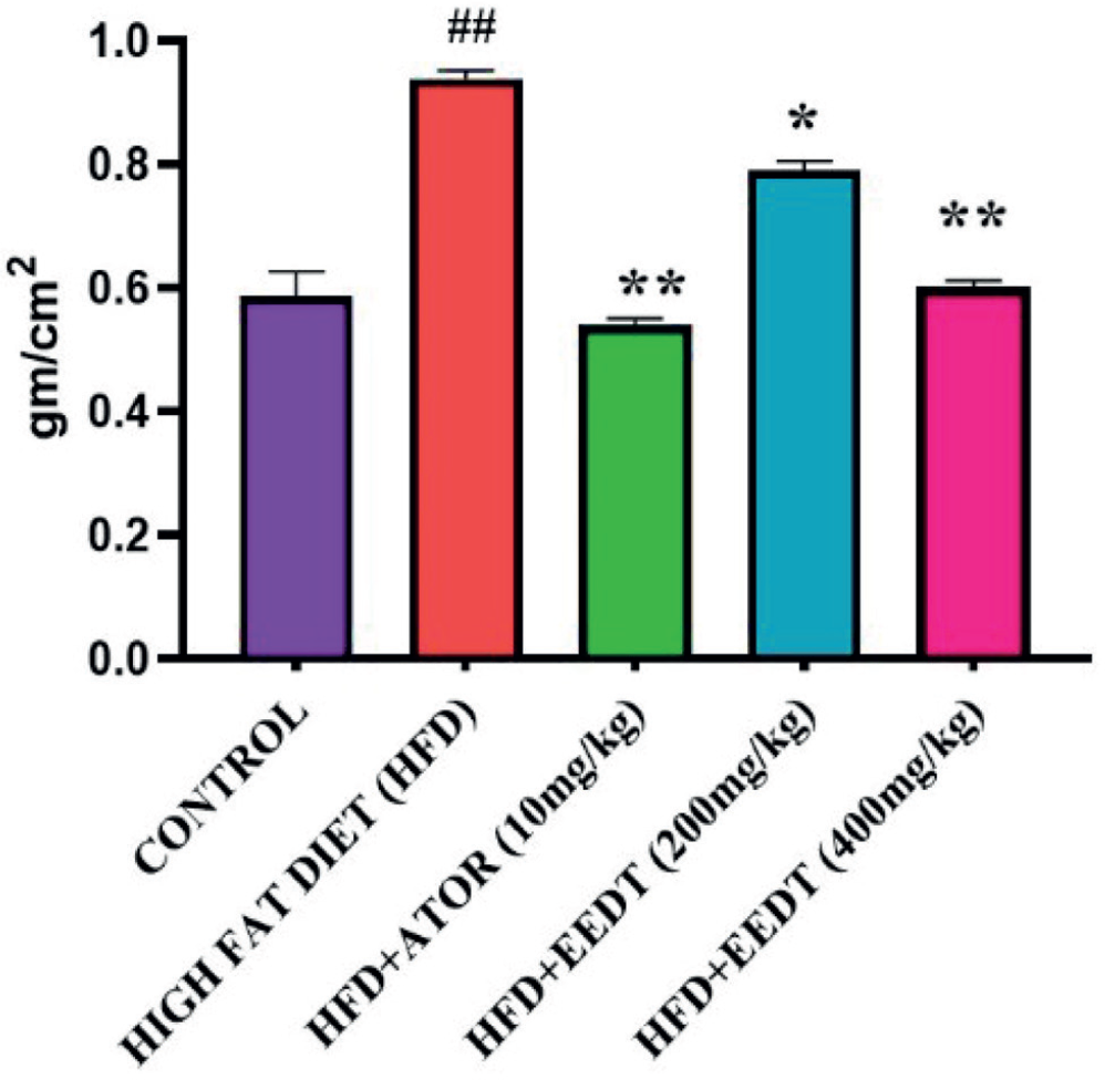
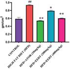
At the end of the experiment, the BMI of rats in all groups was measured. The HFD group showed a significant increase in BMI compared to the normal control group. In contrast, a reduction in BMI was observed in the 200 mg/kg (p < 0.05) and 400 mg/kg (p < 0.01) extract-treated groups compared to the HFD group, as shown in Table VII and Figure 3.
Effect of EEDT on Lipid Profiles
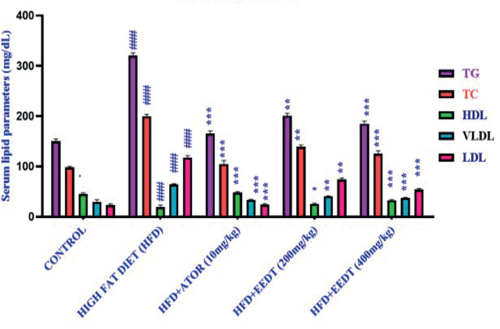
The serum levels of total cholesterol (TC), triglycerides (TG), high-density lipoprotein cholesterol (HDL-C), very-low-density lipoprotein cholesterol (VLDL-C), and low-density lipoprotein cholesterol (LDL-C) were assessed across different treatment groups, as shown in Table VIII and Figure 4. The HFD group exhibited elevated cholesterol (200 mg/dL), triglycerides (320.57 mg/dL), and reduced HDL-C (19.74 mg/dL). Atorvastatin (10 mg/kg) lowered cholesterol (105 mg/dL) and triglycerides (165.50 mg/dL) while increasing HDL-C (48.56 mg/ dL). EEDT at 200 mg/kg and 400 mg/kg also reduced cholesterol, triglycerides, and LDL-C, with the 400 mg/kg dose showing similar efficacy to atorvastatin in improving lipid levels and raising HDL-C (32.67 mg/dL).

Effect of EEDT on Lipid Profiles (Total cholesterol (TC), triglycerides (TG), high-density lipoprotein cholesterol (HDL-C), very-low-density lipoprotein cholesterol (VLDL-C), and low-density lipoprotein cholesterol (LDL-C)). Values are presented as mean ± SEM. ###p<0.001(Compared to control group),*p<0.05, **p<0.01, ***p<0.001(Compared to HFD group).
Effect of EEDT on Atherogenic indices
These indices are valuable in assessing the risk of cardiovascular diseases. In this study, the atherogenic index of plasma (AIP), atherogenic coefficient (AC), and cardiac risk ratio (CRR) were significantly higher in all HFD groups compared to the control, while cardioprotective index (CPI) was lower. Treatment with EEDT at 200 mg/kg and 400 mg/kg significantly reduced AIP, AC, and CRR, while improving CPI levels, as shown in Table IX.
Assessment of liver enzyme activities
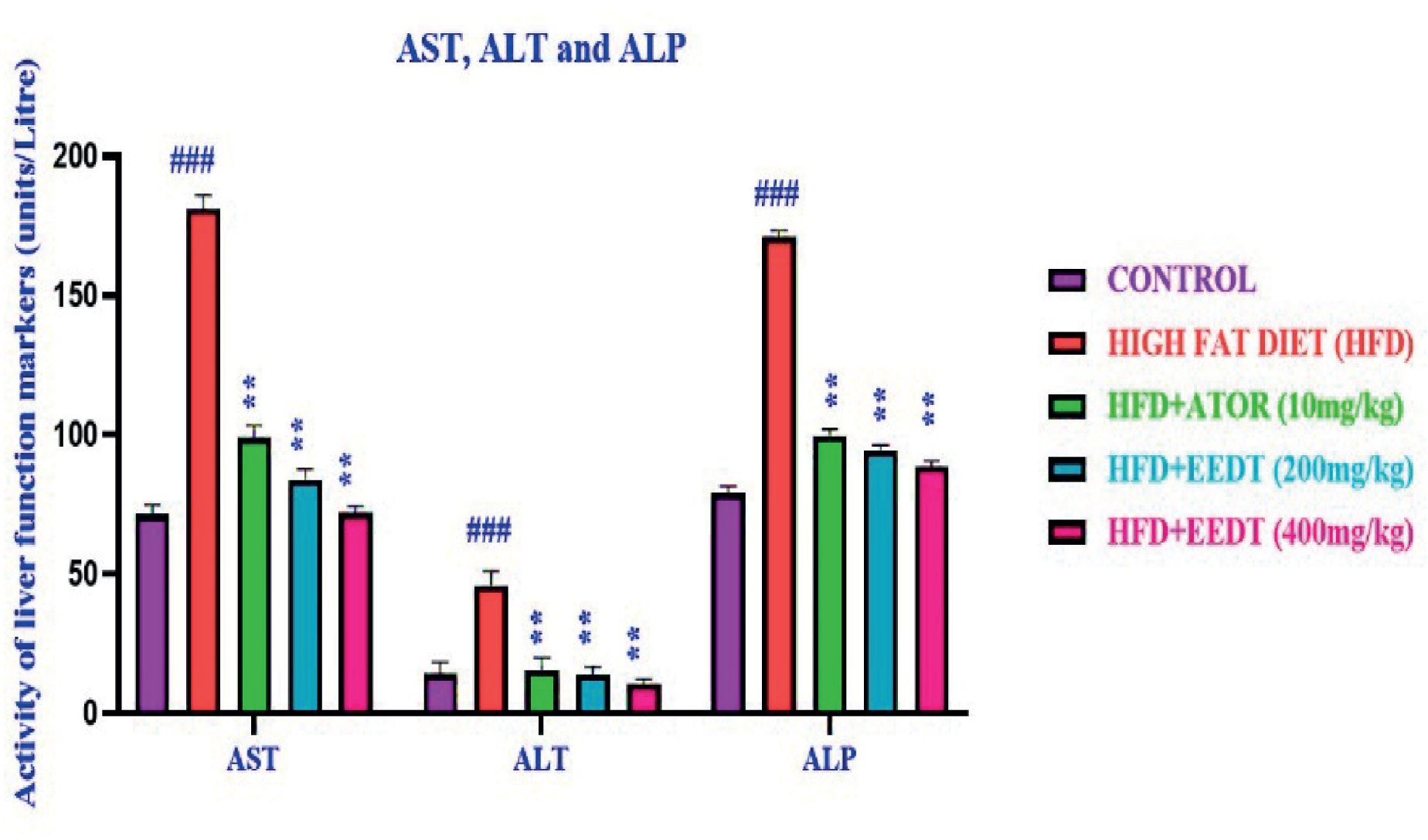
The assessment of hepatic enzyme activities, including aspartate aminotransferase (AST), alanine aminotransferase (ALT), and alkaline phosphatase (ALP), is crucial for evaluating liver function and integrity. These enzymes serve as important indicators for diagnosing liver toxicity and monitoring hepatic health, as they are released into the bloodstream when liver cells are damaged or stressed. Figure 5 and Table X highlight the effect of EEDT on liver enzyme activities in various treatment groups. While the control group exhibited normal enzyme levels, the HFD group showed significantly elevated levels, indicating liver damage. Treatment with EEDT at 200 mg/kg and 400 mg/kg significantly reduced enzyme levels in a dose-dependent manner. This reduction suggests a protective effect of EEDT against liver damage induced by the HFD, highlighting its potential as a therapeutic agent for maintaining hepatic integrity and function. The 400 mg/ kg dose of EEDT was particularly effective, restoring enzyme levels near normal, comparable to atorvastatin, demonstrating its hepatoprotective potential against HFD-induced liver damage.

Effect of EEDT on liver enzyme activities (AST, ALT & ALP). Values are presented as mean ± SEM.### p<0.001(Compared to control group), *p<0.05, **p<0.01(Compared to HFD group).
Assessment of the effect of EDDT on MDA, CAT, and GSH levels
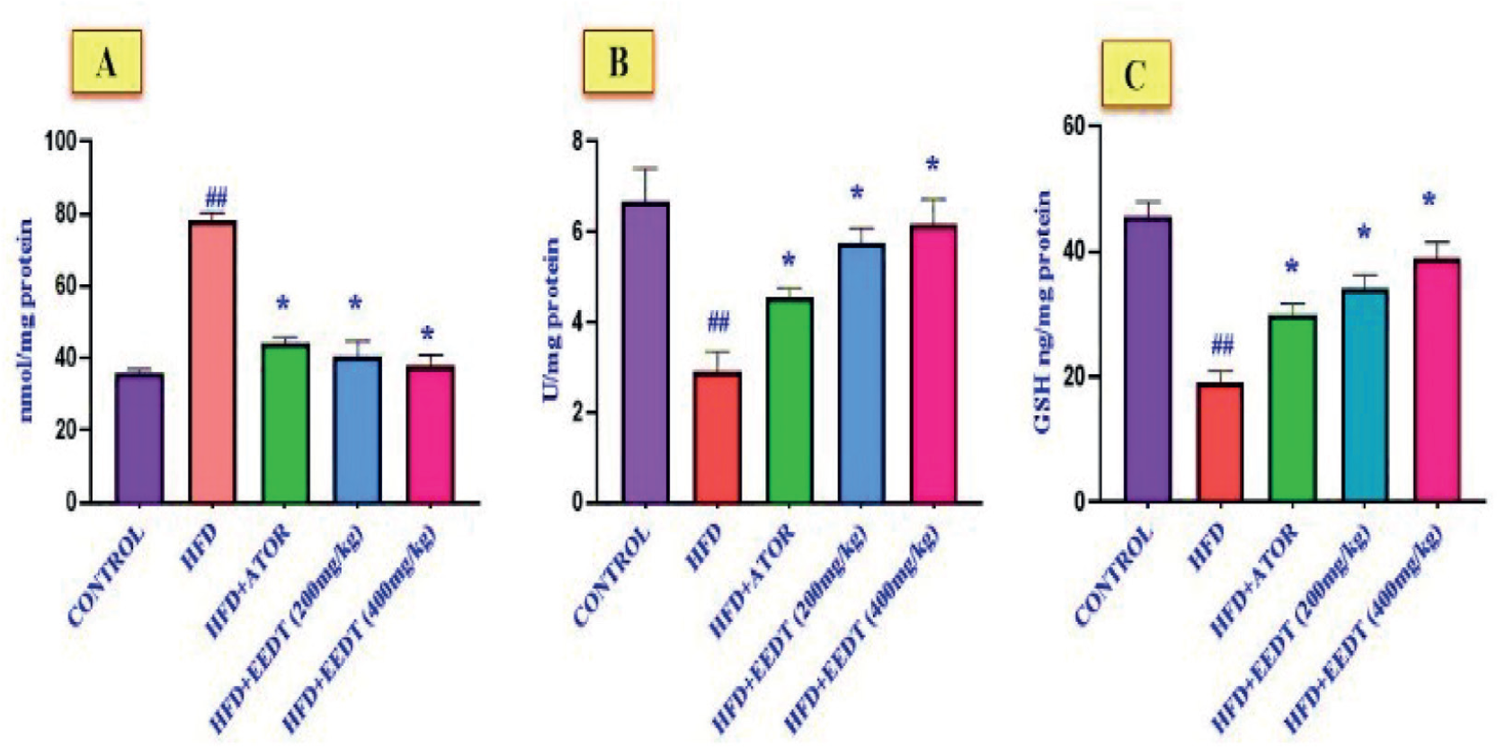
The production of malondialdehyde (MDA) was used as a marker to evaluate lipid peroxidation, with elevated MDA levels indicating greater lipid peroxidation severity. In the high-fat diet (HFD) group, MDA levels were significantly higher compared to the normal control group, reflecting increased oxidative stress. Treatment with EEDT at 200 mg/kg and 400 mg/kg led to a dose-dependent reduction in MDA levels, with the 400 mg/kg dose demonstrating a more pronounced decrease. In the hyperlipidemic control group, glutathione (GSH) and catalase levels in liver homogenates were significantly reduced compared to the normal control group, indicating impaired antioxidant defence mechanisms. However, treatment with EEDT at 200 mg/kg and 400 mg/kg resulted in a significant increase in GSH and catalase levels compared to the hyperlipidemic control group, highlighting the potent antioxidant potential of EEDT at these dosages, as illustrated in Figure 6 and detailed in Table XI.

Effect of EDDT on (A) MDA, (B) CAT, and (C) GSH Levels.Values are presented as mean ± SEM.#p<0.05, ## p<0.01(Compared to control group), *p<0.05 (Compared to HFD group).
Histopathological studies
The histopathological examination of liver tissues revealed normal hepatocyte morphology in the control group and HFD + Atorvastatin (10 mg/kg) group, with no fatty changes or damage. The HFD group exhibited moderate microvesicular and macrovesicular steatosis (34-66%) and hepatocyte ballooning, indicating fatty degeneration and cellular injury. Treatment with HFD + EEDT (200 mg/kg) showed mild steatosis (5-33%) and ballooning, reflecting partial improvement. In contrast, HFD + EEDT (400 mg/kg) demonstrated normal hepatocyte morphology, similar to the control group, indicating a dose-dependent restoration of liver health depicted in Figure 7.

DISCUSSION
Cardiovascular risk assessment is essential for preventing and managing various cardiovascular diseases (CVDs), with hyperlipidemia being a major risk factor for coronary heart disease. This highlights the importance of addressing dyslipidemia, and one promising approach is the use of plant-based treatments due to their diverse pharmacological effects. This study explores the phytochemical composition and potential therapeutic benefits of the ethanolic extract of D. trifasciata, traditionally used in folk medicine to treat conditions such as earaches, ulcers, jaundice, and skin irritations. The plant has also been valued for its analgesic and antipyretic effects.
In this study, ethanol extraction of D. trifasciata from powdered leaves yielded 10.7%, with a total phenolic content of 776.33 mg GAE/g and a total flavonoid content of 284 mg RT/g. Phenolic compounds and flavonoids are well-known for their potent antioxidant properties, which play a critical role in neutralizing free radicals and mitigating oxidative stress; a key contributor to chronic diseases such as cardiovascular disorders, diabetes, and neurodegenerative conditions (Halliwell, Gutteridge, 2015). The high concentration of these bioactive compounds in D. trifasciata underscores its potential as a rich natural source of antioxidants, which could offer cardioprotective benefits by reducing oxidative damage to lipids, proteins, and DNA. This aligns with existing literature that highlights the therapeutic potential of plant-derived antioxidants in managing oxidative stress-related diseases (Pandey, Rizvi, 2009).
The study represents the first investigation into the antihyperlipidemic properties and acute toxicity of D. trifasciata, addressing a critical gap in the scientific literature. Ethical considerations were rigorously followed, adhering to the 3Rs principle (Replacement, Reduction, and Refinement) to ensure the humane treatment of animals and minimize their use. For antihyperlipidemic evaluation, five groups of six animals each were utilized, while acute toxicity testing involved two groups of five animals each. Acute toxicity tests were performed following OECD 420 guidelines, with a dose of 2000 mg/kg body weight administered to rats. No toxicity or adverse health effects were observed, and the dose was well-tolerated, with a 100% survival rate, demonstrating the safety and tolerability of D. trifasciata extract at this dose. These findings provide a strong foundation for further exploration as a therapeutic agent (OECD, 2001).
The study also investigated the effects of D. trifasciata extracts on high-fat diet (HFD)-induced hyperlipidemia in Wistar rats. After 30 days on the HFD, all rats exhibited weight gain, but those treated with the extract showed a significant reduction in body weight compared to the HFD control group. This suggests that D. trifasciata may possess anti-obesity properties, potentially mediated by its ability to modulate lipid metabolism or reduce fat absorption. Body mass index (BMI) evaluations further supported this finding, with EEDT-treated groups displaying significantly lower BMI values than the hyperlipidemic control group. These results highlight the extract’s potential role in managing obesity, a major risk factor for hyperlipidemia and cardiovascular diseases (Grundy et al., 2004).
Lipid profile analysis in this study revealed that treatment with D. trifasciata extract significantly reduced total cholesterol (TC), triglycerides (TG), low-density lipoprotein (LDL), and very low-density lipoprotein (VLDL) levels, while concurrently increasing high-density lipoprotein (HDL) levels. These findings are of considerable clinical importance, as elevated levels of TC, TG, LDL, and VLDL are well-established risk factors for the development of atherosclerosis and cardiovascular diseases (CVDs) (Grundy et al., 2004). LDL and VLDL are particularly atherogenic, as they facilitate the deposition of cholesterol in arterial walls, leading to plaque formation and subsequent narrowing of blood vessels. Conversely, HDL is considered cardioprotective due to its role in reverse cholesterol transport, whereby it removes excess cholesterol from peripheral tissues and transports it to the liver for excretion (Rader, Hovingh, 2014). The observed increase in HDL levels following D. trifasciata treatment further underscores its potential to mitigate cardiovascular risk. In addition to the improvements in lipid parameters, the EEDT-treated groups exhibited a significant reduction in atherogenic indices, including the Atherogenic Index of Plasma (AIP), Atherogenic Coefficient (AC), and Cardiac Risk Ratio (CRR). These indices are robust predictors of cardiovascular risk and provide a comprehensive assessment of the balance between pro-atherogenic and anti-atherogenic lipoproteins. The AIP, calculated as the logarithm of the ratio of TG to HDL, is a strong indicator of small, dense LDL particles, which are highly atherogenic (Dobiásová, Frohlich, 2001). The AC, derived from the ratio of non-HDL cholesterol to HDL cholesterol, reflects the burden of atherogenic lipoproteins relative to protective HDL. Similarly, the CRR, calculated as the ratio of TC to HDL, is a widely used marker for assessing the risk of coronary artery disease (Fernandez, Webb, 2008). The reduction in these indices following D. trifasciata treatment suggests a favourable shift in the lipid profile, reducing the overall risk of atherosclerosis and its associated complications, such as myocardial infarction and stroke.
The lipid-modulating effects of D. trifasciata may be attributed to its bioactive constituents, including flavonoids, polyphenols, and terpenoids. Flavonoids, for instance, have been reported to inhibit 3-hydroxy-3-methylglutaryl coenzyme A (HMG-CoA) reductase, a key enzyme involved in cholesterol biosynthesis, thereby reducing endogenous cholesterol production. Studies on plant extracts, such as Zingiber officinale and Cajanus cajan, have demonstrated that flavonoid-rich extracts suppress cholesterol synthesis through competitive inhibition of HMG-CoA reductase (Wresdiyati et al., 2023; Sudirman, Janna, Widiastuti, 2022). Polyphenols, another major class of bioactive compounds, are known to enhance the expression of low-density lipoprotein (LDL) receptors on hepatocytes, facilitating the clearance of LDL from circulation (Pandey, Rizvi, 2009). Clinical studies on bergamot-derived polyphenols have shown a reduction in LDL cholesterol levels by up to 27%, a mechanism potentially mediated through peroxisome proliferator-activated receptor alpha (PPARα) activation and sterol regulatory element-binding protein-1c (SREBP-1c) modulation (Musolino et al., 2020). Additionally, these polyphenolic compounds have been shown to upregulate the activity of lipoprotein lipase, an enzyme responsible for hydrolyzing triglyceride (TG)-rich lipoproteins, leading to a decrease in circulating TG and very-low-density lipoprotein (VLDL) levels (Kersten, 2001).Furthermore, the increase in HDL levels observed with polyphenol intake may be attributed to the activation of PPAR-dependent pathways, which enhance the synthesis of apolipoprotein A-I, the primary structural protein of HDL (Goldstein, Brown, 1990). Notably, polyphenols from bergamot have been reported to elevate HDL cholesterol by approximately 22% via PPAR-mediated mechanisms (Musolino et al., 2020). These findings suggest that the bioactive compounds in D. trifasciata may contribute to lipid homeostasis through multiple molecular pathways, highlighting its potential as a natural lipid-lowering agent.
The study also demonstrated the hepatoprotective effects of D. trifasciata, as evidenced by the significant reduction in liver enzyme levels: aspartate aminotransferase (AST), alanine aminotransferase (ALT), and alkaline phosphatase (ALP) in EEDT-treated rats compared to the HFD group. Elevated levels of these enzymes are markers of liver damage, often observed in hyperlipidemic conditions due to fat accumulation and oxidative stress (Kim et al., 2014). The reduction in these enzymes indicates that D. trifasciata may protect against HFD-induced hepatic injury, likely through its antioxidant and anti-inflammatory properties.
Additionally, the extract restored antioxidant enzyme levels, including glutathione (GSH) and catalase, while reducing malondialdehyde (MDA) levels. GSH and catalase are critical components of the cellular antioxidant defense system, and their restoration suggests enhanced protection against oxidative stress. The reduction in MDA, a marker of lipid peroxidation, further supports the extract’s ability to mitigate oxidative damage. These findings collectively suggest that D. trifasciata exerts its antihyperlipidemic effects through antioxidant mechanisms and hepatoprotective properties, making it a promising candidate for further research in the management of hyperlipidemia and associated metabolic disorders. The results of this study suggest that the antihyperlipidemic effect of D. trifasciata extract is likely attributed to its bioactive compounds, including alkaloids, flavonoids, saponins, steroids, and phenolic compounds. These findings highlight its potential as a therapeutic agent for managing hyperlipidemia and reducing cardiovascular risk.
CONCLUSION
The ethanolic extract of D.trifasciata demonstrated significant antihyperlipidemic properties, attributed to its rich phytochemical composition, including alkaloids, flavonoids, saponins, steroids, and phenolic compounds. In hyperlipidemic Wistar rats, the extract reduced body weight, improved lipid profiles (lowering TC, TG, LDL, and VLDL while increasing HDL), and decreased atherogenic indices (AIP, AC, and CRR), indicating reduced cardiovascular risk. It also exhibited hepatoprotective effects by lowering liver enzyme levels (AST, ALT, ALP) and enhanced antioxidant activity by increasing GSH and catalase while reducing MDA. These findings suggest D. trifasciata is a promising natural agent for managing hyperlipidemia and oxidative stress-related complications. Future studies should focus on elucidating molecular mechanisms, isolating bioactive compounds, and conducting pharmacokinetic and clinical trials to evaluate its safety, efficacy, and potential synergies with conventional therapies.
ACKNOWLEDGEMENT
The authors are thankful to RBVRR Women’s College of Pharmacy for providing the necessary facilities to carry out this research work.
DATA AVAILABILITY STATEMENT
The data supporting the findings of this study are available from the corresponding author upon reasonable request.
REFERENCES
- Anees S, Manzoor I, Fatima K, Hamid R, Ganie SA. GC-MS analysis and potential therapeutic efficacy of extracts from Allium humile Kunth in lowering dyslipidemia in Wistar rat models. J Ethnopharmacol. 2024;320:117478.
- Azwanida NN. A review on the extraction methods used in medicinal plants: principles, strengths, and limitations. Med Aromat Plants. 2015;4(196):2167-0412.
- Babu K, Prabhu DS. Dracaena trifasciata (Prain) Mabb.-Traditional use, pharmacognosy, phytochemistry and pharmacology: A comprehensive review. J Phytopharm. 2024;13(3):235-241.
- Bahmani M, Mirhoseini M, Shirzad H, Sedighi M, Shahinfard N, Rafieian-Kopaei M. A review on promising natural agents effective on hyperlipidemia. J Evid Based Complement Altern Med. 2015;20(3):228-238.
- Banu Z, Samreen S, Mujeeb SA. Evaluation of antiparkinsonism activity of Manilkara zapota (L.) P. Royen leaf extract in haloperidol-induced parkinsonism in Swiss albino mice. Indian J Pharm Educ Res. 2025;59(1s):s179-s189.
- Banu Z, Fatima SJ, Fatima A, Fatima S, Zohra SF, Sultana T. Phytochemical evaluation and pharmacological screening of antiparkinson’s activity of Allium sativum in Swiss albino mice. IOSR J Pharm. 2016;6(6):1-12.
- Dobiásová M, Frohlich J. The plasma parameter log (TG/HDL-C) as an atherogenic index: correlation with lipoprotein particle size and esterification rate in apoB-lipoprotein-depleted plasma (FER(HDL)). Clin Biochem. 2001;34(7):583-588.
- Duraipandiyan V, Al-Dhabi NA, Irudayaraj SS, Sunil C. Hypolipidemic activity of friedelin isolated from Azima tetracantha in hyperlipidemic rats. Rev Bras Farmacogn. 2016;26(1):89-93.
- Fernandez ML, Webb D. The LDL to HDL cholesterol ratio as a valuable tool to evaluate coronary heart disease risk. J Am Coll Nutr. 2008;27(1):1-5.
- Goldstein JL, Brown MS. Regulation of the mevalonate pathway. Nature. 1990;343(6257):425-430.
- Gupta A, Guyomard V, Zaman MJ, Rehman HU, Myint PK. Systematic review on evidence of the effectiveness of cholesterol-lowering drugs. Adv Ther. 2010;27(6):348-364.
- Grundy SM, Cleeman JI, Merz CN, Brewer HB Jr, Clark LT, et al. National Heart, Lung, and Blood Institute; American College of Cardiology Foundation; American Heart Association. Implications of recent clinical trials for the National Cholesterol Education Program Adult Treatment Panel III guidelines. Circulation. 2004 13;110(2):227-39.
- Halliwell B, Gutteridge JM. Free radicals in biology and medicine. Oxford University Press; 2015.
- Hasani-Ranjbar S, Nayebi N, Moradi L, Mehri A, Larijani B, Abdollahi M. The efficacy and safety of herbal medicines used in the treatment of hyperlipidemia; a systematic review. Curr Pharm Des. 2010;16(26):2935-47.
- Jeyabalan S, Palayan M. Antihyperlipidemic activity of Sapindus emarginatus in Triton WR-1339 induced albino rats. Res J Pharm Technol. 2009;2(2):319-323.
- Jing X, Sarker MM, Gifari MA, Maruf MR, Alam S, Khan F, et al. Improvement of lipid profile and hepatic oxidative stress in high-fat-diet induced hyperlipidemic Swiss albino rats by Piper betle juice: evidences from in vivo and in silico studies. Cell Mol Biol. 2022;68(9):1-3.
- Kersten S. Mechanisms of nutritional and hormonal regulation of lipogenesis. EMBO Rep. 2001;2(4):282-286.
- Kim EJ, Kim BH, Seo HS, Lee YJ, Kim HH, Son HH, et al. Cholesterol-induced non-alcoholic fatty liver disease and atherosclerosis aggravated by systemic inflammation. PloS one. 2014;9(6):e97841.
- Kumar D, Parcha V, Maithani A, Dhulia I. Effect and evaluation of antihyperlipidemic activity guided isolated fraction from total methanol extract of Bauhinia variegata (Linn.) in Triton WR-1339 induced hyperlipidemic rats. Asian Pac J Trop Dis. 2012;2:S913.
-
Lala V, Zubair M, Minter DA. Liver Function Tests [Updated 2023 July 30]. StatPearls. 2024. Available from: https://www.ncbi.nlm.nih.gov/books/NBK482489/
» https://www.ncbi.nlm.nih.gov/books/NBK482489/ - Mannu GS, Zaman MJ, Gupta A, Rehman HU, Myint PK. Evidence of lifestyle modification in the management of hypercholesterolemia. Curr Cardiol Rev. 2013;9(1):2-14.
- Meunier L, Larrey D. Drug-induced liver injury: biomarkers, requirements, candidates, and validation. Front Pharmacol. 2019;10:1482.
- Mishra PR, Panda PK, Chowdary KA, Panigrahi S. Evaluation of acute hypolipidemic activity of different plant extracts in triton WR-1339 induced hyperlipidemia in albino rats. Pharmacologyonline. 2011;3:925-934.
- Modanisi EI, Aboua YG, Chegou NN, Oguntibeju OO. Hepatoprotective, antihyperlipidemic, and anti-inflammatory activity of Moringa oleifera in diabetic-induced damage in male Wistar rats. Pharm Res. 2017;9(2):182-187.
-
Moriles KE, Zubair M, Azer SA. Alanine Aminotransferase (ALT) Test. [Updated 2024 Feb 27]. In: StatPearls [Internet]. Treasure Island (FL): StatPearls Publishing; 2024. Available from: https://www.ncbi.nlm.nih.gov/books/NBK559278/
» https://www.ncbi.nlm.nih.gov/books/NBK559278/ - Mueller S, Riedel HD, Stremmel W. Direct evidence for catalase as the predominant H2O2-removing enzyme in human erythrocytes. Blood. 1997;90(12):4973-4978.
- Musolino V, Gliozzi M, Scarano F, Bosco F, Scicchitano M, Nucera S, et al. Bergamot polyphenols improve dyslipidemia and pathophysiological features in a mouse model of non-alcoholic fatty liver disease. Sci Rep. 2020;10(1):2565.
- Naaz S, Banu Z. Antidepressant, antiamnesic activity, and toxicity profile of the ethanolic extract of Gerbera jamesonii Adlam flowers. J Phytonanotech Pharm Sci. 2024;4(4):1-8.
- Naaz S, Banu Z, Nikilitha G, Mujeeb SA. Pharmacological benefits of Gerbera jamesonii Adlam flower: Qualitative and quantitative analysis of the extract. J Phytonanotechnol Pharm Sci. 2024;4(2):30-36.
- Nelson RH. Hyperlipidemia as a risk factor for cardiovascular disease. Prim Care Clin Off Pract. 2013;40(1):195-211.
- Qomariyah N. Antidiabetic effects of a decoction of leaves of Sansevieria trifasciata in alloxan-induced diabetic white rats (Rattus norvegicus L.). ITB Sci. 2012;44:308-316.
- Oršolić N, Landeka Jurčević I, Đikić D, Rogić D, Odeh D, Balta V, et al. Effect of propolis on diet-induced hyperlipidemia and atherogenic indices in mice. Antioxidants. 2019;8(6):156.
- OECD. (2001). Guideline 420: Acute Oral Toxicity - Fixed Dose Procedure. OECD Publishing.
- Pandey KB, Rizvi SI. Plant polyphenols as dietary antioxidants in human health and disease. Oxid Med Cell Longev. 2009;2(5):270-278.
- Rader DJ, Hovingh GK. HDL and cardiovascular disease. Lancet. 2014;384(9943):618-625.
- Rwawiire S, Tomkova B. Morphological, thermal, and mechanical characterization of Sansevieria trifasciata fibers. J Nat Fibers. 2015;12:201-210.
- Shattat GF. A review article on hyperlipidemia: types, treatments, and new drug targets. Biomed Pharmacol J. 2015;7(1):399-409.
- Stafford GI, Pedersen ME, Van Staden J, Jäger AK. Review on plants with CNS-effects used in traditional South African medicine against mental diseases. J Ethnopharmacol. 2008;119:513-537.
- Sudirman S, Janna M, Widiastuti I. In vitro inhibitory HMG-CoA reductase activity of purified polyphenol compounds from water lettuce (Pistia stratiotes) leaf extract. Trop J Nat Prod Res. 2022;6(7):1245-1250.
- Tabassum H, Parvez S, Rehman H, Banerjee BD, Raisuddin S. Catechin as an antioxidant in liver mitochondrial toxicity: inhibition of tamoxifen-induced protein oxidation and lipid peroxidation. J Biochem Mol Toxicol. 2007;21(3):110-117.
- Trease GE, Evans WC. A Textbook of Pharmacognosy. 11th ed. Brailliar Tindall Ltd, London. 1989;45-50.
- Trease GE, Evans WC. Pharmacognosy. Alden Press, Oxford. 1996:213-232.
- Wresdiyati T, Papilaya MC, Lailia SR, Darawati M, Sadiah S, Astawan M. 3-hydroxy-3-methylglutaryl-coenzyme A (HMG-CoA) reductase inhibitory activity of Indonesian Cajanus cajan leaves and Zingiber officinale extracts. Food Res. 2023;7:139-144.
Edited by
-
Associate Editor:
Daniel Demarque