 Microemulsion-based dual delivery of malkangani and phalsa: formulation, optimization and in-vivo pharmacodynamics in rats
Abstract
Alzheimer’s Disease is a prevalent form of dementia typically characterized by neurodegeneration, memory loss and cognition loss. Celastrus paniculatus (malkangani) is a well-known brain tonic in Ayurveda, while Grewia asiatica (phalsa) is reported to improve memory and cognition. This study was focused on the development and evaluation of a combined microemulsion of phalsa extract and malkangani oil. The microemulsion was prepared using the water titration method and optimized using Box Behnken Design. The optimized formulation showed particle size of 14.91±0.20 nm, 0.323±0.03 polydispersity index (PDI) and % transmittance of 96.31±0.04 %. The in-vitro drug release was estimated by media change method in 0.1 N HCl and phosphate buffer (pH 6.8) and it found to follow Higuchi order kinetics with about 90 % release up to 8 hrs. Microemulsion significantly improved inflexion ratio in elevated plus maze test; indicating improvement in learning, memory and recognition in scopolamine-induced amnesia in rat as compared to single treatment of oil or extract, however statistically non-significant effect was seen in novel object recognition test. The biochemical tests and histopathological examinations of rat brains also showed the effect of microemulsion when compared to the disease control group. This combination in microemulsion can be used as a prophylactic treatment to prevent neurodegeneration.
Keywords:
Microemulsion; Memory enhancement;
Celastrus paniculatus
;
Grewia asiatica
; Amnesia; Optimization
INTRODUCTION
In the current scenario, neurodegenerative diseases are becoming one of the serious causes of concern. Multiple factors contribute to neurodegeneration, such as oxidative stress, neurotransmitter levels in brain, etc. Alzheimer’s disease (AD), the most common form of dementia, is marked by memory loss, declined cognition and difficulties in performing daily tasks (Ovais et al., 2018). In 2020, more than 55 million people all over the world were living with dementia. This number is expected to double annually, reaching 78 million by 2030 and 139 million by 2050. It is continuously increasing clinical concern in the elderly population (Nichols et al., 2022; Gustavsson et al., 2023).
The treatments for AD are limited. Moreover, the complex treatment regimen of medicines and non-pharmacological interventions makes patients difficult to adhere the prescribed therapies. The available treatments for AD are limited and have serious side effects such as nausea, vomiting, diarrhoea, dizziness, and insomnia (Ferreira, Lopes, Bergamaschi, 2020). These side effects can affect patient compliance and quality of life, especially in the geriatric population, who may be more sensitive to medication-related adverse effects. There is a need for pharmacologically active compounds that can act on multiple targets. Even though the disease-modifying agents are still not available, the disease progression can be slowed down, and patients with mild-to-moderate AD can live with the disease for several years with a good quality of life. Hence, early intervention is important (Rasmussen, Langerman, 2019). Currently, scientists are exploring the plethora of natural compounds supported by traditional systems of medicine as well as those identified from new findings.
Natural compounds and their derivatives are a vast untapped reservoir for discovering new therapeutically active molecules. Many medicinal plants possess a nootropics and neuroprotective effect that can potentially be used in AD (Farzaei et al., 2019; Mehla et al., 2020). Using this ancient knowledge, early-stage intervention of disease medicinal properties of plants can be utilized effectively.
Celastrus panculatus is a well-known medicinal plant, famous for its memory enhancement activity in the traditional medicinal system of India, i.e., Ayurveda (Ayurvedic Pharmacopoeia of India, 2016; Tandon, Sharma, 2011). Its seed oil has been reported to reduce scopolamine-induced impairments in learning and memory models (Gattu et al., 1997; Bhanumathy et al., 2010; Alama, Haque, 2011). Polyphenols are known for their wide therapeutic activities such as antioxidant, neuroprotective, anti-inflammatory, and anti-cancer. Wild berries are a rich source of polyphenolic compounds. Grewia asiatica is one of the famous summer wild berries in India and has an abundant amount of polyphenolic compounds such as anthocyanins, flavonoids, phenolics, flavonols, isoflavonols etc. (Talpur, 2017; Koley et al., 2020). Various extracts of phalsa are reported to improve cognition across multiple animal models of amnesia (Paul et al., 2020; Imran et al., 2021).
Moreover, the phytoconstituents present in these berries have been included in excellent reviews by Mehla et al. (2020) and Lange, Nakamura (2022), detailing the potentially beneficial effects of various plant polyphenols for improving memory and AD.
Due to multi-component nature, plant extracts have the potential to act on multiple therapeutic targets, often synergistically, and can therefore be tested to prevent a complex disease like AD (Ovais et al., 2018).
Despite the strong therapeutic activity, the main limiting factor of these natural compounds is the inability to cross the blood-brain barrier (BBB). However, a novel drug delivery system can resolve this limitation (Ovais et al., 2018; Moradi et al., 2020).
Hence, the main goal of this study was to formulate and optimize a microemulsion of a combination of Celastrus paniculatus seed oil (malkangani oil) and polyphenol rich extract of Grewia asiatica (phalsa extract) employing design of experiments (DoE)) technique. The secondary objective was to study the effects of the above two components in the scopolamine-induced amnesia model in rats to identify synergistic, additive, antagonistic or no effect of combination.
MATERIAL AND METHODS
Material
The cold-pressed Celastrus paniculatus seed oil (malkangani oil) was purchased from Oilcure Ltd. Grewia asiatica (phalsa) berries were collected from Gandhinagar, Gujarat, India and authenticated at Agarkhar Research Institute, Pune (Report No. - AUTH 21-52). All the solvents utilized were of analytical grade. Epigallocatechin gallate (EGCG) and gallic acid (GA) were procured from Yucca Enterprises, Mumbai, Maharashtra, India. Squalene was purchased from TCI chemicals, India. Gift sample of Acconon E was received from Gattefose India Pvt Ltd. Propylene glycol, Polyethylene glycol 400, Span 20, Span 80, Tween 20 and tween 80 were purchased from Loba Chemie, India.
Experimental Animals
Male Wistar rats aged 6-8 weeks, weighing approximately 180-200 g each, were obtained from Global Bioresearch Laboratory, Pune and housed at the animal house facility at AISSMS College of Pharmacy, Pune. The animals were housed in polyacrylic cages in groups of six under laboratory conditions. They were maintained at a temperature of 22-24 °C and 5060% humidity in a controlled animal facility with a 12-hour light-dark cycle. The rats were acclimatized for approximately 7 days before behavioral studies commenced. All experiments were conducted during the daytime from 8:00 am to 12:00 noon. The animals had access to standard food pellets and potable drinking water ad libitum. The study protocol was approved by the Institutional Animal Ethics Committee (Project Approval Number - CPCSEA/IAEC/PT-08/01-2K23).
Extraction of Polyphenols from Phalsa
About 5 g of fruit pulp was extracted in 10 ml acidified alcohol for about 2 h by maceration. Constant stirring was maintained using a magnetic stirrer at room temperature, protected from light. The polyphenol extract was separated from marc, concentrated and stored in amber colored bottle at -20 °C.
Characterization of malkangani oil and polyphenol fraction of phalsa
Cold-pressed malkangani oil was sourced from the manufacturer, characterized and standardized for its squalene content as per our previously published data (Jarande, Damle, 2024) using High Performance-Thin Layer Chromatography (HPTLC). The polyphenol fraction from phalsa was standardized for its EGCG and GA content using optimized HPTLC conditions only (unpublished data).
Preparation of Microemulsion
Ternary Phase Diagram
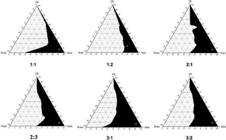
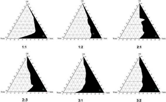
Depending upon the hydrophilic-lipophilic balance (HLB) propylene glycol, tween 80 (HLB value 15) were selected as microemulsion components. Microemulsion system was prepared by water titration method at ambient temperature by varying concentration of mixture of acconon E and malkangani oil constituted the oil phase, tween 80 and propylene glycol - as surfactant and as a co-surfactant respectively. The acconon E was used as an emulsifier. A pseudo-ternary phase diagram was generated to examine the concentration of constituents for the predominant range of microemulsion formation or to examine the microemulsion area. The selected surfactants and co-surfactants were combined at seven weight ratios as 1:1, 1:2, 1:3, 2:1, 2:3, 3:1, 3:2 to set up various surfactant/co-surfactant mixture (Smix) (Patil et al., 2022). This was followed by the addition of malkangani oil and acconon E mixture (1:2) in different volumes to get different oil: Smix ratios (1:9, 2:8, 3:7, 4:6, 5:5, 6:4, 7:3, 8:2 and 9:1 by weight). The mixture of oil and Smix was continuously stirred on a magnetic stirrer for about 30 min, after which during water was incorporated dropwise till turbidity was observed as the endpoint. Ternary phase diagrams were constructed using CHEMIX School software v 10 (Arne Standnes, Bergen, Norway).
Optimization of Microemulsion using a Design of experiments (DoE) approach
Different response variables were optimized utilizing 33 Box-Behnken design (BBD) using design of experiments (DoE) software (version 13). The experimental design had three independent variables, i.e. oil (A), Smix (B) and water (C), studied at 3 levels, i.e., -1 (low), 0 (intermediate) and +1 (high), which are equidistant from each other. Particle size (nm), polydispersity index (PDI) and % transmittance were measured as the response variables (dependent variables) to optimize the formulation with optimal physicochemical characteristics. In this study, levels of independent variables were chosen following a preliminary assessment of oil and Smix concentration as well as the pseudo-ternary phase diagram (Table I).
The initial design comprised a total of 17 runs. The software assessed various mathematical models of the mixture design, including linear, quadratic, cubic and other specialized models. The significance of each model was evaluated through comparisons of statistical parameters such as R2, adjusted R2, predicted R2, standard deviation (SD), and predicted residual error sum of squares (PRESS).
Preparation of Optimized batches
Seventeen formulation batches were designed and developed from the design space generated by DoE software. The quantities of excipients were according to the formulation composition mentioned earlier, while the Smix proportion was selected based on the maximum microemulsion area in the pseudo-ternary phase diagram. The mixture of malkangani oil and Acconon E (oil phase) was mixed with the surfactant (tween 80) and stirred on a magnetic stirrer. A mixture of propylene glycol, i.e. co-surfactant and phalsa extract, was added to it. The whole mixture was vortexed and then stirred on a magnetic stirrer for about 30 min, then water was added dropwise to get a limpid microemulsion. About 5 mL batches of microemulsion were prepared.
In-vitro Characterization/Formulation Evaluation
Drug-Excipient Compatibility Study
FTIR analysis of malkangani oil, phalsa extract other excipients, and microemulsion was performed (IRspirit FT-IR, Shimadzu, Kyoto, Japan). The spectra were recorded using attenuated total reflectance (ATR) over a 4000-400 cm-1 range.
Determination of particle size, polydispersity index (PDI) and zeta potential (Patil et al., 2022)
Particle size, PDI and zeta potential were determined by using Malvern Zetasizer (M/s Malvern, Worcestershire, UK). The optimized formulation was diluted 100 times with distilled water and then scanned for particle size, PDI and zeta potential. The study was performed in triplicates.
Quantification of Marker Compounds
The squalene, EGCG and GA content of the microemulsion was determined by method reported in section of characterization of malkangani oil and polyphenol fraction of phalsa.
In-vitro release of malkangani oil and phalsa extract and its Kinetics
The release behavior of malkangani oil and phalsa extract was determined using dialysis bag method. The release was performed in 0.1 N HCl and phosphate buffer pH 6.8 (Mitrevska et al., 2019). Prior to start of experiment dialysis bags were activated in 0.1 N HCl and phosphate buffer pH 6.8 for 24 hrs. About 4 mL microemulsion as well as blank microemulsion were filled in a (2.5 cm) dialysis bag separately and both ends of bag were tied by thread and fitted to the paddle of dissolution apparatus (Electrolab USP II). The paddle was transferred to a vessel containing 500 mL medium volume and speed set at 75 rpm, with temperature maintained at 37±0.5 °C. For first 2 h, dissolution was performed in 0.1 N HCl, later it was replaced by phosphate buffer and release study was continued till total 8 h. The 5 mL sample was withdrawn from each vessel and replenished with same volume of fresh media respectively at 15, 30, 45, 60,75, 90, 120, 180, 240, 300, 360,420 and 480 min time points. The absorbance was determined at 217 and 276 nm using UV-visible spectrophotometer (Jasco V730, Kyoto, Japan). The absorbance of blank microemulsion was used to correct the errors in absorbance of microemulsion. The % cumulative release was plotted vs time. Data from release study was fitted in different kinetics models of drug release such as first order, Korsemeyers Peppas, Hixon Crowell and Higuchi (Ramteke, Dighe, Kharat, 2014). Best fitted model was identified from regression coefficient (r2) value from all evaluated models (Dash et al., 2010).
In-vivo Evaluation in Scopolamine-induced amnesia model in Rats
Thirty-six healthy male Wistar rats were chosen and randomly allocated into six groups of six animals each to assess their response to the exteroceptive behavior model. The rats were administered either vehicle, phalsa extract, malkangani oil, microemulsion or donepezil daily for 14 days as per the treatment plan in Table II. One hour after the last dose on day 14, all groups except the control group (Group 1) received scopolamine 1 mg/kg by intraperitoneal group. The control group received 1 mL/kg physiological saline instead of scopolamine. Then, 45 min after the administration of scopolamine, behavioral tests were conducted to test the nootropic effects of each treatment (Bohra, Kale, 2018; El-Marasy, Abd-Elsalam, Ahmed-Farid, 2018).
Behavioral Tests
Elevated Plus Maze (EPM)
EPM test serves as the exteroceptive behavioral model for assessing learning and memory in laboratory animals. It is utilized to test learning and memory function as a nootropic activity of the treatment. The test was conducted following a previously reported method (Bohra, Kale, 2018; Paul et al., 2020). The apparatus consisted of two open arms (50*10) and two closed arms (50*10*40) cm (L*W*H) extending from a central platform, elevated to a height of 75 cm from the floor. On the 14th day of study, each rat was placed on the edge of an open arm, facing away from the central platform, and the duration of time taken by the rats to move from the open arm to either of the closed arms, referred to as transfer latency (TL), was recorded. This phase corresponds to the acquisition phase, indicative of learning. Subsequently, on day 15, TL was measured again, representing retention memory, which corresponds to the index of memory retrieval. The inflexion ratio was calculated from the recorded transfer latency using equation 1 (Bohra, Kale, 2018).
Where, L1 is TL measured in seconds on day 14, L0 is TL measured in seconds on day 15 after 24 h.
Novel Object Recognition Test
This test was conducted in an apparatus consisting of open field box (40x40x40 cm) made of acrylic with transparent side walls. The object recognition test was performed as per previously reported methods (Lueptow, 2017; Bhuvanendran et al., 2018). During the treatment, on day 12, each rat was habituated to the apparatus for 5 minutes. On day 13, during the training phase (T1), two identical objects (red and round shaped) were positioned in two diagonal corners of the open field, each 10 cm away from the sidewall. Each rat was placed in the center of the open field and allowed to explore these two identical objects for 5 minutes. Afterwards, they were returned to their cages.
After 24 h of T1, on day 14, the test phase (T2) was conducted. During T2, a new object (an orange coloured and oval) was introduced, and the rats were re-exposed to both the familiar (F) and the new (N) objects for 5 minutes. The preference of each rat towards both objects was recorded via camera. The time spent by the rat exploring each object during T2 was recorded, and the discrimination index (DI) was calculated using equation (2).
TN = time spent with novel object; TF = time spent with familiar object
Biochemical estimation
After evaluation of behavioral tests, on the 15th day, the animals were sacrificed by cervical dislocation. The whole brain was removed from the skull and weighed. Further acetylcholinesterase activity and lipid peroxidation level were measured as per the methods mentioned in literature (Ellman et al., 1961; Rajashri et al., 2020).
Histopathology
After biochemical estimation, the remaining brain was kept in a 10 % formalin solution. The hippocampus of the rat brain was stained with eosin solution and observed under a microscope for changes (Pattanashetti et al., 2017).
Statistical Analysis
The Graphpad Instat for 32-bit Windows, version 3.10, was used for statistical assessment. One-way ANOVA followed by the Tukey-Kramer Multiple Comparisons Test was used for the calculation of statistical analysis of the data. The data was represented as mean ± SEM values and n = 6 per group.
RESULTS
Characterization of phalsa extract and malkangani oil
The phalsa extract was exhibited to contain 13.95 mg/g and 2.13 mg/g of EGCG and GA, respectively. The squalene content in the malkangani oil was estimated at 3.71 mg/g of oil.
Preparation of Microemulsion
Pseudo-ternary Phase Diagram
A pseudo-ternary phase diagram was constructed to determine the ratio of surfactant and co-surfactant (Smix) that forms larger microemulsion area. The different Smix ratios and oil were titrated with water to form a microemulsion. We observed that an increase in Smix ratio from 1:1 w/w to 3:2 w/w is forming a stable microemulsion and the Smix ratio of 3:1 w/w formed largest microemulsion area (Figure 1). Hence, it was finalized for formulation development.
Formulation Optimization
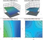
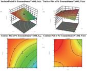
The software generated 17 experimental runs, and the responses obtained experimentally are displayed in Table III. As per the composition given by the software, microemulsion batches were prepared by water titration method and allowed to equilibrate for 24 h. The responses viz., particle size, PDI and % transmittance were determined after equilibration. The results are depicted as contour plots and 3D response surface plots using Design Expert Software, version 13.
Model Fit Summary
In statistics, the R2 value, also known as the coefficient of determination, indicates how well the independent variables explain the variability of the dependent variables. A higher R2 value implies a stronger fit of the model to the data, indicating that the independent variables can better explain the variation observed in the dependent variable. Predicted R2 is a measure of how well the model predicts outcomes for new observations. A higher predicted R2 indicates that the model is likely to perform well in predicting outcomes for new observations, suggesting good generalization ability. Adjusted R2 is a modified version of the regular R2 that accounts for the number of predictors in the model. A higher adjusted R2 signifies a better balance between model complexity and explanatory power, suggesting a more reliable model. The optimal model for optimizing the microemulsion was determined based on the higher values of adjusted R2, predicted R2 and lower PRESS value.
For each variable, different model was best fitted. The 2FI model showed an excellent fit for particle size. For PDI and % transmittance, superior fit was exhibited by a mean and linear model exhibited superior fit, Table IV.
Influence of factors on responses
Influence on Particle size
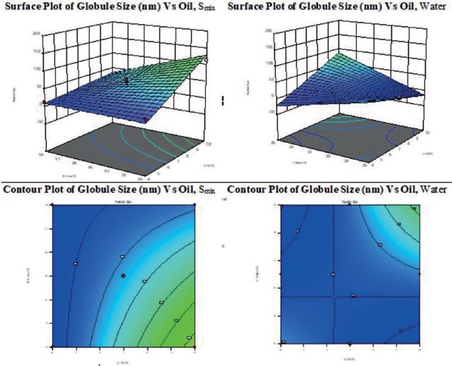
Examining effect of oil and Smix on the particle size is crucial as particle size directly affects drug absorption in body. Smaller particles generally enhance surface area available for dissolution which significantly improves the bioavailability and absorption rate of the drug. It was observed that increasing the oil proportion while keeping Smix constant leads to an increase in particle size. Conversely, increasing the Smix proportion while keeping the oil constant results in a decrease in particle size. Optimizing particle size is essential for ensuring efficient drug delivery and maximizing therapeutic efficacy. The equation for particle size is given below.
Particle size (nm) = 37.07*A-22.85*B+28.13*C-23.55*A+41.52*B*C
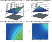
The linear, 2FI and quadratic model (p-value <0.05) were found to be significant for particle size. The particle size ranged from a maximum of 197 nm to a minimum of 12.16 nm, both within the desired range. The 3D surface response plots and contour plots of particle size are shown in Figure 1S of supplementary information.
Influence on PDI
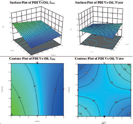
The square model was a good fit for PDI, indicating a non-linear relationship between the factors and PDI. It suggests that the interaction between factors may have a quadratic effect on PDI, implying that changes in factor level may not have a linear/proportional impact on PDI (Figure 2S, supplementary information). PDI value less than 0.5 indicates the uniform and homogenous droplet size distribution (John et al., 2022; Jusril et al., 2022). The equation is, PDI = -18.49*B*C
Influence on % Transmittance
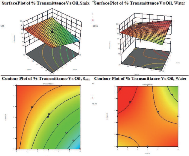
The linear model (p-value <0.05) was significant for % Transmittance. An increase in Smix concentration and dilution of microemulsion with water increased % Transmittance whereas oil concentration had a negative correlation with % transmittance (Figure 3S, supplementary information). The equation for % Transmittance is given below.
% Transmittance = -1.63*A+1.33*B+0.0402*C
Validation and Formulation of Optimized Batch
The optimized formula was selected on the basis of range of variables given as an input. Solutions for microemulsion batches of desirability 1 are mentioned in Table S2 of supplementary information. The composition, which showed comparable predicted values and actual values, is mentioned in Table V. Hence, this composition was considered as an optimized, validated batch and taken for further evaluation in animal model.
In-vitro Characterization/Formulation Evaluation - FTIR, Particle size, Zeta potential and % content of identified compounds etc.
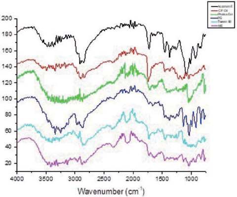
The superimposed FTIR spectra of malkangani oil, phalsa extract, excipients and microemulsion indicate absence of interactions between excipients, Figure 2. The absence of change in absorption spectra of the malkangani oil and phalsa extract indicate their compatibility with the excipients.
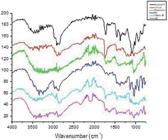
Stack of FTIR Spectra of excipients, CP (Malkangani) oil, Phalsa extract and microemulsion.
The clear, transparent, and yellow-coloured microemulsion did not break and separate after centrifugation, indicating its physical stability. Malkangani oil was standardized for its squalene content and phalsa extract for EGCG and GA presence, as herbal drug standardization is the primary quality control measure for herbal drugs. In microemulsion, squalene was found to be 0.13 % whereas EGCG and GA were found to be 0.17 % and 0.026 % using HPTLC. The particle size and PDI were found to be 14.91±0.20 nm and 0.323±0.03 with a % Transmittance of96.31±0.04 % and zeta potential of-12.33±1.37 mV. Through meticulous formulation and optimization processes, we achieved a stable micro-emulsion system that effectively co-delivers both active compounds.
In-vitro Drug Release Study of microemulsion and its Kinetics
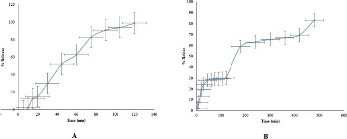
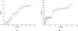
The in-vitro dissolution study of optimized batch of microemulsion using a dialysis bag and media change method was carried out in 0.1 N HCl for first 2 h and phosphate buffer pH 6.8 upto 8 h. Within 2 h, percentage amount of phalsa extract released from dialysis bag was 99.00±0.860 %, whereas the release of malkangani oil was 29.90 ± 0.42 %. However, it increased up to 84.83±1.86 % in 8 h. (Figure 4S, Supplementary information).
During initial trials to finalize the dissolution media, phalsa extract and malkangani oil were separately tested in 0.1 N HCl and phosphate buffer (pH 6.8). The release of malkangani oil was observed in both media. Therefore, media change was employed. We found that the media change method enhanced the release extent of malkangani oil. To understand the release kinetics, the results of the in-vitro drug release study were fitted into different models such as zero order, first order, Korsmeyer peppas, Hixon Crowell and Higuchi, given in Table VI. The best fitted kinetic model was evaluated by comparing correlation coefficient (r2) values. It was deduced that microemulsion followed the Higuchi kinetic model as we got the highest r2, see Table VI, demonstrating that the drug release is proportional to the square root of time and occurred through the mechanism of Fickian diffusion. The Higuchi model assumes Fickian diffusion, which means both phalsa extract and malkangani oil diffuse from regions of higher concentration within the microemulsion matrix to regions of lower concentration in the surrounding media (Elsevier, 2015).
In-vivo Evaluation of Microemulsion
Behavioral Evaluation on Scopolamine Induced Amnesia Rat Model
Scopolamine is a muscarinic antagonist that disrupts the central cholinergic system, leading to memory impairments. It induces degeneration of cortical cholinergic neurons, which is strongly linked to the cognitive deficits observed in AD. Hence, the scopolamine-induced amnesia model is widely used to study the anti-AD effects of therapeutics in pre-clinical.
Effect on Transfer Latency in EPM
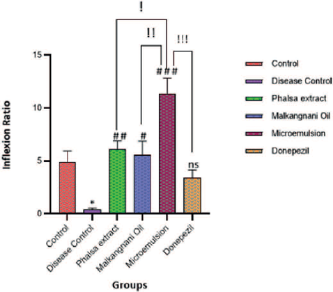
In the disease control group, the inflexion ratio significantly decreased i.e. 0.056 ± 0.0067, compared to 4.938 ± 1.023 of the control group. We observed a significant increase in inflexion ratio in all the treatment and donepezil groups compared to a disease control group. It increased significantly in the microemulsion group to 11.388 ±1.446 compared to the phalsa extract, malkangani oil and donepezil treated groups, Figure 3. This indicates improvement in memory impairments induced by scopolamine.
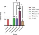
Elevated Plus Maze - inflexion ratio. [Data is represented in as mean± SEM (n=6/group)] Significant difference is denoted by * P <0.05 as compared to control group I; # # P < 0.01, # P < 0.05, # # # < 0.001, ns P > 0.05 as compared to Disease Control group II; ! P <0.05, !! P < 0.01, !!! P < 0.001 as compared to Microemulsion treated Group
Effect on Discrimination Index in Novel Object Recognition (NOR) Test
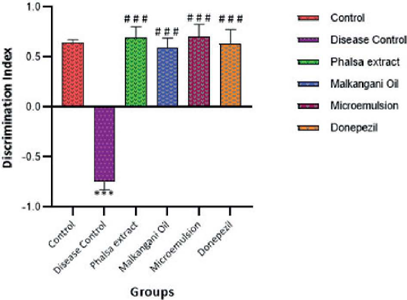
The animals in scopolamine-induced groups (disease control) could not discriminate between novel and familiar object. We observed a drop in the DI of the familiar object, which is -0.742 ± 0.088 in the disease control group compared to the 0.6483± 0.025 of the control groups. It improved in the phalsa extract, malkangani oil, microemulsion and donepezil groups compared to the disease control group. The highest DI was observed in the microemulsion group, i.e. 0.7060 ± 0.1212, however statistically insignificant difference between the DI of the microemulsion group and individual treatment of malkangani oil and phalsa extract groups was observed. Figure 4 represents the graph plot of the discrimination index for the scopolamine-induced amnesia model.
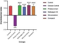
Novel Object Recognition - discrimination Index. [Data is represented in as mean± SEM (n=6/group)] Significant difference is denoted by *** P <0.001 as compared to control group I; # # # P < 0.001, as compared to Disease Control Group II
Biochemical Tests: Effect on acetylcholinesterase activity (AChE activity)
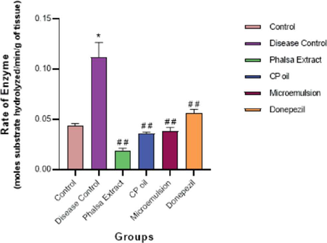
The AChE activity is represented as the rate in a mole of substrate hydrolyzed/min/g of wet tissue. It was increased significantly in Group II, i.e., the disease control group (0.112±0.015) compared to the control group (0.043±0.003). All the treatments lowered the acetylcholinesterase activity. Groups III to VI showed significant decrease in AChE activity compared to Group II, refer Table 1S of supplementary information. The results are represented in Figure 5.
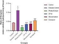
AchE activity (Rate in moles substrate hydrolyzed per min per g of tissue). [Data is represented in as mean± SEM (n=6/group)] Significant difference is denoted by *P <0.001 as compared to control group I; # # P < 0.001, as compared to Disease Control Group II (Disease Control)
Lipid Peroxidation
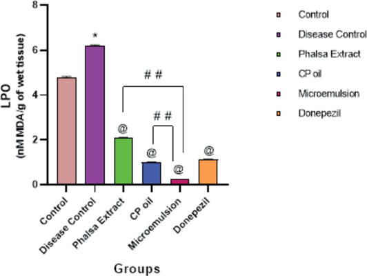
The lipid peroxidation was represented as nanomoles of malondialdehyde (MDA) species formed. Compared to the control group, the MDA levels were significantly higher in Group II, i.e., the disease control group. Groups III to VI exhibited significantly lower MDA concentrations than Group II. Groups III to VI exhibited significantly lower MDA concentration than Group II. Though all treatments lowered lipid peroxidation, Group V, i.e. microemulsion treated showed the lowest level of MDA, i.e. 0.235±0.002 nM MDA/g of wet tissue, refer to Figure 6 and Table 1S (supplementary information).
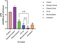
Lipid Peroxidation Level (nM MDA/g of wet tissue). [Data is represented in as mean± SEM (n=6/group)] Significant difference is denoted by *P <0.001 as compared to control group I; @ P < 0.001, as compared to Disease Control Group II; # # P <0.001 as compared to Microemulsion group IV
Histopathological Studies
The control group showed normal brain hippocampus, neuronal cells and brain parenchyma. In the disease control group, histology of the hippocampus showed significant edema, severe neurodegeneration and pyknotic nuclei and distorted pyramidal cells (Pattanashetti et al., 2017; Rajashri et al., 2020). The histopathological images are represented in Figure 7. The phalsa extract, malkangani oil group and microemulsion groups (group II to group VI) showed mild oedema, neuronal damage and normal arrangement of pyramidal cells. This showed the ameliorating effect of the tested samples.
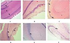
Representative photomicrographs of hippocampal sections stained with hematoxylin and eosin (HE) Histopathological changes in the hippocampal CA1 region.
DISCUSSION
AD is a multifaceted disease with many possible targets. Plant-derived materials contain a mixture of bioactive compounds that have the potential to modulate multiple pathways involved in oxidative stress, inflammation and neuroprotection. Early-stage intervention with scientifically proven plant based formulation could offer significant neuroprotection and halt or slow down the progress of the disease. In the current study, malkangani oil and phalsa extract were selected due to their reported efficacy and high polyphenol content, respectively.
Microemulsion offers a distinct advantage in pharmaceutical formulations due to their ability to encapsulate high dose drugs and facilitate targeted drug delivery, thereby enhancing pharmacological efficacy (Pires, Paiva-Santos, Veiga, 2022). Moreover, microemulsion systems have demonstrated efficacy in achieving optimal drug concentrations within the brain following oral administration (Etman et al., 2018; Guo et al., 2019). Additionally, the ability to tailor microemulsion formulations allows fine-tuning of drug release kinetics, further optimizing therapeutic efficacy while minimizing potential adverse effects. Thus, the utilization of microemulsion-based drug delivery systems holds significant promise in advancing treatment strategies for various neurological ailments.
In the current study, a microemulsion based formulation of phalsa extract and malkangani oil was prepared and optimized using the DoE approach. Proof of concept was established by evaluating the formulation in the scopolamine-induced amnesia model in rats. The identified active constituents of phalsa extract (EGCG and GA) and malkangani oil (squalene) were quantified by HPTLC. DoE is a powerful statistical approach that enables systematic investigation of the relationships between factors affecting a formulation and its performance - thus optimizing formulations in a robust and efficient manner. It also ensures that the final product performs consistently and meets the standards. In this study, the microemulsion was successfully optimized using a Box-Behnken design, considering oil, Smix, and water as independent variables (factors). The results indicate that the selected responses (critical parameters) such as particle size, PDI and % transmittance were effectively optimized to achieve a stable and homogenous microemulsion system suitable for further applications. These parameters significantly influence stability and drug absorption, enhancing bioavailability and efficacy (Trivedi, Siriah, Puranik, 2014). For a thermodynamically stable microemulsion, it is desirable to have a particle size < 100 nm, PDI < 0.5, zeta potential ±30 mV or more and % transmittance near 100. Smaller particle sizes lead to a higher surface area, which enhances drug solubilization and absorption. The optimized formulation had a PDI of 0.323±0.03 indicating the uniform distribution of particles (Butt et al., 2018; Shen, Zhang, 2021) and a zeta potential of -12.33±1.37 mV indicating good electrostatic stability. This helps to prevent aggregation of the microemulsion droplets both at formulation as well as reduces the chances of particle aggregation before reaching the mucosal surface. Hence, its evaluation is crucial during the formulation stage as well as the development stage. It also suggests the consistent drug release from microemulsion as well as better interaction with mucosal epithelium, enhancing the permeability of the drug across the intestinal barrier (Vyas et al., 2006; Zhang et al., 2015; Wen et al., 2021). In-vitro drug release studies aid in predicting the amount of drug release with respect to time. Further, release kinetics highlight whether the system is capable of demonstrating sustained or controlled release behavior as well as underlying mechanisms governing the particular release pattern. The results of the study indicate that the optimized microemulsion facilitates Fickian diffusion. This ensures that both hydrophilic and lipophilic components can be effectively delivered, making it a promising candidate for oral drug delivery systems (Elsevier, 2015).
In this study, the individual treatments viz. - phalsa extract and malkangani oil as well as microemulsion exhibited memory enhancing effect in the scopolamine-induced amnesia model in rats. These findings align with previous studies where the different extracts of phalsa fruits decreased the AChE activity, reduced lipid peroxidation, and enhanced memory in different cognitive models (Paul et al., 2020; Imran et al., 2021). Similarly, malkangani oil is also reported to improve memory and lower lipid peroxidation and improve levels of different neurotransmitters associated with memory impairments in laboratory animals (Gattu et al., 1997; Bhanumathy et al., 2010; Alama, Haque, 2011). Thus, the memory enhancing effects of the developed microemulsion can be explained by the inhibition of AchE and lipid peroxidation in the scopolamine-induced amnesia model in rats. EPM is a widely accepted behavioral test used to study learning and memory in rodents (‘Elsevier, 2015; Bhuvanendran et al., 2018). The reduced transfer latency and increased inflexion ratio in the EPM indicate improvement in memory, thus nootropic activity.
The NOR test evaluates recognition memory and gives useful insights on short-term memory, intermediate and long-term memory in basic research (Yadang et al., 2020). Although the rats in the microemulsion group demonstrated higher DI than those treated with the individual components, this effect was not found to be statistically insignificant. In the AChE assay, phalsa extract, malkangani oil as well as microemulsion significantly inhibited AChE activity in the brain. In this regard, the most prominent effect was observed with phalsa extract. The interesting finding in this study was the significantly reduced lipid peroxidation in a microemulsion-treated group compared to the single treatments of phalsa extract and malkangani oil (# #P <0.001), revealing the neuroprotective activity of the combination. Lipid peroxidation is one of the pathways in disease progression that leads to the formation of amyloid plaques, tau protein aggregation and neuronal death. Reduced lipid peroxidation is indicative of the combination’s antioxidant activity.
These findings are correlated to the identified phyconstituents in the microemulsion, i.e. EGCG, GA and squalene. EGCG is a type of catechin shown to have neuroprotective properties, mainly in the context of AD (Nan et al., 2021). Several vital pathways can explain the plausible mechanism of action by which EGCG exerts its neuroprotective effects. Firstly, EGCG is an established effective antioxidant, which is essential as oxidative stress is one of the fundamental reasons associated with neurodegenerative disorders like AD. Studies have shown that EGCG can reduce oxidative damage by scavenging free radicals and activating the nuclear factor erythroid 2-related factor 2 (Nrf2) pathway, thus enhancing the cellular antioxidant defence mechanism (Khalatbary, Khademi, 2018; Ahadi et al., 2019). The nrf2 pathway activates upregulated antioxidant enzymes, protecting neurons from oxidative injury (Khalatbary, Khademi, 2018). EGCG also enhances cholinergic function, which is often impaired in AD. Research indicates that this effect is mediated through inhibition of AChE, the enzyme responsible for the breakdown of ACh, thus increasing ACh levels in the synaptic cleft and improving cholinergic transmission (Nan et al., 2021). This mechanism is particularly relevant to the cholinergic hypothesis of AD, which suggests that a deficit in ACh contributes to cognitive decline.
GA, a phenolic compound, also demonstrates multifaceted neuroprotective actions (Shabani, et al., 2020). It has been identified as an effective antioxidant that neutralizes free radicals and reduces oxidative stress, which is crucial in preventing neuronal death in AD as well as stabilizing a transcription factor, i.e. nuclear factor kappa B (NF-ΚB)- involved in inflammatory processes, thereby protect against neuroinflammation (Sani, Hokmabadi, 2023).
Squalene is a triterpene found in various plant sources and human sebum and has also garnered attention for its neuroprotective properties. It possesses antioxidant capabilities that can mitigate oxidative stress in neuronal cells (Yellamma, Pradesh, 2023). Squalene enhances the fluidity of cell membranes, which is crucial for maintaining neuronal integrity and function (Kabuto et al., 2013). Squalene is reported to reduce neuroinflammation and regulate the neurotransmitter system in depression-induced ICR mice (Kazunori et al., 2019). The findings in the current research confirm that a combination of malkangani oil and a polyphenol rich fraction of phalsa give additive action to improve cognitive functions and behaviors in a scopolamine-induced amnesia model in rats. Thus, the combined microemulsion can be used as prophylactic to prevent further neurodegeneration and disease progression. We could develop more effective and safer treatments for neurodegenerative diseases by harnessing the combination of phytochemicals and advanced delivery systems.
ACKNOWLEDGEMENTS
The authors extend their gratitude to the Principal and Management of All India Shri Shivaji Memorial Society’s College of Pharmacy, Pune, for providing the necessary facilities to conduct this research. Corresponding author, Suvarna Jarande is appreciative to the Chhatrapati Shahu Maharaj Research, Training and Human Development Institute (SARTHI), an autonomous institute of Govt. of Maharashtra, India, for providing a doctoral fellowship under CSMNRF2021.
SUPPLEMENTARY MATERIAL
AVAILABILITY OF DATA AND MATERIAL
All data is included in manuscript and supplementary document.
REFERENCES
- Ahadi S, Zargari M, Khalatbary AR, Ahadi S, Zargari M, Khalatbary AR. reperfusion injury in rats Assessment of the neuroprotective effects of (-) -epigallocatechin-3-gallate on spinal cord ischemia-reperfusion injury in rats. J Spinal Cord Med. 2019;0(0):1-8.
- Alama B, Haque E. Anti-alzheimer and antioxidant activity of celastrus paniculatus seed. Iran J Pharm Sci. 2011;7(1):49-56.
- Ayurvedic Pharmacopoeia of India. Ghaziabad: Pharmacopoeia Commission for Indian Medicine & Homeopathy, Ministry of Ayush, Government of India. 2016. Part I Vol.2. 60 -61.
- Bhanumathy M, Harish MS, Shivaprasad HN, Sushma G. Nootropic activity of Celastrus paniculatus seed. Pharm Biol. 2010;48(3):324-7.
- Bhuvanendran S, Kumari Y, Othman I, Shaikh MF. Amelioration of cognitive deficit by embelin in a scopolamine-induced Alzheimer’s disease-like condition in a rat model. Front Pharmacol. 2018; 9:1-12.
- Bohra BS, Kale PP. Evaluation of nootropic effects of galantamine and sildenafil as a combination in mice. Indian J Exp Biol. 2018;56(1):60-5.
- Butt U, Elshaer A, Snyder LAS, Alany RG. Fatty Acid Based Microemulsions to Combat Ophthalmia Neonatorum Caused by Neisseria gonorrhoeae and Staphylococcus aureus. Nanomaterials. 2018;8(51):1-22.
- Dash S, Murthy PN, Nath L, Chowdhury P. Kinetic modeling on drug release from controlled drug delivery systems. Acta Pol Pharm - Drug Res. 2010;67(3):217-23.
- El-Marasy SA, Abd-Elsalam RM, Ahmed-Farid OA. Ameliorative effect of silymarin on scopolamine-induced dementia in rats. Open Access Maced J Med Sci. 2018;6(7):1215-24.
- Ellman GL, Courtney KD, Andres V, Featherstone RM. A new and rapid colorimetric determination of acetylcholinesterase activity. Biochem Pharmacol. 1961;7(2):88-95.
- Etman SM, Elnaggar YSR, Abdelmonsif DA, Abdallah OY. Oral Brain-Targeted Microemulsion for Enhanced Piperine Delivery in Alzheimer’s Disease Therapy: In Vitro Appraisal, In Vivo Activity, and Nanotoxicity. AAPS PharmSciTech. 2018;19(8):3698-711.
- Elsevier. Mathematical models of drug release. Strategies to Modify the Drug Release from Pharmaceutical Systems. In: Marcos Luciano Bruschi. Strategies to Modify the Drug Release from Pharmaceutical Systems. 2015. p.63-86.
- Farzaei MH, Bahramsoltani R, Abbasabadi Z, Braidy N, Nabavi SM. Role of green tea catechins in prevention of age-related cognitive decline: Pharmacological targets and clinical perspective. J Cell Physiol. 2019;234(3):2447-59.
- Ferreira TR, Lopes LC, Bergamaschi C de C. Frequency and Severity of Adverse Drug Reactions to Medications Prescribed for Alzheimer’s Disease in a Brazilian City: Cross-Sectional Study. Front Pharmacol. 2020; 11:1-11.
- Gattu M, Boss KL, Terry AV, Buccafusco JJ. Reversal of scopolamine-induced deficits in navigational memory performance by the seed oil of Celastrus paniculatus. Pharmacol Biochem Behav. 1997;57(4):793-9.
- Guo Y, Mao X, Zhang J, Sun P, Wang H, Zhang Y, et al. Oral delivery of lycopene-loaded microemulsion for brain-targeting: preparation, characterization, pharmacokinetic evaluation and tissue distribution. Drug Deliv. 2019;26(1):1191-205.
- Gustavsson A, Norton N, Fast T, Frölich L, Georges J, Holzapfel D, et al. Global estimates on the number of persons across the Alzheimer’s disease continuum. Alzheimer’s Dement. 2023;19(2):658-70.
- Imran I, Javaid S, Waheed A, Rasool MF, Majeed A, Samad N, et al. Grewia asiatica Berry Juice Diminishes Anxiety, Depression, and Scopolamine-Induced Learning and Memory Impairment in Behavioral Experimental Animal Models. Front Nutr. 2021;7:1-19.
- Jarande S, Damle M. Gas chromatography-mass spectrophotometric profile of cold-pressed Celastrus paniculatus seed oil and its high-performance thin-layer chromatographic analysis emphasizing squalene content. Sep Sci Plus. 2024;7(3):1-10.
- John OO, Amarachi IS, Chinazom AP, Adaeze E, Kale MB, Umare MD, et al. Phytotherapy: A promising approach for the treatment of Alzheimer’s disease. Pharmacol Res - Mod Chinese Med. 2022;2(October 2021):100030.
- Jusril NA, Abu Bakar SI, Khalil KA, Md Saad WM, Wen NK, Adenan MI. Development and Optimization of Nanoemulsion from Ethanolic Extract of Centella asiatica (NanoSECA) Using D-Optimal Mixture Design to Improve Blood-Brain Barrier Permeability. Evidence-based Complement Altern Med. 2022;2022.
- Kabuto H, Tomoko T. Yamanushi, Najma Janjua FT and, Mankura M. Effects of squalene / squalane on dopamine levels , antioxidant enzyme activity , and fatty acid composition in the striatum of Parkinson ‘ s disease mouse model. 2013;28(1):21-7.
- Kazunori S, Mahmoud BO, Farhana F, Masaki Y, Makoto KT, Hiroko I. Modulation ofthe neurotransmitter systems through the anti-inflammatory and antidepressant-like effects of squalene from Aurantiochytrium sp. 2019;1-15.
- Koley TK, Khan Z, Oulkar D, Singh B, Bhatt BP, Banerjee K. Profiling of polyphenols in phalsa (Grewia asiatica L) fruits based on liquid chromatography high resolution mass spectrometry. J Food Sci Technol. 2020;57(2):606-16.
- Khalatbary AR, Khademi E. Epigallocatechin gallate and neuroprotection The green tea polyphenolic catechin epigallocatechin gallate and neuroprotection. Nutr Neurosci. 2018;0(0):1-14.
- Lange KW, Lange KM, Nakamura Y. Green tea, epigallocatechin gallate and the prevention of Alzheimer’s disease: Clinical evidence. Food Sci Hum Wellness. 2022;11(4):765-70.
- Lueptow LM. Novel object recognition test for the investigation of learning and memory in mice. J Vis Exp. 2017;2017(126):1-9.
- Mehla J, Gupta P, Pahuja M, Diwan D, Diksha D. Indian medicinal herbs and formulations for Alzheimer’s disease, from traditional knowledge to scientific assessment. Brain Sci. 2020;10(12):1-31.
- Mitrevska I, Achkoska T, Brezovska K, Toshev K, Dimitrovska A, Ugarkovic S. Development and Validation of Discriminative Dissolution Method for Metformin Immediate-Release Film-Coated Tablets. J Anal Methods Chem. 2019;2019.
- Moradi SZ, Momtaz S, Bayrami Z, Farzaei MH, Abdollahi M. Nanoformulations of Herbal Extracts in Treatment of Neurodegenerative Disorders. Front Bioeng Biotechnol. 2020; 8:1-20.
- Nan S, Wang P, Zhang Y, Fan J. Epigallocatechin-3-Gallate Provides Protection Against Alzheimer’s Disease-Induced Learning and Memory Impairments in Rats. Drug Des Devel Ther. 2021;2013-24.
- Nichols E, Steinmetz JD, Vollset SE, Fukutaki K, Chalek J, Abd-Allah F, et al. Estimation of the global prevalence of dementia in 2019 and forecasted prevalence in 2050: an analysis for the Global Burden of Disease Study 2019. Lancet Public Heal. 2022;7(2):105-25.
- Ovais M, Zia N, Ahmad I, Khalil AT, Raza A, Ayaz M, et al. Phyto-Therapeutic and Nanomedicinal Approaches to Cure Alzheimer’s Disease: Present Status and Future Opportunities. Front Aging Neurosci. 2018; 10:1-23.
- Patil V, Mhamane S, More S, Pawar A, Arulmozhi S. Exploring the protective effect exhibited by curcumin-loaded coconut oil microemulsion in the experimental models of neurodegeneration: an insight of formulation development, in vitro and in vivo study. Futur J Pharm Sci. 2022;8(1).
- Pattanashetti LA, Taranalli AD, Parvatrao V, Malabade RH, Kumar D. Evaluation of neuroprotective effect of quercetin with donepezil in scopolamine-induced amnesia in rats. Indian J Pharmacol. 2017;49(1):60-4.
- Paul S, Sharma S, Paliwal SK, Kasture SB. Protective action of Grewia asiatica (phalsa) berries against scopolamine-induced deficit in learning and memory using behavior paradigms in rats. Adv Tradit Med. 2020;20(2):243-53.
- Pires PC, Paiva-Santos AC, Veiga F. Nano and Microemulsions for the Treatment of Depressive and Anxiety Disorders: An Efficient Approach to Improve Solubility, Brain Bioavailability and Therapeutic Efficacy. Pharmaceutics. 2022;14(12).
- Rajashri K, Mudhol S, Serva Peddha M, Borse BB. Neuroprotective Effect of Spice Oleoresins on Memory and Cognitive Impairment Associated with Scopolamine-Induced Alzheimer’s Disease in Rats. ACS Omega. 2020;5(48):30898.
- Ramteke K.H., Dighe P.A., Kharat A. R. PS. Mathematical Models of Drug Dissolution: A Review. Sch Acad J Pharm. 2014;3(5):388-396.
- Rasmussen J, Langerman H. Alzheimer’s Disease - Why We Need Early Diagnosis. Degener Neurol Neuromuscul Dis. 2019; 9:123-130.
- Sani M, Hokmabadi ME. The effect of gallic acid as a plant polyphenol compound on oxidative stress induced in alzheimer’s neurodegenerative disease. New Armen Med J. 2023;17(3):40-50.
- Shabani S, Rabiei Z, Amini-khoei H. Exploring the multifaceted neuroprotective actions of gallic acid : a review. Int J Food Prop. 2020;23(1):736-52.
- Shen J, Zhang D. In vitro and in vivo evaluation of a water - in - oil microemulsion of platycodin D. Arch der Pharm - Chem Life Sci. 2021;(March):1-7.
- Talpur FN. Analysis and Characterization of Anthocyanin from Phalsa (Grewia asiatica). MOJ Food Process Technol. 2017;5(3).
- Tandon N, Sharma P. Celastrus paniculatus Wild (Jyotismati) in Quality standards of Indian medicinal plants. Medicinal Plants Unit, Indian Council of Medical Research, New Delhi. 2011; 6:81-91.
- Trivedi HR, Siriah TM, Puranik PK. Experimental design approach for development of novel microemulsion system and immediate release self microemulsifying tablet of nebivolol HCl. Brazilian J Pharm Sci. 2014;1-20.
- Vyas TK, Babbar AK, Sharma RK, Singh S, Misra A. Intranasal Mucoadhesive Microemulsions of Clonazepam : Preliminary Studies on Brain Targeting. J Pharm Sci. 2006;95(3):570-80.
- Wen MM, Ismail N, Ismail K, Nasra MMA, El-kamel H. Repurposing ibuprofen-loaded brain microemulsion for the management of Alzheimer ‘ s disease : evidence of potential intranasal targeting. Drug Deliv. 2021;28(1):1188-203.
- Yellamma K, Praveen K. Squalene: a potential anti- alzheimer’s compound in albino rat. IJPSR. 2023;14(1):505-11.
- Yadang FSA, Nguezeye Y, Kom CW, Betote PHD, Mamat A, Tchokouaha LRY, et al. Scopolamine-Induced Memory Impairment in Mice: Neuroprotective Effects of Carissa edulis (Forssk.) Valh (Apocynaceae) Aqueous Extract. Int J Alzheimers Dis. 2020;2020.
- Zhang J, Lv Y, Wang B, Zhao S, Tan M, Lv G, et al. Influence of Microemulsion-Mucin Interaction on the Fate of Microemulsions Diffusing through Pig Gastric Mucin Solutions. Mol Pharm. 2015;12(3):695-705.
Edited by
-
Associated Editor:
Carlota Oliveira Rangel Yagui

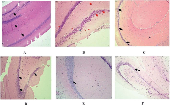 A: Control group, B: Disease control group, C: Phalsa extract group, D: malkangani oil group, E: Microemulsion group, F: Donepezil group. Observed Parameters - Neuronal Damage, Hippocampal Edema, Arrangement of pyramidal cells and polymorphic cells, Pyknotic cel