 Gas chromatography-mass spectrometry profiling and assessment of antiulcer and antioxidant activities of methanol extract from Dioscorea praehensilis rhizomes
Abstract
Peptic ulcer is a common disease in gastroenterology clinics. This study aimed to investigate the chemical composition, antiulcer, and antioxidant activities of methanol extract of Dioscorea praehensilis rhizomes. The rhizomes were extracted by macerating in 80% methanol and subjected to chemical profiling using Gas Chromatography-Mass Spectrometry (GC-MS). The assessment of anti-ulcer activity was done using ethanol and indomethacin-induced gastric ulcer models in rats while the antioxidant studies were conducted using quantitative analysis of phenolic (total phenols and total flavonoid) compounds, 1,1-diphenyl-2-picrylhydrazyl (DPPH), total antioxidant capacity (TAC) and ferric ion reducing antioxidant power (FRAP) assays. D. praehensilis extracts at 125, 250, and 500 mg/kg ‘per os’ (PO) exhibited a significant (p<0.01) dose-dependent decrease in gastric lesions with the percentage inhibition of 65.16, 68.20, and 79.64% respectively in the ethanol model and 81.89, 90.44, and 95.35% in the indomethacin model compared with the control group. D. praehensilis extract contained high phenolic and flavonoid content. The extract also showed considerable antioxidant activity in the DPPH, TAC and FRAP assays. Furthermore, D. praehensilis contains bioactive constituents such as Pentadecanoic acid, 9-Eicosene, n-Hexadecanoic acid, 4-tert-butylphenol and Pentacosane. In conclusion, D. praehensilis possesses antiulcer and antioxidant activity, supporting its traditional use in treating ulcers and dyspepsia.
Keywords:
Antiulcer; Gastric ulcer; Dioscorea praehensilis; Antioxidant; GC-MS
INTRODUCTION
Peptic ulcer; either gastric or duodenum ulcer, is caused by an imbalance between the damaging factors, including hydrochloric acid and pepsin, and protective factors such as bicarbonate buffer, mucus secretion, antioxidants (vitamins and other compounds), and synthesis of prostaglandins in the stomach (Harold, Grant, Mitchel, 2007; Tytgat, 2011; Abdoulrahman, 2023). It is one of the most common diseases in gastroenterology clinics, with a rising global prevalence (Ren et al., 2022). For instance, in 2019, Peptic Ulcer Diseases (PUD) had a global prevalence of 8.09 million, which shows a 25.82% increase compared to the 6.4 million reported in 1990 (Ren et al., 2022; Xie et al., 2022).
PUD has various causes, including Helicobacter pylori, excessive use of non-steroidal anti-inflammatory drugs (NSAID), reactive oxygen species produced via oxidative stress, smoking, and excess alcohol consumption (Byeon et al., 2018; Abdoulrahman, 2023). NSAID use ranks second only to H. pylori infection as the most common cause of PUD (Lanas et al., 2015). The rising prevalence of PUD is associated with changes in some of the risk factors related to its aetiology, especially the use of non-steroidal antiinflammatory drugs (Xie et al., 2022).
Current pharmacological management of gastric ulcers, such as proton pump inhibitors, prostaglandin analogues, cytoprotective agents, and histamine type 2 antagonists’ aims to offset the imbalance between gastric acid production and the defensive mechanisms. Although effective, these agents are expensive and associated with reoccurrence, drug interactions, and a variety of side effects (Yismaw et al., 2020). This has made herbal medicines popular as they are considered acceptable, economical, effective, relatively safe, and readily available (Onyeka et al., 2020). According to Yismaw et al. (2020), extracts from medicinal plants and other natural products have become widely accepted sources of therapeutic agents for treating peptic ulcers.
D. praehensilis Benth belongs to the family Dioscoreaceae. Traditional healers use its tubers to treat various diseases, including stomach aches, diarrhoea, dyspepsia, and dysentery, and apply them to ulcers (Ngelinkoto et al., 2021), inflammatory conditions, and scorpion bites (Adebisi et al., 2018a). However, no literature has documented the scientific basis for these ethnomedicinal uses. This study aims to investigate the antiulcer and antioxidant abilities of D. praehensilis and ascertain the bioactive compounds.
MATERIAL AND METHODS
Material
Drugs, chemicals, solvents and equipment
The 1,1-diphenyl-2-picrylhydrazyl (DPPH), ethanol, diethyl ether, formalin, and ascorbic acid (AA), were obtained from Sigma-Aldrich Chemical Co (Darmstadt Germany), methanol (Guangdong Guanghua Sci-Tech Co., Ltd. Guangdong, China), Omeprazole (Surmount Laboratories Private Limited, Gujarat India), normal saline (Unique Pharmaceutical Limited, Lagos Nigeria) and Indomethacin (Krishat Pharm. Industries Ltd. Ibadan Nigeria), were obtained from a local pharmacy premise, All other reagents, chemicals, and solvents used for this study were of analytical grade. The equipment includes a UV-Vis Spectrophotometer (Thermo Fisher), Agilent Intuvo 9000 GC, a Hot water bath (Genlab Ltd), a Microscope (Zeiss) and a weighing balance (Mettler Toledo).
Collection, identification, and extraction of plant material
D. praehensilis rhizomes were collected from Wammako, Sokoto Nigeria in August 2022. It was identified and authenticated in the herbarium unit of the Department of Pharmacognosy and Ethnopharmacy Usmanu Danfodiyo University where a voucher specimen (PCG/ UDUS/ Dios/ 0002) was deposited. The rhizomes were allowed to dry under shade and powdered using a pestle and mortar. Seven hundred and fifty grams (750 g) of the dried plant material was extracted by maceration with 2.3 litres of 80% methanol. It was allowed to stand for 3 days, after which the mixture was filtered with Whatman filter paper. The resulting filtrate was evaporated to dryness in a water bath at 45°C.
In vivo antiulcer studies
Experimental animals
Fifty Sprague Dawley rats of both sexes aged 8-10 weeks old, weighing 160-180 g were obtained and housed at the animal house facility located at the Department of Pharmacology and Toxicology, Usmanu Danfodiyo University, Sokoto. The animals were maintained under uniform husbandry conditions having natural photoperiod, humidity, temperature (26 ± 2°C), and free access to water and standard commercial chow (Chikun®), manufactured by Crown Flour Mill Ltd Lagos Nigeria, containing crude protein (15.00%), crude fat (4.00%), crude fiber (6.00%), calcium (1.00%), phosphorous (0.45%), Lysine (0.70%), methionine (0.35%), metabolizable energy (2600 kcal/kg) and coccidiostats (60 mg/kg diet). The institution’s guidelines for the care and use of laboratory animals in research with approval number NHREC/UDU-HREC/25/06/2023 were followed in all experiments.
Ethanol-induced gastric ulcer model
Twenty-five Sprague Dawley rats were divided into five groups of five rats each and fasted for 24 hours with free access to water until 2 hours before the commencement of the experiment. Group 1 received the vehicle, distilled water 5 mL/kg (negative control). Group 2 received omeprazole 20 mg/kg (positive control) while groups 3-5 received 125, 250, and 500 mg/kg of methanol extract of D. praehensilis respectively. All treatments were given by oral gavage. One hour after treatment, absolute ethanol (8 mL/kg) was used to induce gastric lesions according to the method of Ugwah et al. (2019). After 2 hours, rats were sacrificed humanely using diethyl ether, and their stomach was removed and opened along the greater curvature. The doses used for this study were selected based on the LD50 value of >2000 mg/kg previously reported by our team (Adebisi et al., 2018b).
Indomethacin-induced gastric ulcer model
Twenty-five rats were similarly pretreated as described above. An hour after treatments, gastric lesions were induced with 100 mg/kg indomethacin. Four hours later, rats were sacrificed and their stomach was excised as described in the ethanol-induced model.
Gross assessments of stomachs
The stomach tissues were rinsed in normal saline. The gastric ulcers were photographically documented. The sum of all lesions in each stomach was designated as the ulcer index and the percentage inhibition was calculated using the expression: % Inhibition = (Ulcer index of control / Ulcer index of the testUlcer index of control X 100
Histopathological analysis
The stomachs of the animals were immediately fixed in a 10% buffered formalin solution and left for at least 24 hours. Sections of the stomach membrane and stomach disc were made from each stomach tissue. All the fixed tissues were dehydrated in alcohol, cleared in xylene, and embedded in paraffin wax melting at 60°C. Serial sections (5 μm thick) were then mounted on 3-aminopropyl trisilane-coated slides and dried at 37°C for 24 hours. Slide sections were deparaffinized, hydrated, and stained with Mayer’s hematoxylin and eosin dyes and examined microscopically at 200 magnification.
In vitro antioxidant study
Quantitative analysis of phenolic compounds
The total flavonoid (TFC) and total phenolic (TPC) contents of the methanol extract of D. praehensilis were measured spectrophotometrically. TFC was determined as described by Miliauskas, Venskutonis and Van Beek (2004). Two millilitres of D. praehensilis extract (1.0 mg/mL) in methanol were added to a 2% AlCl3 in ethanol. After one hour of incubation at room temperature, the absorbance was measured at 420 nm. The positive control consisted of similar concentrations of quercetin. The TFC was calculated as mg quercetin equivalent per gram of extract.
The method of Folin and Ciocalteau (1927) was used to determine the TPC. Briefly, gallic acid or extract concentrations ranging from 200 to 1000 µg/mL were prepared in methanol. Afterwards, 0.5 mL of these was mixed with 4.5 mL of distilled water followed by 0.5 mL of a ten-fold diluted Folin-Ciocalteau reagent. Next, five millilitres of 7% sodium carbonate followed by an additional 2 mL of distilled water were then added to the tubes. After ninety minutes of incubation at room temperature, the absorbance was measured at 760 nm. All experiments were performed in triplicates with gallic acid as the positive control. TPC was measured as gallic acid equivalent.
Determination of antioxidant activity by DPPH assay
The ability of D. praehensilis to scavenge free radicals was investigated using a DPPH assay as described by Hussen and Endalew (2023). Four milligrams of DPPH crystalline solid were dissolved in 100 mL of methanol to make a 0.1 mM solution. Similarly, a 500 μg/mL stock solution of the extract in methanol was prepared by dissolving 5 mg in 10 mL. Subsequently, the necessary concentrations (100, 200, 300, 400, 500, and 600 μg/mL) were prepared by serial dilution with methanol. Two millilitres of the plant extract from each concentration were then taken into a test tube, and 3 mL of DPPH solution was added. After thirty minutes of dark incubation, the absorbance was recorded at 517 nm using a spectrophotometer. Methanol was used as the blank, while the control consisted of a mixture of three millilitres of 0.1 mM DPPH and 100 μL methanol. The same concentrations of ascorbic acid was used as the standard. All measurements were performed in triplicate and the 50% inhibitory concentration (IC50) was estimated from the plot of percentage inhibition against concentration. This percentage inhibition was calculated using the expression:
DPPH % Inhibition = (ABS of control – ABS of sample) / ABS of control X 100
ABS of control is the absorbance of methanol and DPPH solution
ABS of sample is the absorbance of the DP and DPPH solution.
Determination of total antioxidant capacity of the extract
Total antioxidant capacity (TAC) of D. praehensilis was investigated using the phosphor molybdenum assay as described by Prieto, Pineda and Aguilar (1999). This assay is based on the reduction of Molybdenum (VI) cation to Molybdenum (V) with a consequent formation of green phosphate Molybdenum (V) complex at acidic pH. In summary, 1 mL each of 0.6 Mtetraoxosulphate (IV) acid, 28 mM sodium phosphate, and 4 mM ammonium molybdate was added into a twenty millilitre of distilled water and the volume made up to fifty millilitres with additional distilled water. Afterwards, 0.3 mL of D. praehensilis extract in different concentrations ranging from 100 to 600 μg/mL were added to different test tubes individually containing three millilitres of the reagent solution and incubated at 95°C for ninety minutes, cooled to room temperature and the absorbance was measured at 695 nm using a spectrophotometer. Methanol was used as the blank, while a mixture of the molybdate solution (0.3 mL) was used as a control. Ascorbic acid was used as the standard. All experiments were done in triplicate, and the antioxidant ability of D. praehensilis was estimated using the expression
Antioxidant effect % = (ABS of control - ABS of sample) / ABS of control X 100
ABS of control is the absorbance of the control.
ABS of sample is the absorbance of the sample.
Ferric reducing antioxidant power assay
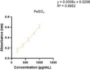
FRAP assay as described by Benzie and Strain (1996) was adopted. The assay assesses the extract’s capacity to convert ferric tripyridyltriazine complex into ferrous tripyridyltriazine at low pH levels. Briefly, One milliliter of the extract at a concentration of 1.0 mg/mL was mixed with 1.5 mL of freshly prepared FRAP solution (Twenty-five milliliter of 300 mM acetate buffer pH 3.6, 2.5 mL of 10 mM 2,4,6-tripyridyls- triazine in 40 mM HCl, and 2.5 mL of 20 mM ferric chloride solution). The reaction mixtures were then incubated at 37oC for thirty minutes and the absorbance was measured at 593 nm using a spectrophotometer. Ferrous sulfate was used for the calibration curve, and the FRAP values (expressed as milligrams of ferrous ion (Fe2+) per gram of the extract) were then extrapolated from the curve.
GC-MS analysis of the extract
Agilent Intuvo 9000 GC system coupled with detector system 5977B MSD with split/splitless injector and equipped with a DB-5 MS fused silica capillary column (5% phenyldimethylsiloxane, 30.0 m × 320 µm, film thickness 0.25 µm) was used. The carrier gas was helium gas, at flow rates of 1.2 mL per minute. The inlet temperature was set at 300°C, MS Source at 230°C, and MS Quad at 150°C while the oven temperature was set as follows: 50°C for 5 minutes, increased to 200°C at 20°C per minute and held for 7 minutes, and thereafter increased to 300°C at 20°C per minute and held for 6 minutes. Using Agilent Automated Liquid Sampler (ALS) G4513A, 1 µL of the extract and standard was injected into the GC system in splitless mode. The relative percent amount was determined by comparing each constituent’s average peak area to the total areas. GCMSD/Enhanced MassHunter Software was used for data acquisition and processed with GCMSD data analysis software incorporated with the 2017 Version of the National Institute of Standards and Technology (NIST) Library. The obtained spectra of the unknown constituents of the D. praehensilis extract were compared with the standard mass spectra of known constituents stored in the NIST library.
Statistical analysis
Data obtained in the antiulcer studies were expressed as mean ± standard error of the mean (SEM). Statistical analysis was performed by One-way analysis of variance followed by the Dunnet test using GraphPad Prism version 8.4.3 (Graph Pad Software Inc., CA, USA). The level of significance was set at p < 0.05. For the antioxidant studies, the IC50 was calculated from the dose-response curves (n=3).
RESULTS
Percentage yield of the plant material
The total weight of the crude methanol extract obtained from 750 g of the dried rhizomes of D. praehensilis was 44 g, hence, the percentage yield of the rhizomes of D. praehensilis after extraction with 80% methanol was 5.87%.
In vivo Antiulcer studies
Ethanol-induced gastric ulcer model
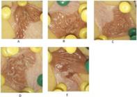
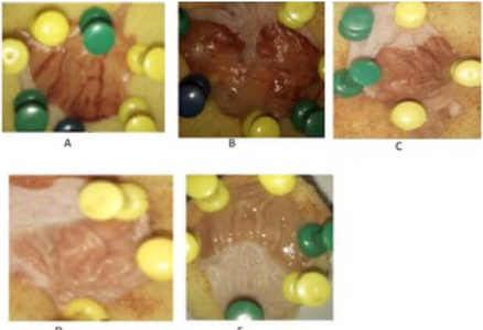
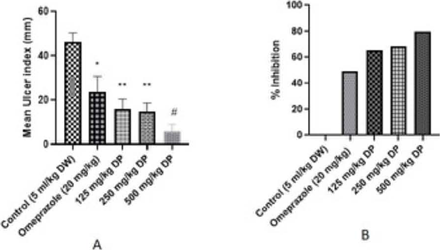
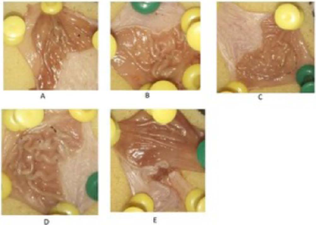
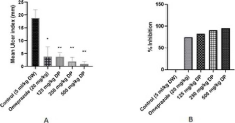
In this study, the administration of absolute ethanol in rats produced damage to the stomach and severe gastric lesions on gross examination as in Figure 1A. However, rats treated with Omeprazole and D. praehensilis extract suffered fewer gastric lesions compared to the untreated animals (Figures 1B, C, D, E). D. praehensilis extract at doses of 125, 250, and 500 mg/kg and omeprazole 20 mg/kg exhibited a significant dose-dependent reduction in the mean ulcer index (Figure 2A) and a percentage inhibition of 65.16, 68.20, 79.64 and 49.01% respectively (Figure 2B).
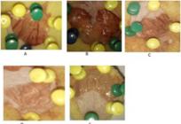
Effect of D. praehensilis on the macroscopic appearance of ethanol-induced stomach damage. A. Ulcer control group displayed extensive haemorrhagic lacerations of the stomach epithelium. B. Omeprazole group demonstrated slight injuries to stomach mucosa. C, D, & E. Experimental groups (125, 250, and 500 mg/kg respectively) showed a reduction in gastric ulcers.
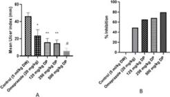
Effect of D. praehensilis on ethanol-induced ulcer showing the mean ulcer index (A) and percentage ulcer inhibition (B). DW=Distilled water, DP= D. praehensilis. Data presented as mean ± SEM, N=5, *p < 0.05, **p < 0.01, #p < 0.0001.
Indomethacin-induced gastric ulcer model
In this study, we observed that indomethacin also induced gastric ulcers, albeit not as severe as those observed in the ethanol model (Figure 3A). Rats treated with Omeprazole and D. praehensilis extract suffered fewer gastric lesions compared to the untreated rats (Figures 3B, C, D, E). D. praehensilis at 125, 250, and 500 mg/kg and omeprazole at 20 mg/kg produced a reduction in mean ulcer index (Figure 4A). The percentage inhibition of ulceration obtained were 81.89, 90.44, 95.35 and 74.78% respectively compared to the vehicle-treated (control) group (Figure 4B).
Effect of D. praehensilis on the macroscopic appearance of indomethacin-induced stomach damage. A. Ulcer control group displayed haemorrhagic lacerations of the stomach epithelium. B. Omeprazole group showed only slight injuries to stomach mucosa. C, D, & E. Experimental groups (125, 250, and 500 mg/kg respectively) showing a decrease in gastric ulceration.
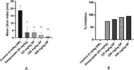
Effect of D. praehensilis on indomethacin-induced ulcer showing mean ulcer index (A) and percentage ulcer inhibition (B). DW= Distilled water, DP =D. praehensilis. Data presented as mean ± SEM, N=5 for all groups.*p < 0.05, **p < 0.01.
Histological analysis
Histological examination of the stomach samples of the rats showed the presence of ulceration in the control groups for both ethanol and indomethacin-induced ulcers. Ethanol and indomethacin-induced gastric damage is seen as a disruption of the surface epithelium and mucosal erosion strands (Figures 5A, 6A). Pretreatment with D. praehensilis methanol extract prevented severe ethanol and indomethacin-induced histological damage with the 250 mg/kg groups showing a healing gastric mucosa with epithelial regeneration and refilling of the ulcer crater (Figures 5C, 6C). The same was observed in the group that received omeprazole 20 mg/kg in both models (5B and 6B). The group that received 500 mg/kg D. praehensilis showed a re-epithelized gastric mucosa (healed ulcer) (Figure 5D, 6D).
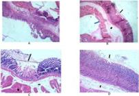
Histological analysis of the stomach of rats treated with methanol extract ofD. praehensilis in the ethanol-induced gastric ulcer model.
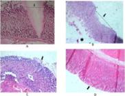
Histological analysis of the stomach of rats treated with methanol extract of D. praehensilis in the indomethacin-induced gastric ulcer model
In vitro antioxidant study
Quantitative analysis of phenolic compounds
Results of the analysis of the phenolic compounds showed that the methanol extract of D. praehensilis contains high flavonoid and total phenolic contents. The TFC was 65.17 ± 11.54 mg of quercetin /g of the extract while that of TPC was 144.28 ±14.94 mg gallic acid equivalent/g of the extract. The standard curves are shown in Figures 7A, 7B.
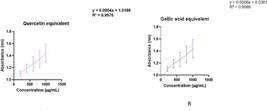
Standard curve of the total flavonoid content of quercetin in methanol extract of D. praehensilis. Values expressed as mean ± SD, N = 3.
DPPH, TAC and FRAP assays
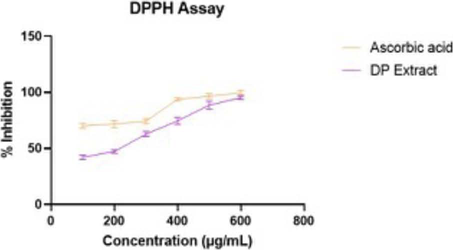
In the DPPH assay D. praehensilis exhibited antioxidant activity at varying concentrations tested (100, 200, 300, 400, 500, and 600 µg/mL) with a percentage inhibition of 42.00 ± 2.37% at 100 µg/mL and 95.23 ± 1.69% at 600 µg/mL and an IC50 of 189.87 µg/mL. The standard ascorbic acid however showed better activity, with a percentage inhibition of 70.05 ± 2.31% and 99.58 ± 2.10% at these same concentrations and IC50 of 76 µg/mL (Figure 8).
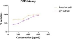
DPPH scavenging property of methanol extract of D. praehensilis represented as percentage inhibition. N=3.
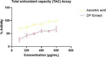
In the phosphomolybdenum assay, the extract showed a good antioxidant capacity though lower than the standard ascorbic acid as observed with the DPPH assay. The percentage inhibition obtained for the D. praehensilis extract was 27.03 ± 7.08% at 100 µg/mL and 66.20 ±9.39% at the highest concentration of 600 µg/mL with an IC50 of 340.05 87 µg/mL while that of ascorbic acid was 69.29 ± 3.41% and 98.63 ± 3.41% with an IC50 of 124.02 μg/mL at these same concentrations (Figure 9).
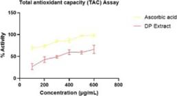
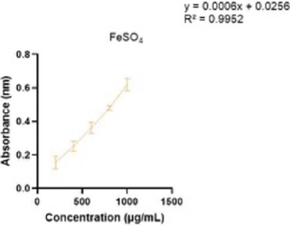
In the FRAP assay, the extract was found to have a remarkable reducing power with a FRAP value of 77.33 ± 13.22 mg ferrous ion per gram of the extract. The standard curve is presented in Figure 10.
GC-MS profiling of the methanol extract of D. praehensilis
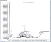
The GC-MS analysis of the methanol extract of D. praehensilis rhizomes yielded a total of twenty-one (21) compounds. Figure 11 depicts the chromatogram while Table I is a list of the chemical constituents with their retention time (RT), percentage quality, peak area (i.e. concentration percent), molecular formula, and molecular weight. The compounds identified include: Benzene, 1,1’-oxybis[3-phenoxy-; 3,4,5-Trimethyldihydrofuran-2-one; Toluene; Hexadecane, 2,6,10,14-tetramethyl-; Cyclohexane, hexyl-; Tridecane; Cyclohexane, 1,1’-(2-methyl-1,3-propanediyl)bis; Cyclooctane, cyclohexyl-; Heptylcyclohexane; Pentacosane; 4-Piperidineacetic acid, 5-ethylidene-2-[3-(2-hydroxyethyl)-1H-indol -2-yl]-.alpha.-methylene-, methyl ester, [2S-(2. alpha.,4.alpha.,5E)]; 9-Eicosene, (E)-; Sulfurous acid butyl dodecyl ester; 1-Octadecene; Pentadecanoic acid, 14-methyl-, methyl ester; n-Hexadecanoic acid; 5-Eicosene, E; 11-Octadecenoic acid, methyl ester; 1,2-Bis(trimethylsilyl)benzene; Tetrasiloxane, decamethyl- and 4-tert-Butylphenol.
DISCUSSION
In this study, ethanol and indomethacin models were employed to investigate the antiulcer properties of D. praehensilis. Ethanol-induced ulcers which mimic acute ulcers in humans and are not affected by gastric acid secretion are used for cytoprotective studies (Mekonnen, Asrade Atnafie, Wahab Atta, 2020). It is used to screen medicinal plants for antiulcer and cytoprotective properties as it causes severe damage to the gastric mucosa by decreasing bicarbonate and mucus secretions, leading to haemorrhage and necrotic lesions. On the other hand, NSAIDs induce gastric ulceration by preventing the synthesis of prostaglandins, leading to an increase in gastric acid production (Dengiz et al., 2007; Musumba, Pritchard, Pirmohamed, 2009).
Our results showed that absolute ethanol caused severe hemorrhagic ulcerative lesions, as seen by the significant increase in ulcer index in the animals in the control group that received the vehicle as compared with those treated with the graded doses of the extract. This was confirmed by the histopathological investigation, showing severe mucosal erosion, acute haemorrhage, and submucosal oedema in the control group compared to the epithelial regeneration with refilling of the ulcer crater that characterized the extract-treated groups. These results demonstrated that the extract effectively prevented ethanol-induced ulcers probably by promoting the growth of gastric mucosa’s epithelial cells leading to increased secretion of bicarbonate and protective mucus (Ugwah et al., 2019). Similarly, animals that received the graded doses of the extract in the indomethacin model also demonstrated a significant reduction in the ulcer index. This may suggest that the extract induces prostaglandin synthesis, inhibiting gastric acid secretion. It is pertinent to mention that the crude extract of D. praehensilis had a higher percentage inhibition of ulceration in the indomethacin model compared to that of ethanol. This is similar to the report of Shams and Eissa (2022) who reported the capacity of ethanol to produce greater levels of gastric ulceration. It is also noteworthy that the percentage inhibition of ulceration obtained with this extract is greater than that obtained with the standard drug in both models.
Oxidative stress, which results from a disruption of the balance between reactive oxygen species and antioxidants is implicated in the pathophysiology of peptic ulcer disease (Mohamed et al., 2024) and studies have shown that the two gastric ulcer models used in this study are related to the generation of free radicals. Ethanol mediates gastric ulcers by inducing oxidative stress with a resultant generation of hydrogen peroxide (H2O2) which is then converted to hydroxyl free radical (OH) which consequently induces lipid peroxidation and Malondialdehyde (MDA) formation that attacks the gastric tissues, eroding the protective layer (Li et al., 2018). Qiu et al. (2020) reported that there is a correlation between an increase in oxidant levels and a decrease in antioxidant levels with the degree of damage to gastric mucosa in an indomethacin model of gastric ulcer. Several recent studies have shown that agents with antioxidant and anti-inflammatory properties may play an important protective role in mitigating the effect of these gastric ulcer-inducing agents (Altuner, Kaya, Suleman, 2020; Shaik, Eid 2022; Mahmoud et al., 2023; Danisman et al., 2023).
In this study, we have reported the presence of a high amount of phenolic and flavonoid contents and antioxidant properties of the methanol extract of D. praehensilis in all the tests performed in a concentration-dependent manner. Plant secondary metabolites which have a range of physiological effects on the human body, are responsible for their medicinal value (Adebisi et al., 2018a) and bioactive compounds and secondary metabolites derived from plants have a significant impact on preventing various human diseases (Wang et al., 2023). For instance, the presence of one or more aromatic rings with varying numbers of hydroxyl groups allows for the good radical scavenging properties of phenolic and flavonoid compounds (Barreto et al., 2020). These phytochemicals have the potential to mitigate free radical accumulation and prevent oxidative gastric mucosal damage; an effect which may be related to their high reactivity as electron donors. Moreover, flavonoids can also boost mucosal prostaglandin, enhance mucus secretion, and prevent the growth of H. pylori (Mekonnen, Asrade Atnafie, Wahab Atta, 2020; Al-Mamary, Moussa, 2021; Beslo et al., 2023). Tannins have been reported to offer protection to the outmost layer of mucosa to resist chemicals and mechanical injury. Saponins on the other hand can mediate increased mucus production. (Mekonnen, Asrade Atnafie, Wahab Atta, 2020; Adane et al., 2021). A previous analysis of the phytochemical constituents of D. praehensilis by our team revealed the presence of flavonoids, tannins, saponins, and steroids (Adebisi et al., 2018a). The antiulcer properties being reported in this study may be due to the presence of these phytochemicals.
Other species of Dioscorea such as Dioscorea bulbifera are used in India to cure ulcers (Dutta, 2015), Dioscorea oppositifolia shows anti-ulcer activity in adult Wistar rats (Singh, 2022). These species have been reported to possess phytochemicals including phenolic acids; flavonoids, phenanthrene derivatives, and flavonoids (Adomeniene, Venskutonis, 2022). In a similar study in DR Congo, Bukatuka et al. (2016) reported the antioxidant properties of D. praehensilis tubers with an IC50 lower than that obtained in our study and the absence of flavonoids as against that reported in this study. This may be due to differences in the geographical locations of the plants as geographical locations have been shown to affect the phytochemical constituents and bioactivity of medicinal plants (Liu et al., 2018; Dewanjee, Saha, Mishra, 2021)
The GC-MS analysis of the methanol extract of D. praehensilis showed the presence of some volatile phytocompounds that have been reported to possess some pharmacological activities. Among the identified compounds, n-Hexadecanoic acid and pentadecanoic acid have been shown to possess antioxidant, Anti-inflammatory, antimicrobial and antifungal properties (Hema, Kumaravel, Alagusundaram, 2011; Akpuaka et al., 2013; Hadadi, Nematzadeh, Ghahari, 2020; Chirumamilla, Dharavath, Taduri, 2022); Octadecenoic acid possesses anti-inflammatory activity (Gomathi et al., 2015); 4-tert-Butylphenol possesses antioxidant properties (Shakira et al. 2022); Pentacosane has shown remyelinating and anti-inflammatory properties (Senol et al., 2023) while 1,2-Bis(trimethylsilyl)benzene possesses antioxidant, antibacterial, anti-inflammatory and antimicrobial effects (Susheela, Rosaline, Radha, 2018; Ali et al., 2021).
The antiulcer activity ofthe extract of D. praehensilis may therefore be attributed to its antioxidant properties and the activities of the phytochemicals present in the rhizome. Further studies are needed to determine the precise mechanism(s) of action of this extract.
CONCLUSION
The data from this study showed that the methanol extract of D. praehensilis significantly decreased gastric lesions in ethanol and indomethacin-induced gastric ulcers, demonstrating its antiulcer properties. The extract also showed considerable antioxidant activity in some in-vitro antioxidant assays and contains bioactive constituents such as n-Hexadecanoic acid, pentadecanoic acid, 4-tert-Butylphenol and 1,2-Bis(trimethylsilyl)benzene that has been previously reported to possess antioxidant, anti-inflammatory and antimicrobial properties. These findings lend support to its use in the folkloric treatment of ulcers and dyspepsia. D. praehensilis may therefore be a source of new phytotherapeutic formulations for treating gastric ulcers.
DATA AVAILABILITY STATEMENT
Data available from the corresponding author upon reasonable request.
REFERENCES
- Abdoulrahman K. Anti-ulcer effect of Ranunculus millefoliatus on absolute alcohol-induced stomach ulceration. Saudi J Biol Sci. 2023;30(8):103711.
- Adane H, Atnafie SA, Kifle ZD, Ambikar D. Evaluation of in vivo antiulcer activity of hydro-methanol extract and solvent fractions of the stem bark of Ficus thonningii (Moraceae) on rodent models. Biomed Res Int. 2021;(1):6685395.
- Adebisi IM, Bello SO, Shehu CE, Musa A, Mshelia HE, Suleiman A. Quantitative phytochemicals and inhibitory effect of methanol extract of Dioscorea prehensilis on platelet aggregation in vitro Niger J Pharm Sci. 2018a;3(1):41-45.
- Adebisi IM, Bello SO, Shehu CE, Abdullahi MI, Ugwah-Oguejiofor CJ, Ndodo N, et al. Dioscorea prehensilis ameliorates the features of preeclampsia in the LN G-nitroarginine methyl ester (L-NAME) preeclamptic model. TJNPR. 2018b;2(9):422-428.
- Adomeniene A, Venskutonis PR. Dioscorea spp.: Comprehensive review of antioxidant properties and their relation to phytochemicals and health benefits. Molecules. 2022;27(8):2530.
- Akpuaka A, Ekwenchi MM, Dashak DA, Dildar A. Biological activities of characterized isolates of n-hexane extract of Azadirachta indica A. Juss (Neem) leaves. Nat Sci. 2013;11(5):141-147.
- Ali S, Sudha KG, Karunakaran G, Kowsalya M, Kolesnikov E, Rajeshkumar MP. Green synthesis of stable antioxidant, anticancer and photocatalytic activity of zinc oxide nanorods from Leea asiatica leaf. J. Biotechnol. 2021;329:65-79.
- Al-Mamary MA, Moussa Z. Antioxidant activity: The presence and impact of hydroxyl groups in small molecules of natural and synthetic origin in: Waisundara, V. (Ed.), Antioxidants: Benefits, sources, mechanisms of action. 2021;13:318-377.
- Altuner D, Kaya T, Suleyman H. The protective effect of lercanidipine on indomethacin-induced gastric ulcers in rats. Braz Arch BiolTechnol. 2020;63:e20190311.
- Barreto SM, Cadavid CO, Moura RA, Silva GM, Araújo SV, Silva Filho JA, et al. In vitro and in vivo antioxidant activity of Agave sisalana agro-industrial residue. Biomolecules. 2020;10(10):1435.
- Benzie IF, Strain JJ. The ferric reducing ability of plasma (FRAP) as a measure of “antioxidant power”: the FRAP assay. Anal Biochem. 1996:239(1):70-76.
- Bešlo D, Golubić N, Rastija V, Agić D, Karnaš M, Šubarić D, et al. Antioxidant activity, metabolism, and bioavailability of polyphenols in the diet of animals. Antioxidants. 2023;12(6): 1141.
- Bukatuka F, Ngombe K, Mutwale K, Moni B, Makengo K, Pambu L, et al. Bioactivity and nutritional values of some Dioscorea species traditionally used as medicinal foods in Bandundu, DR Congo European J Med Plants. 2016;14(1):1-11.
- Byeon S, Oh J, Lim JS, Lee JS, Kim JS. Protective effects of Dioscorea batatas flesh and peel extracts against ethanol-induced gastric ulcer in mice. Nutrients. 2018;10(11):1680.
- Chirumamilla P, Dharavath SB, Taduri S. GC–MS profiling and antibacterial activity of Solanum khasianum leaf and root extracts. Bull Pharm Res Inst. 2022:46(1):127.
- Danisman B, Cicek B, Yildirim S, Bolat I, Kantar D, Golokhvast KS, et al. Carnosic acid ameliorates indomethacin-induced gastric ulceration in rats by alleviating oxidative stress and inflammation. Biomedicines. 2023;11(3):829.
- Dengiz GO, Odabasoglu F, Halici Z, Cadirci E, Suleyman H. Gastroprotective and antioxidant effects of montelukast on indomethacin-induced gastric ulcer in rats. J Pharmacol Sci. 2007;105(1):94-102.
-
Dewanjee D, Saha S Mishra HN. Effect of geographical locations on qualitative and quantitative phytochemical evaluation, evaluation of antioxidant activity of H. difformis leaf and stem collected from different sites in India. Ann Rom Soc Cell Biol. 2021;25(6):18070-18079. doi: 10.5772/intechopen.95616.
» https://doi.org/10.5772/intechopen.95616 - Dutta B. Food and medicinal values of certain species of Dioscorea with special reference to Assam. J Pharmacogn Phytochem. 2015;3(5):15-18.
- Folin O, CiocalteuV. On tyrosine and tryptophane determinations in proteins. J Biol Chem. 1927;73(2):627-650.
- Gomathi D, Kalaiselvi M, Ravikumar G, Devaki K, Uma C. GC-MS analysis of bioactive compounds from the whole plant ethanolic extract of Evolvulus alsinoides L. J Food Technol. 2015;52:1212-1217.
- Hadadi Z, Nematzadeh GA, Ghahari S. A study on the antioxidant and antimicrobial activities in the chloroformic and methanolic extracts of 6 important medicinal plants collected from North of Iran. BMC Chem. 2020;14:1-11.
- Harold K, Grant DM, Mitchel J. Principles of medical pharmacology, Seventh Ed. Elsevier Canada 2007.
- Hema R, Kumaravel S, Alagusundaram K. GC/MS determination of bioactive components of Murraya koenigii. J Am Sci. 2011;7(1):80-83.
- Hussen EM, Endalew SA. In vitro antioxidant and free-radical scavenging activities of polar leaf extracts of Vernonia amygdalina. BMC Complement Med Ther. 2023;23(1):146.
- Lanas Á, Carrera-Lasfuentes P, Arguedas Y, García S, Bujanda L, Calvet X. et al. Risk of upper and lower gastrointestinal bleeding in patients taking nonsteroidal anti-inflammatory drugs, antiplatelet agents, or anticoagulants. Clin Gastroenterol Hepatol. 2015;13(5):906-912.
- Li Q, Hu X, Xuan Y, Ying J, Fei Y, Rong J, et al. Kaempferol protects ethanol-induced gastric ulcers in mice via pro-inflammatory cytokines and NO. Acta Biochim Biophys Sin. 2018;50(3):246-253.
- Liu Y, Chen P, Zhou M, Wang T, Fang S, Shang X, et al. Geographic variation in the chemical composition and antioxidant properties of phenolic compounds from Cyclocarya paliurus (Batal) Iljinskaja leaves. Molecules. 2018;23(10):2440.
- Mahmoud MF, Abdo W, Nabil M, Drissi B, El-Shazly AM, Abdelfattah MA, et al. Apple (Malus domestica Borkh) leaves attenuate indomethacin-induced gastric ulcers in rats. Biomed Pharmacother. 2023;160:114331.
- Mekonnen AN, Asrade Atnafie S, Wahab Atta MA. Evaluation of the antiulcer activity of 80% methanol extract and solvent fractions of the root of Croton macrostachyus Hocsht: Ex Del.(Euphorbiaceae) in rodents. Evid Based Complement Alternat Med. 2020;11:2809270.
- Miliauskas G, Venskutonis PR, Van Beek TA. Screening of radical scavenging activity of some medicinal and aromatic plant extracts. Food Chem. 2004;85(2):231-237.
- Mohamed AO, Abd-Elghaffar SK, Mousa RA, Kamel AA. Aloe vera gel confers therapeutic effect by reducing pyroptosis in ethanol-induced gastric ulcer rat model: Role of NLRP3/GSDMD signaling pathway. Mol Biol Rep. 2024;51(1):401.
- Musumba C, Pritchard DM, Pirmohamed M. Cellular and molecular mechanisms of NSAID-induced peptic ulcers. Aliment Pharmacol Ther. 2009;30(6):517-531.
- Ngelinkoto PM, Lokassa AMK, Bulumuka B, Maliani JK, Ngondo MM Mbuli RL, et al.. Phytochemical study and anti-nutritional factors in stems of Dioscorea praehensilis Benth (Dioscoreaceae). J Botanical Res. 2021;3(4):1-10.
- Onyeka IP, Bako SP, Suleiman MM, Onyegbule FA, Morikwe UC, Ogbue CO. Antiulcer effects of methanol extract of euphorbia hirta and honey combination in rats. Biomed Res Int. 2020:e6827504.
- Prieto P, Pineda M, Aguilar M. Spectrophotometric quantitation of antioxidant capacity through the formation of a phosphomolybdenum complex: specific application to the determination of vitamin E. Anal Biochem. 1999;269(2):337-341.
- Qiu L, Yao L, Fang Y, Wang L, Xue M, Lin Z, et al. Effect of cuttlebone on healing of indomethacin-induced acute gastric mucosal lesions in rats. Evid Based Complement Alternat Med. 2020;(1):9592608.
- Ren J, Jin X, Li J, Li R, Gao Y, Zhang J, et al. The global burden of peptic ulcer disease in 204 countries and territories from 1990 to 2019: a systematic analysis for the global burden of disease study 2019. Int J Epidemiol. 2022;51(5):1666-1676.
- Senol H, Ozgun-Acar O, Dag A, Eken A, Guner H, Aykut ZG, et al. Synthesis and comprehensive in vivo activity profiling of olean-12-en-28-ol, 3β-pentacosanoate in experimental autoimmune encephalomyelitis: a natural remyelinating and anti-inflammatory agent. J Nat Prod. 2023;86(1):103-118.
- Shaik RA, Eid BG. Piceatannol affects gastric ulcers induced by indomethacin: association of antioxidant, anti-inflammatory, and angiogenesis mechanisms in rats. Life. 2022;12(3):356.
- Shakira RM, Abd Wahab MK, Nordin N, Ariffin A. Antioxidant properties of butylated phenol with oxadiazole and hydrazone moiety at ortho position supported by DFT study. RSC Adv. 2022;12(27):17085-17095.
- Shams SGE, Eissa RG. Amelioration of ethanol-induced gastric ulcer in rats by quercetin: implication of Nrf2/HO1 and HMGB1/TLR4/NF-κB pathways. Heliyon. 2022;8(10):e11159.
- Singh P. Bioactive components, food applications and health benefits of yam (Dioscorea spp.): A review. Pharma Innovation. 2022;11(6):092-2102.
- Susheela P, Rosaline M, Radha R. Gas chromatography and mass spectrometry of the ethanolic extract of nest material of mud dauber wasp, sceliphron caementarium. Asian J Pharm Clin Res. 2018;11:234-236.
- Tytgat GNJ. Etiopathogenetic principles and peptic ulcer disease classification. Dig Dis. 2011;29(5):454-458.
- Ugwah MO, Ugwah-Oguejiofor CJ, Etuk EU, Bello SO, Aliero AA. Evaluation of the antiulcer activity of the aqueous stem bark extract of Balanites aegyptiaca L Delile in Wistar rats. J Ethnopharmacol. 2019;239:111931.
- Wang Z, Zhao S, Tao S, Hou G, Zhao F, Tan S, et al. Dioscorea spp.: Bioactive Compounds and Potential for the Treatment of Inflammatory and Metabolic Diseases. Molecules. 2023;28(6):2878.
- Xie X, Ren K, Zhou Z, Dang C, Zhang H. The global, regional and national burden of peptic ulcer disease from 1990 to 2019: a population-based study. BMC Gastroenterol. 2022;22(1):1-13.
- Yismaw YE, Abdelwuhab M, Ambikar DB, Yismaw AE, Derebe D, Melkam W. Phytochemical and antiulcer activity screening of seed extract of Cordia africana lam (Boraginaceae) in pyloric ligated rats. Clin Pharmacol Adv Appl. 2020;12:67.
Edited by
-
Associate Editor:
Severino Matias de Alencar

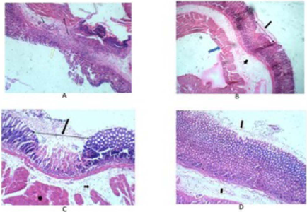 A: Histological slide of the control group in the ethanol-induced model showing: Gastric ulcer (white arrow), Sub mucosa (short black arrow), and muscularis propria (long black arrow) H & E X 200.B: Histological slide of the omeprazole group in the ethanol-induced model showing: Healing ulcer edge (Black long arrow), Sub mucosa (Short black arrow), and muscularis propria (Blue arrow) H & E X200.C: Histological slide of the 250 mg/kg group in the ethanol-induced model showing: Epithelial regeneration (Black arrow) with refilling of the ulcer crater, Sub mucosa (short black arrow), and muscularis propria (Black star) H & E X 400.D: Histological slide of the 500 mg/kg in the ethanol-induced model showing: Re-epithelized gastric mucosal (Long black arrow), Sub mucosa (Short black arrow) H & E X 400.
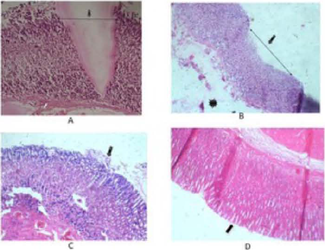 A: Histological slide of the control group in the indomethacin-induced model showing: Ulcer crater (Black arrow), and Muscularis mucosa (White arrow) H & E X 200.B: Histological slide of the omeprazole 20 mg/kg group in the indomethacin-induced model showing healing gastric ulcer (black arrow) Sub mucosa (Black star) H & E X 200.C: Histological slide of the 250 mg/kg group in the indomethacin-induced model showing: Healing gastric mucosa ulcer (Black arrow) H & E X 400.D: Histological slide of the 500 mg/kg group in the indomethacin-induced model showing: Healed gastric mucosa ulcer (Black arrow) H & E X 400.
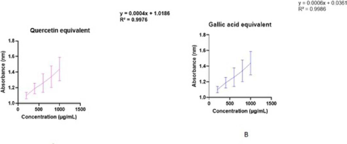 FIGURE 7B - Standard Curve of the total phenolic content of gallic acid in methanol extract of D. praehensilis. Values expressed as mean ± SD, N = 3.