 DNA lipoplexes for gene therapy: a review
Abstract
Gene therapy holds substantial promise for the treatment of a wide range of genetic and acquired diseases. Despite its potential, efficient and safe delivery of therapeutic genes into target cells remains a significant challenge. Lipoplexes, lipid-base formulations complexed with nucleic acids, have emerged as an attractive non-viral vector for gene delivery due to their ability to encapsulate and protect nucleic acids and facilitate their delivery into cells. This review focuses on the design, mechanism of action, and structure of lipoplexes for gene therapy and discusses the role of various lipids used in the formation of lipoplexes. Furthermore, we explore the application of lipoplexes in various disease models, cystic fibrosis, and cancers, highlighting recent advances. We conclude that while challenges remain, lipoplexes represent a promising strategy for safe and effective gene therapy.
Keywords:
Lipoplexes; Gene therapy; Non-viral vectors; Cationic liposomes; Cationic lipids
INTRODUCTION
Gene therapy has evolved to be an innovative approach to treat, circumvent or potentially cure certain inherited genetic diseases, cancer, and even infections. (National Heart, Lung, Blood Institute, 2022). Gene therapy involves delivery of suitable nucleic acids such as plasmid DNA, small interfering RNA (siRNA), mRNA, CRISPR systems to cells either by in vivo or ex vivo techniques. (Pan et al., 2021). Essentially, in vivo technique entails direct transfer of genes to target cells whereas the ex vivo approach involves the removal of target cells, their genetic modification, and subsequent reinfusion back into the host. (Mout et al., 2017; Alhakamy, Curiel, Berkland, 2021; Sprugel et al, 1987).
Gene therapy was first tested in a human with adenosine deaminase-deficient severe combined immunodeficiency (SCID) in 1990 (Anderson, 1992). However, due to the death of a patient receiving gene therapy due to a strong immune response, various safety concerns were raised regarding gene therapy and its potential clinical translation (Shinkuma, 2021). As a result, concentrated efforts have been taken to improve the safety of gene therapies and has led to a substantial increase in approved products (Ma et al., 2020). Additionally, a major challenge is ensuring that genes are specifically targeted to tumor cells while avoiding normal cells. To address this, gene delivery systems known as “vectors” have been utilized in gene therapy (Gonçalves, Paiva, 2017; Witlox et al., 2007).
Vectors are one of the widely used gene-transferring technologies. Genetic material carries a negative charge and is susceptible to degradation by extracellular nucleases, which makes it crucial to employ vectors during gene delivery. These vectors are responsible for delivering genetic material to the target cell, by the process known as “transfection.” They are broadly classified into non-viral and viral vectors. (Al-Dosari, Gao, 2009; Gao, Hui, 2001; Patil et al, 2019). Viral vectors are essentially modified viruses that contain genes of interest that are required to be delivered to the target cells or tissues. These viral vectors possess high transfection efficiency but are also responsible for inducing immunogenic responses in the host. (Warnock, Daigre, Al-Rubeai, 2011). Attempts to minimize these immune responses led to the development of non-viral vectors. Non-viral vectors are synthesized with chemical or natural compounds and are less immunogenic as compared to their viral counterpart thus making them an attractive alternative to viral vectors. In the past few years, various novel drug delivery systems such as liposomes, solid lipid nanoparticles, and polymeric nanoparticles amongst many others, have emerged as acceptable non-viral vectors. (Luiz et al, 2022).
Among these non-viral vectors, lipoplexes i.e. lipid-based nucleic acid complexes have garnered attention for their potential to enhance gene delivery, offering advantages such as improved stability and reduced immunogenicity. Table I provides a list of lipoplex formulations under the clinical trials. The objective of this review is to highlight the use of DNA lipoplexes as a strategy for enhanced gene therapy, and its applications. (Y.P. Patil, Jadhav, 2014; Luiz et al., 2022; Al-Dosari, Gao, 2009).
LIPOPLEXES FOR GENE DELIVERY
In gene therapy, liposomes are used as non-viral gene transfer vectors due to their dynamic properties that can be modulated according to their applications. (Balazs, Godbey, 2011) These properties include size (small and large), charge (anionic, cationic, or neutral), bilayer structure (unilamellar and multilamellar), and type of lipids used. Additionally, they possess relatively lower toxicity and immunogenicity profile. (Akbarzadeh et al, 2013). Liposomes that are prepared by using cationic lipids are better candidates to form successful complexes with negatively charged genetic material. These stable structures formed as a result of electrostatic interaction between cationic lipids and genetic material are termed as lipoplexes. (Bansal et al., 2020). The utilization of cationic lipids-DNA complex for gene transfer was introduced by Felgner et al. (1987).
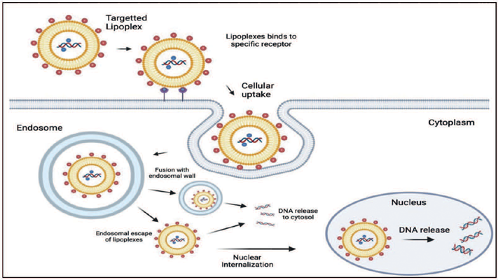
For successful gene delivery, lipoplexes should overcome certain intracellular barriers. These include cellular binding of cationic lipoplexes to negatively charged cell membranes typically via ionic interactions. (Zhou, Huang, 1994).
Following this, the lipoplexes are internalized by the cell via endocytosis. Once inside the cell, the lipoplexes are sequestered within endosomes, where they escape to avoid degradation in lysosomes. The ability of a lipoplex to disrupt the endosomal membrane and release its DNA cargo into the cytoplasm is critical for successful gene delivery. After escaping the endosome, the DNA or lipoplex must navigate the cytoplasmic environment, which is filled with barriers such as the cytoskeleton, that can impede its movement toward the nucleus (Figure 1). Furthermore, once the DNA reaches the nucleus, it must be released from the lipoplex to be accessible for transcription.
STRUCTURE OF LIPOPLEXES
Two different types of models are described in order to elucidate the structure of lipoplexes, an “internal” model, and an “external” model. In the internal model, the DNA is coated by a lipid envelope whereas, in the external model, the DNA is adsorbed onto the surface of the cationic liposome. (Eastman et al., 1997).
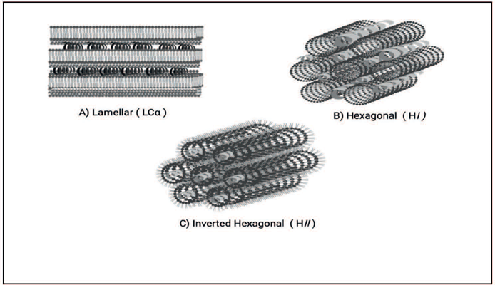
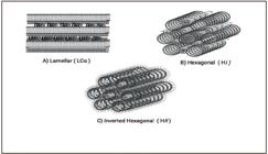
The DNA is bound electrostatically to the surface of the cationic lipid vesicles, i.e., it is adsorbed onto the vesicle, representing beads on a string structure. Such a structure was also explained by Felgner et al. (1987) in their work. Lipoplexes are generally extremely ordered tubular structures when they are endocytosed by cells, and they assume perinuclear localization in these endosomes. Cationic lipoplexes chiefly have multi-lamellar structures (LCα) (Figure 2) with DNA monolayers sandwiched between cationic membranes, and hexagonal structures (non-lamellar) structures such as HII and HI. (Hirlekar et al., 2010; Koltover et al., 1998). The HII phase (i.e. the inverted hexagonal phase) is assumed when the polar headgroup of lipids is aligned in the direction of the internal space of the cylinder, which is aqueous in nature. On the other hand, when the internal space is packed with hydrocarbon chains, the phase is known as micellar hexagonal HI (Seddon, 1990). Koltover et al. (1998), in their study, identified that the commonly used helper lipid (DOPE) is considered to facilitate the conversion from inverted hexagonal structure LCα to HII and further enhance the transfection efficiency of lipoplexes.
LIPIDS EMPLOYED IN LIPOPLEX FORMATION
Mainly two types of lipids are employed for the formation of lipoplexes and other lipid-based gene delivery platforms- a cationic lipid and a neutral/helper lipid. (Cheng, Lee, 2016).
Cationic lipids
Cationic lipids are essentially amphiphilic molecules containing a hydrophobic as well as a hydrophilic domain, which are connected via a linker. (Rietwyk, Peer, 2017). DOTMA (1,2-dioleoyloxy-3-trimethylammonium-propane) and DOTAP (1,2-dioleoyl-3-trimethylammonium-propane) are the two most explored cationic lipids for gene delivery. Felgner et al. (1987), in their seminal work, demonstrated that DOTMA can directly interact with plasmid DNA and form lipid/DNA complexes with 100% entrapment. However, DOTMA is a cationic lipid that is known to be cytotoxic in nature. (Farhood et al, 1992; Kim et al, 2016; Zelphati, Szoka, 1996). To overcome this challenge, researchers attempted to introduce a biodegradable ester bond in the cationic lipid structure. This led to the formation of DOTAP which turned out to be relatively less toxic than its ether homologue- DOTMA. (Leventis, Silvius, 1990).
Later, Song et al. (2008) assessed the transfection efficiency of DOTMA and DOTAP in vivo and observed that DOTMA showed 10-fold higher transfection efficiency than DOTAP in contrast to the in vitro results which concluded similar efficiency for both lipids. To further evaluate the basis of high transfection efficiency in, in vivo conditions, Ren et al. (2000) developed 11 structural analogues of DOTMA and DOTAP. It was concluded that DOTMA’s higher transfection efficiency is attributed to its structural characteristics wherein the hydrocarbon chains are adjacent to the cationic head group, the presence of two ether bonds, and paired oleyl chains as the hydrophobic anchor. Like DOTAP, other cationic lipids like cetyltrimethylammonium bromide (CTAB), didodecyldimethylammonium bromide (DDAB), and dioctadecylamidoglycylspermine (DOGS) were formed by attaching quaternary ammonium headgroups. (Niculescu-Duvaz, Heyes, Springer, 2005; Zhou et al., 2012). However, evidence suggests that tertiary amine-based lipids like DC-Chol (3β-[3beta-[N-(N’,N’-Dimethylaminoethane)-carbamoyl]cholesterol) and DODMA (1,2-dioleyloxy-3-dimethylaminopropane) have better efficiency in transfection process than other amines as a result of their lipid buffering capacity which is helpful in endosomal escape. (Mo et al., 2013; Habrant et al., 2016). In this light, novel lipoplexes based on tertiary amine-based lipid and DOPE (dioleoylphosphatidylethanolamine)-a helper lipid in 1:1 ratio, containing siRNA were formulated. These liposomes proved to be more effective in silencing genes in various cell lines as well as normal mice as against the commercially available gene delivery vector Lipofectamine™. (Lin et al., 2019).
Another important aspect of cationic lipids is the nature of linkers that join the hydrophobic and hydrophilic domains of the lipid as they are crucial for lipid stability, biodegradability, transfection efficiency, and cytotoxicity. (Luiz et al, 2022).
Neutral/helper lipids
To further improve the transfection efficiency of cationic lipids, helper lipids (also known as co-lipids or auxiliary lipids) are added to the formulation. These lipids are zwitterionic such as DOPE (Dioleoylphosphatidylethanolamine) and DOPC (1,2-dioleoylsn-glycerol-3-phosphatidylcholine), to name a few(Ponti et al., 2021). As the mechanism of action of the release of genetic material from endosomes depends on pH change, DOPE is commonly employed as it exhibits structural transitions with changes in pH and thus also acts as a fusogenic lipid. (Zuidam, Barenholz, 1998; Lonez et al, 2010).Similarly, CHOL (cholesterol) plays an important role in the composition of lipoplexes, by facilitating their interaction with biological membranes and imparting stability to their structure and is thus used as a helper lipid along with cationic lipids (Briuglia et al, 2015). Cholesterol-containing cationic liposomes were found to be structurally more stable in physiologic media, thereby helping the lipoplexes to succeed in reaching their target tissue intact, they also help to protect the DNA from degradation. (Tros de Ilarduya, Sun, Düzgünes., 2010).
Given the crucial role of lipid composition in determining the stability and efficiency of lipoplexes, it becomes essential to consider the structure-mechanism relationship in lipoplexes to achieve successful gene delivery.
STRUCTURE-MECHANISM RELATIONSHIP
The interactions of lipoplexes with the cell membrane, their absorption, intracellular trafficking, and ultimately the effectiveness of gene delivery are significantly influenced by their structure and composition. (Elouahabi, Ruysschaert, 2005). For instance, the charge ratio between cationic lipids and nucleic acids affects lipoplex stability and cellular uptake. A higher positive charge density enhances interaction with negatively charged cellular membranes, increasing uptake but potentially leading to cytotoxicity. (Elouahabi, Ruysschaert, 2005).
Lipid packing parameters and phase behavior further impact the ability of lipoplexes to fuse with endosomal membranes; lipids that form an inverted hexagonal phase (HII) may enhance membrane fusion and facilitate nucleic acid release. (Wasungu, Hoekstra, 2006).
High-resolution synchrotron small-angle X-ray scattering (SAXS) studies have been instrumental in revealing these structural transitions. Rädler et al. (1997) observed that changes in the cationic lipid (CL)/DNA molar ratio could induce a topological shift from a multilayer “onion-like” lamellar structure to a more fusogenic inverted hexagonal phase. These studies further highlighted that lipofection is governed by a complex interplay of multiple parameters, rather than a single factor. A key finding was the importance of the entropic gain resulting from lipid mixing, which drives the formation of lipoplexes (Pozzi, Caracciolo, 2023). This mixing is crucial as it facilitates the transition from lamellar phases to non-lamellar structures, such as the inverted hexagonal phase, which has been shown to universally enhance transfection efficiency by promoting better interaction with cellular membranes (Xu, Szoka, 1996).
The ability to induce such phase transitions can be modulated by specific triggers like pH or temperature changes, allowing for controlled and efficient intracellular cargo release. (Pozzi, Caracciolo, 2023).
METHOD OF PREPARATION
As lipids are water insoluble in nature, lipoplexes must be pre-formed as liposomes, and the aqueous suspension of liposomes is combined with the aqueous solution of DNA to generate lipoplexes. (Felgner et al., 1987; Li, Szoka, 2007). This technique is called the lipid film hydration method is reported for the preparation of liposomes and as well as lipoplexes. (Rasoulianboroujeni et al., 2017; Bangham, Standish, Watkins, 1965). The ethanol injection method has also been reported for the formation of liposomes and subsequent transfection. (Batzri, Korn, 1973; Campbell, 1995). However, these methods require an initial formation of liposomes or liposomal intermediates and the consequent addition of DNA solution. To eliminate this step, Jeffs et al. (2005) and Hayes et al. (2006) proposed methods that led to the spontaneous formation of lipoplexes without the use of preformed liposomes however it required specialized equipment like a T-shaped mixing chamber to form lipoplexes. Thus, Meisel and Gokel (2016) suggested a fast and simple method wherein lipids dissolved in organic solvents, which may be added directly to aqueous DNA to form a lipoplex.
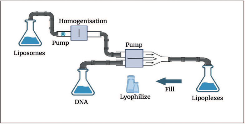
For large-scale manufacturing of lipoplexes with longer shelf-life and reproducible characteristics a simple continuous mixing method, followed by lyophilization is represented in Figure 3. (Clement et al., 2005).
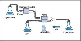
First, the cationic lipid and plasmid DNA are diluted separately in the transfection medium. After dilution, the extrusion process follows with the lipid being extruded through a membrane with a defined pore size of 800 nm and then constantly mixed with equal volumes of plasmid DNA in a second pump system via a Y-connector. The manufactured lipoplexes are bottled and then lyophilized. The lyophilized lipoplexes can be stored for 18 months at 4-8 °C and the transfection efficiency ofthe stored lyophilized powder is comparable to the initial value obtained after preparation. (Clement et al., 2005).
CHARACTERIZATION OF LIPOPLEXES
Much effort has been taken to completely characterize lipoplexes. This is often the only way to improve, understand, and control their transfection potency. The size and zeta potential, DNA accessibility to DNase I after complexation with lipids, and encapsulation potency of the lipid vector are some of the parameters thought about in the characterization of lipoplexes. (Madeira et al., 2011). The foremost and widely used techniques for the characterization of lipoplexes are fluorescence-based techniques, and their main benefits are associated with the possibility of using lipid and DNA concentrations like those used to transfect cells in vitro, or in vivo. This technique makes it possible to monitor the behavior of these complexes under physiological conditions. (Madeira et al., 2011). The utilization of various techniques in lipoplex characterization is vital to know their formation, the ability of DNA condensation, microscopic and macroscopic structure, and in specific cases, to predict their behavior in vivo. Table II depicts methods utilized for the physicochemical characterization of lipoplexes in solution, with similar DNA concentrations to those utilized in transfection protocols.
TOXICITIES
The toxicity related to lipoplexes is dependent on the dose and route of administration. Lipoplexes are observed to be toxic and may cause acute inflammation and even tissue damage at relatively higher doses. (Yew, Scheule, 2005). Intravenous and intrapulmonary are the most explored routes of administration wherein, toxicities related to the pulmonary route ofadministration began emerging during clinical trials conducted for managing cystic fibrosis, wherein patients suffered from flu-like symptoms which resolved within 36h. (Alton et al., 1999). Systemic administration of lipoplexes in mice led to hepatotoxicity as a result of high levels of transaminases in addition to previously documented high production of proinflammatory cytokines (Loisel et al., 2001; Tan et al., 2004). Various methods are developed for overcoming the inflammatory toxicity of lipoplexes. Some of these methods include elimination and inhibition of unmethylated CpG dinucleotides in the plasmid DNA, (Yew et al., 2000) targeting gene delivery to lung endothelium for effective therapeutic response in case of gene delivery to lungs, (Li et al., 2000) and use of immunosuppressive agents (Tan et al., 2004).
APPLICATIONS
Currently, lipoplexes are being investigated in a wide range of diseases. Some of the applications of lipoplexes will be discussed in depth hereafter.
Cystic fibrosis (CF)
CF is a life-threatening monogenic disorder that occurs due to mutations in cystic fibrosis transmembrane conductance regulator (CFTR) and subsequent chloride transport defect leading to accumulation of mucus at the surface of the epithelium. (Clunes, Boucher, 2007). CF primarily affects the lungs and patients with this condition die as a result of lung disease, but it also affects other organs such as the pancreas and intestine. In 1995, the first clinical trial (NCT00004471) (ClinicalTrials.gov, 2011) was initiated in 9 CF patients to evaluate the deliverability of cationic liposome-mediated gene delivery. In the following years, human trials were conducted to assess the clinical efficacy, safety, tolerability, and gene expression of CFTR gene-liposome complexes after single (NCT00789867) (Alton et al., 2015) and repeated application (NCT01621867) (ClinicalTrials. gov, 2015). Lindberg et al. (2012) developed cationic lipophosphoramidates (a class of cationic lipids) and assessed there in vivo and in vitro gene transfection efficiency in mice lungs. The lipoplexes were made of phosphoramidates attached with two phytanyl chains (BSV18) and plasmid DNA and exhibited efficient gene transfection in both in vitro and in vivo conditions with reduced cytotoxicity. Thus, this BSV18-mediated gene transfection may prove to be of great potential for use in CF. (Lindberg et al., 2012). Another study employed receptor-targeted nanocomplexes (RTN)- a combination of peptides, cationic liposomes, and plasmid DNA for the delivery of genes through nebulization. The results highlighted the capabilities of RTNs to deliver genes via nebulization in vitro and in vivo and thus displaying attributes that are advantageous for CF gene therapy. (Manunta et al., 2011).
Cancer
The non-viral gene delivery systems like lipoplexes are especially beneficial for use in cancer therapy as they have low immunogenicity and pathogenicity. It is possible to condense genetic material of various sizes owing to better packing capacity than their viral alternatives. Phase I/II clinical trials of lipoplex-based delivery systems have been conducted for cancers including lung cancer, recurrent/refractory solid tumors in children, metastatic pancreatic cancer, advanced malignant solid neoplasm, Ewing’s sarcoma, and melanoma (Luiz et al., 2022).
The study done by Buck et al. (2020) explored the enhancement of DNA vector delivery using lipoplexes composed of the cationic lipid DOTAP combined with newly synthesized short-chain aminolipids. These lipoplexes are designed to improve gene delivery efficiency while minimizing toxicity, a common challenge with non-viral gene delivery systems. The researchers synthesized a library of short-chain aminolipids and incorporated them into DOTAP lipoplexes. These modified lipoplexes were tested in vitro (in a human liver-derived cell line, HuH7) and in vivo (in zebrafish embryos) to assess their gene delivery performance and toxicity. The lead aminolipid (AL-A12) demonstrated a twofold increase in gene delivery efficiency and reduced toxicity compared to traditional DOTAP lipoplexes. This was achieved through the incorporation of short-chain aminolipids that optimize lipoplex formation and stability. The modified lipoplexes showed lower cytotoxicity across various concentrations, making them safer for potential clinical applications. The best-performing formulation, AL-A12, achieved higher transfection efficiency with lower doses of the potentially toxic transfection reagent. (Buck et al., 2020).
In the zebrafish embryo model, the AL-A12 lipoplexes enabled significantly enhanced gene expression compared to the conventional DOTAP lipoplexes. This result suggested that these modified lipoplexes can improve the delivery and expression of therapeutic genes in vivo, highlighting their potential for clinical translation. The study concluded that the new short-chain aminolipid-containing lipoplexes offer a promising strategy for DNA-based gene delivery in cancer therapy, potentially overcoming the limitations of conventional non-viral vectors by enhancing delivery efficiency while reducing toxicity. (Buck et al., 2020).
Overall, these findings suggest that lipoplexes prove to be an efficient strategy for gene therapy in various cancers.
Above mentioned applications are only some of the applications of lipoplexes that can be described within the scope of this article and various other diseases and conditions are investigated with the advancement in lipoplex technology.
CONCLUSION
In conclusion, lipoplexes present a promising frontier in gene therapy, with the potential to revolutionize the treatment landscape for various genetic disorders and diseases. However, the intracellular fate of lipoplexes is not yet clearly understood and increased efforts are required to improve the transfection efficiency of lipoplexes. Thus, as the field of gene therapy continues to evolve, it is crucial for researchers, clinicians, and regulatory bodies to collaborate and address these challenges to ensure the safe and effective implementation of lipoplexes in therapeutic interventions. Preclinical studies and well-controlled clinical trials are imperative in elucidating the long-term safety, efficacy, and optimal dosing regimens of lipoplex-based therapies. By harnessing the capabilities of liposomal-based delivery systems, we can look forward to a future where targeted and efficient gene therapies become a reality, offering hope for patients and their families worldwide. As we move forward, the advancement of lipoplex technology holds the key to unlocking the full potential of gene therapy and reshaping the paradigm of modern medicine.
DATA AVAILABILITY STATEMENT
All data is available within the article or its supplementary materials.
REFERENCES
- Akbarzadeh A, Rezaei-Sadabady R, Davaran S, Joo SW, Zarghami N, Hanifehpour Y, et al. Liposome: classification, preparation, and applications. Nanoscale Res Lett. 2013;8:102.
- Al-Dosari MS, Gao X. Nonviral gene delivery: principle, limitations, and recent progress. AAPS J. 2009;11(4):671-81.
- Alhakamy NA, Curiel DT, Berkland CJ. The era of gene therapy: from preclinical development to clinical application. Drug Discov Today. 2021;26(7):1602-19.
- Alton EWFW, Boyd AC, Porteous DJ, Davies G, Davies JC, Griesenbach U, Higgins TE, et al. A Phase I/IIa safety and efficacy study of nebulized liposome-mediated gene therapy for cystic fibrosis supports a multidose trial. Am J Respir Crit Care Med. 2015;192(11):1389-92.
- Alton EWFW, Stern M, Farley R, Jaffe A, Chadwick SL, Phillips J, et al. Cationic lipid-mediated CFTR gene transfer to the lungs and nose of patients with cystic fibrosis: a double-blind placebo-controlled trial. Lancet. 1999;353(9157):947-54.
- Anderson WF. Human gene therapy. Science. 1992;256(5058):808-13.
- Balazs DA, Godbey WT. Liposomes for use in gene delivery. J Drug Deliv. 2011;12.
- Bangham AD, Standish MM, Watkins JC. Diffusion of univalent ions across the lamellae of swollen phospholipids. J Mol Biol. 1965;13(1):238-52.
- Bansal SK, Rajpoot K, Sreeharsha N, Youngren-Ortiz SR, Anup N, Tekade R. Endosomal escape tendency of drug delivery systems to mediate cytosolic delivery of therapeutics. Future Pharm Prod Dev Res. 2020;227-58.
- Barreleiro PCA, May RP, Lindman B. Mechanism of formation of DNA-cationic vesicle complexes. Faraday Discuss. 2002; 122:191-201.
- Batzri S, Korn ED. Single bilayer liposomes prepared without sonication. Biochim Biophys Acta. 1973;298(4):1015-19.
- Briuglia M-L, Rotella C, McFarlane A, Lamprou DA. Influence of cholesterol on liposome stability and on in vitro drug release. Drug Deliv Transl Res. 2015;5(3):231-42.
- Buck J, Mueller D, Mettal U, Ackermann M, Grisch-Chan HM, Thöny B, et al. Improvement of DNA vector delivery of DOTAP lipoplexes by short-chain aminolipids. ACS Omega. 2020;5(38):24724-32.
- Campbell MJ. Lipofection reagents prepared by a simple ethanol injection technique. Biotechniques. 1995;18(6):1027-32.
- Cheng X, Lee RJ. The role of helper lipids in lipid nanoparticles (LNPs) designed for oligonucleotide delivery. Adv Drug Deliv Rev. 2016;99:129-37.
- Choosakoonkriang S, Wiethoff CM, Koe GS, Koe JG, Anchordoquy TJ, Middaugh CR. An infrared spectroscopic study of the effect of hydration on cationic lipid/DNA complexes. J Pharm Sci. 2003;92(1):115-30.
- Clement J, Kiefer K, Kimpfler A, Garidel P, Peschka-Süss R. Large-scale production of lipoplexes with long shelf-life. Eur J Pharm Biopharm. 2005;59(1):35-43.
-
ClinicalTrials.gov. Phase I pilot study of gene therapy for cystic fibrosis using cationic liposome mediated gene transfer [Internet]. 2011 [cited 2011 Mar 29]. Available from: https://classic.clinicaltrials.gov/ct2/show/NCT00004471
» https://classic.clinicaltrials.gov/ct2/show/NCT00004471 -
ClinicalTrials.gov. Repeated application of gene therapy in CF patients [Internet]. 2015 [cited 2015 Oct 22]. Available from: https://classic.clinicaltrials.gov/ct2/show/NCT01621867
» https://classic.clinicaltrials.gov/ct2/show/NCT01621867 - Clunes MT, Boucher RC. Cystic fibrosis: the mechanisms of pathogenesis of an inherited lung disorder. Drug Discov Today Dis Mech. 2007;4(2):63.
- Eastman SJ, Siegel C, Tousignant J, Smith AE, Cheng SH, Scheule RK. Biophysical characterization of cationic lipid complexes. Biochim Biophys Acta Biomembr. 1997;1325(1):41-62.
- Elouahabi A, Ruysschaert JM. Formation and intracellular trafficking of lipoplexes and polyplexes. Mol Ther. 2005;11(3):336-47.
- El-Zahaby SA, Kaur L, Sharma A, Prasad AG, Wani AK, Singh R, et al. Lipoplexes’ structure, preparation, and role in managing different diseases. AAPS PharmSciTech. 2024;25(5).
- Even-Chen S, Barenholz Y. DOTAP cationic liposomes prefer relaxed over supercoiled plasmids. Biochim Biophys Acta. 2000 Dec 20;1509(1-2):176-88.
- Farhood H, Bottega R, Epand RM, Huang L. Effect of cationic cholesterol derivatives on gene transfer and protein kinase C activity. Biochim Biophys Acta Biomembr. 1992;1111(2):239-46.
- Felgner PL, Gadek TR, Holm M, Roman R, Chan HW, Wenz M, et al. Lipofection: A highly efficient, lipid-mediated DNA-transfection procedure. Proc Natl Acad Sci U S A. 1987;84(21):7413.
- Gao H, Hui KM. Synthesis of a novel series of cationic lipids that can act as efficient gene delivery vehicles through systematic heterocyclic substitution of cholesterol derivatives. Gene Ther. 2001;8(11):855-63.
- Gershon H, Ghirlando R, Minsky A, Guttman SB. Mode of formation and structural features of DNA-cationic liposome complexes used for transfection. Biochemistry. 1993;32(28):7143-51.
- Gonçalves GAR, Paiva RM. Gene therapy: Advances, challenges and perspectives. Einstein (São Paulo). 2017;15(3):369-75.
- Habrant D, Peuziat P, Colombani T, Dallet L, Gehin J, Goudeau E, et al. Design of ionizable lipids to overcome the limiting step of endosomal escape: application in the intracellular delivery of mRNA, DNA, and siRNA. J Med Chem. 2016;59(7):3046-62.
- Hayes ME, Drummond DC, Kirpotin DB, Zheng WW, Noble IV CO, Park JW, et al. Genospheres: Self-assembling nucleic acid-lipid nanoparticles suitable for targeted gene delivery. Gene Ther. 2006;13(7):646-51.
- Hirlekar R, Jain S, Patel M, Garse H, Kadam V. Hexosomes: A novel drug delivery system. Curr Drug Deliv. 2010;7(1):28-35.
- Jeffs LB, Palmer LR, Ambegia EG, Giesbrecht C, Ewanick S, MacLachlan I. A scalable, extrusion-free method for efficient liposomal encapsulation of plasmid DNA. Pharm Res. 2005;22(3):362-72.
- Kim N, Duncan GA, Hanes J, Suk JS. Barriers to inhaled gene therapy of obstructive lung diseases: A review. J Control Release. 2016;240:465-88.
- Koltover I, Salditt T, Rädler JO, Safinya CR. An inverted hexagonal phase of cationic liposome-DNA complexes related to DNA release and delivery. Science. 1998;281(5373):78-81.
- Kral T, Leblond J, Hof M, Scherman D, Herscovici J, Mignet N. Lipopolythiourea/DNA interaction: A biophysical study. Biophys Chem. 2010;148(1-3):68-73.
- Lai E, Van Zanten JH. Real-time monitoring of lipoplex molar mass, size and density. J Control Release. 2002;82(1):149-58.
- Leventis R, Silvius JR. Interactions of mammalian cells with lipid dispersions containing novel metabolizable cationic amphiphiles. Biochim Biophys Acta Biomembr. 1990;1023(1):124-32.
- Li S, Tan Y, Viroonchatapan E, Pitt BR, Huang L. Targeted gene delivery to pulmonary endothelium by anti-PECAM antibody. Am J Physiol Lung Cell Mol Physiol. 2000;278(3).
- Li W, Szoka FC. Lipid-based nanoparticles for nucleic acid delivery. Pharm Res. 2007;24(3):438-49.
- Lin Z, Bao M, Yu Z, Xue L, Ju C, Zhang C. The development of tertiary amine cationic lipids for safe and efficient siRNA delivery. Biomater Sci. 2019;7(7):2777-92.
- Lindberg MF, Carmoy N, Le Gall T, Fraix A, Berchel M, Lorilleux C, et al. The gene transfection properties of a lipophosphoramidate derivative with two phytanyl chains. Biomaterials. 2012;33(26):6240-53.
- Liu J, Guo S, Li Z, Liu L, Gu J. Synthesis and characterization of stearyl protamine and investigation of their complexes with DNA for gene delivery. Colloids Surf B Biointerfaces. 2009;73(1):36-41.
- Loisel S, Le Gall C, Doucet L, Ferec C, Floch V. Contribution of plasmid DNA to hepatotoxicity after systemic administration of lipoplexes. Hum Gene Ther. 2001;12(6):685-96.
- Lonez C, Lensink MF, Kleiren E, Vanderwinden JM, Ruysschaert JM, Vandenbranden M. Fusogenic activity of cationic lipids and lipid shape distribution. Cell Mol Life Sci. 2010;67(3):483-94.
- Luiz MT, Dutra JAp, Tofani LB, Araújo JTC, Di Filippo LD, Marchetti JM, Chorilli M. Targeted liposomes: A nonviral gene delivery system for cancer therapy. Pharmaceutics. 2022;14(4):821.
- Ma CC, Wang ZL, Xu T, He ZY, Wei YQ. The approved gene therapy drugs worldwide: From 1998 to 2019. Biotechnol Adv. 2020;40:107502.
- Madeira C, Loura LMS, Aires-Barros MR, Prieto M. Fluorescence methods for lipoplex characterization. Biochim Biophys Acta Biomembr. 2011;1808(11):2694-2705.
- Manunta MDI, McAnulty RJ, Tagalakis AD, Bottoms SE, Campbell F, Hailes HC, et al. Nebulisation of receptor-targeted nanocomplexes for gene delivery to the airway epithelium. PLoS ONE. 2011;6(10):e26768.
- Meisel JW, Gokel GW. A simplified direct lipid mixing lipoplex preparation: Comparison of liposomal-, dimethylsulfoxide-, and ethanol-based methods. Sci Rep. 2016;6(1):27662.
- Mo R, Sun Q, Li N, Zhang C. Intracellular delivery and antitumor effects of pH-sensitive liposomes based on zwitterionic oligopeptide lipids. Biomaterials. 2013;34(11):2773-86.
- Mok KWC, Cullis PR. Structural and fusogenic properties of cationic liposomes in the presence of plasmid DNA. Biophys J. 1997;73(5):2534-45.
- Mout R, Ray M, Lee YW, Scaletti F, Rotello VM. In vivo delivery of CRISPR/Cas9 for therapeutic gene editing: Progress and challenges. Bioconjug Chem. 2017;28(4):880-84.
-
National Heart, Lung, and Blood Institute. Benefits and risks of genetic therapies [Internet]. Bethesda (MD): National Institutes of Health; 2022 [cited 2023 Oct 30]. Available from: https://internet-prod.nhlbi.nih.gov/health/genetic-therapies/benefits-risks
» https://internet-prod.nhlbi.nih.gov/health/genetic-therapies/benefits-risks - Niculescu-Duvaz D, Heyes J, Springer C. Structure-activity relationship in cationic lipid mediated gene transfection. Curr Med Chem. 2005;10(14):1233-61.
- Pan X, Veroniaina H, Su N, Sha K, Jiang F, Wu Z, et al. Applications and developments of gene therapy drug delivery systems for genetic diseases. Asian J Pharm Sci. 2021;16(6):687-703.
- Patil S, Gao YG, Lin X, Li Y, Dang K, Tian Y, et al. The development of functional non-viral vectors for gene delivery. Int J Mol Sci. 2019;20(21):5491.
- Patil YP, Jadhav S. Novel methods for liposome preparation. Chem.Phys.Lipids. 2014;177:8-18.
- Ponti F, Campolungo M, Melchiori C, Bono N, Candiani G. Cationic lipids for gene delivery: Many players, one goal. Chem Phys Lipids. 2021;235:105032.
- Pozzi D, Caracciolo G. Looking back, moving forward: Lipid nanoparticles as a promising frontier in gene delivery. ACS Pharmacol Transl Sci. 2023;6(11):1561-73.
- Rädler JO, Koltover I, Salditt T, Safinya CR. Structure of DNA-cationic liposome complexes: DNA intercalation in multilamellar membranes in distinct interhelical packing regimes. Science.1997;275(5301):810-14.
- Rasoulianboroujeni M., Kupgan G, Moghadam F, Tahriri M, Boughdachi A, Khoshkenar P, et al. Development of a DNA-liposome complex for gene delivery applications. Mater Sci Eng C Mater Biol Appl.2017;75:191-197.
- Ren T, Song YK, Zhang G, Liu D. Structural basis of DOTMA for its high intravenous transfection activity in mouse. Gene Ther. 2000;7(9):764-68.
- Rietwyk S, Peer D. Next-generation lipids in RNA interference therapeutics. ACS Nano. 2017;11(8):7572-86.
- Ross PC, Hui SW. Lipoplex size is a major determinant of in vitro lipofection efficiency. Gene Ther. 1999;6(4):651-9.
- Salvati A, Ciani L, Ristori S, Martini G, Masi A, Arcangeli A. Physico-chemical characterization and transfection efficacy of cationic liposomes containing the pEGFP plasmid. Biophys Chem. 2006;121(1):21-9.
- Seddon JM. Structure of the inverted hexagonal (HII) phase, and non-lamellar phase transitions of lipids. Biochim Biophys Acta Rev Biomembr. 1990;1031(1):1-69.
- Shinkuma S. Advances in gene therapy and their application to skin diseases: a review. J Dermatol Sci. 2021;103(1):2-9.
- Simberg D, Danino D, Talmon Y, Minsky A, Ferrari ME, Wheeler CJ, et al. Phase behavior, DNA ordering, and size instability of cationic lipoplexes: relevance to optimal transfection activity. J Biol Chem. 2001;276(50):47453-9.
- Simberg D, Weisman S, Talmon Y, Faerman A, Shoshani T, Barenholz Y. The role of organ vascularization and lipoplex-serum initial contact in intravenous murine lipofection. J Biol Chem. 2003;278(41):39858-39865.
- Song YK, Liu F, Chu S, Liu D. Characterization of cationic liposome-mediated gene transfer in vivo by intravenous administration. Hum Gene Ther. 1997;8(13):1585-94.
- Sprugel KH, McPherson JM, Clowes AW, Ross R. Effects of growth factors in vivo. I. Cell ingrowth into porous subcutaneous chambers. Am J Pathol. 1987;129(3):601.
- Tan Y, Li S, Pitt BR, Huang L. The inhibitory role of CpG immunostimulatory motifs in cationic lipid vector-mediated transgene expression in vivo. Hum Gene Ther. 2004;10(13):2153-61.
- Tarahovsky YS, Rakhmanova VA, Epand RM, MacDonald RC. High temperature stabilization of DNA in complexes with cationic lipids. Biophys J. 2002;82(1 Pt 1):264.
- Teagle AR, Birchall JC, Hargest R. Gene therapy for pyoderma gangrenosum: optimal transfection conditions and effect of drugs on gene delivery in the HaCaT cell line using cationic liposomes. Skin Pharmacol Physiol. 2016;29(3):119-29.
- Tros de Ilarduya C, Sun Y, Düzgünes. N. Gene delivery by lipoplexes and polyplexes. Eur J Pharm Sci. 2010;40(3):159-70.
- Warnock JN, Daigre C, Al-Rubeai M. Introduction to viral vectors. Methods Mol Biol. 2011;737:1-25.
- Wasungu L, Hoekstra D. Cationic lipids, lipoplexes and intracellular delivery of genes. J Control Release. 2006;116(2):255-64.
- Weisman S, Hirsch-Lerner D, Barenholz Y, Talmon Y. Nanostructure of cationic lipid-oligonucleotide complexes. Biophys J. 2004;87(1):609-14.
- Witlox MA, Lamfers ML, Wuisman PIJM, Curiel DT, Siegal GP. Evolving gene therapy approaches for osteosarcoma using viral vectors: Review. Bone. 2007;40(4):797-812.
- Xu Y, Szoka FC. Mechanism of DNA release from cationic liposome/DNA complexes used in cell transfection. Biochemistry. 1996;35(18):5616-23.
- Yew NS, Scheule RK. Toxicity of cationic lipid-DNA complexes. Adv Genet. 2005;53PA:189-214.
- Yew NS, Zhao H, Wu IH, Tousignant JD, Przybylska M, Cheng SH. Reduced inflammatory response to plasmid DNA vectors by elimination and inhibition of immunostimulatory CpG motifs. Mol Ther. 2000;1(3):255-62.
- Zelphati O, Szoka FC. Mechanism of oligonucleotide release from cationic liposomes. Proc Natl Acad Sci USA. 1996;93(21):11493.
- Zhou J, Liu J, Cheng CJ, Patel TR, Weller CE, Piepmeier JM, et al. Biodegradable poly(amine-co-ester) terpolymers for targeted gene delivery. Nat Mater. 2012;11(1):82.
- Zhou X, Huang L. DNA transfection mediated by cationic liposomes containing lipopolylysine: Characterization and mechanism of action. Biochim Biophys Acta Biomembr. 1994;1189(2):195-203.
- Zuidam NJ, Barenholz Y. Electrostatic and structural properties of complexes involving plasmid DNA and cationic lipids commonly used for gene delivery. Biochim Biophys Acta. 1998;1368:115-28.
Edited by
-
Associate Editor:
Taís Gratieri